A distinct pattern among sclerosing bone disorders
Camurati–Engelmann disease = TGFB1-related progressive diaphyseal dysplasia.
Bilateral symmetric diaphyseal cortical hyperostosis + bone pain + proximal muscle weakness + waddling gait
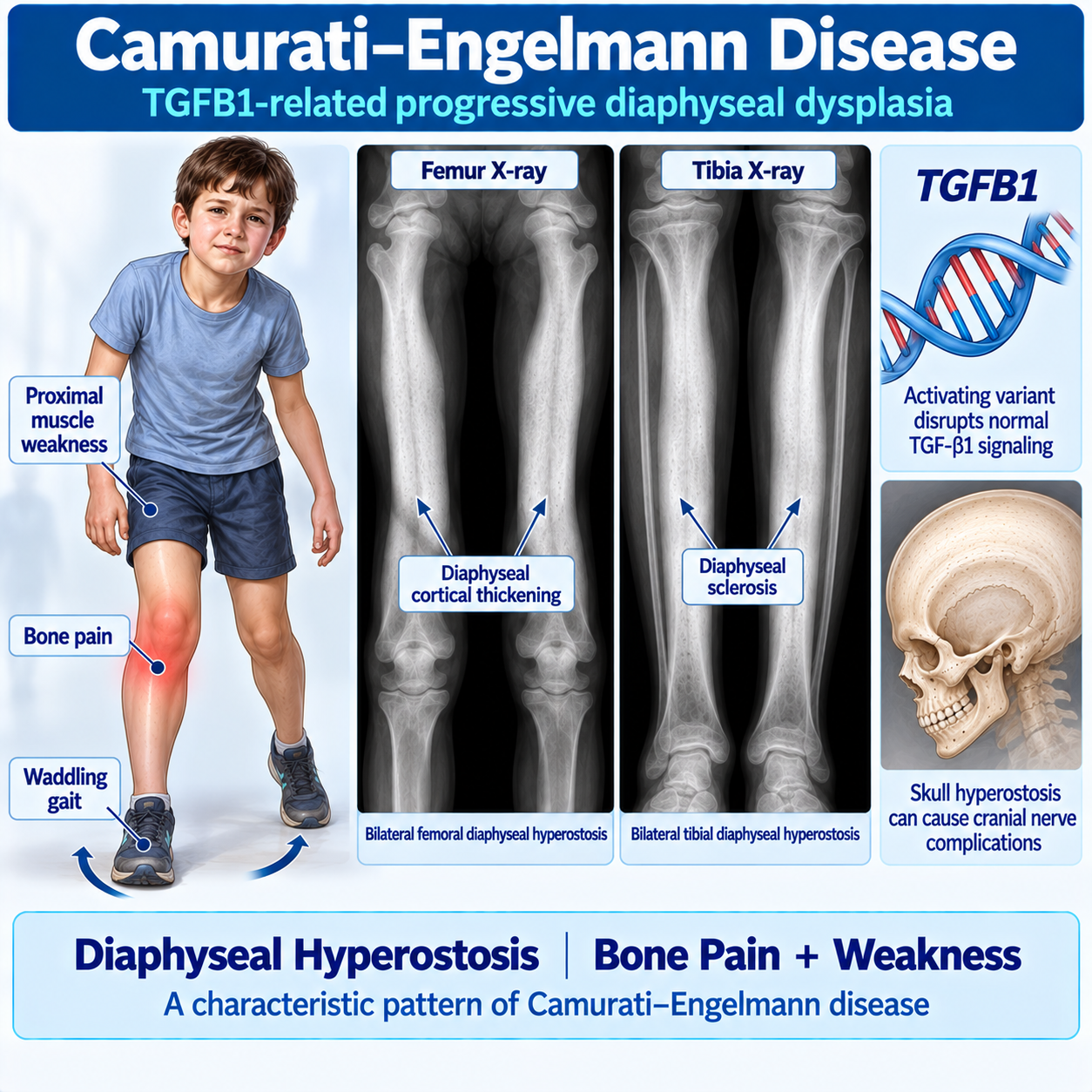
What Is Camurati–Engelmann Disease?
Core Teaching Question
How do TGFB1 pathogenic variants alter bone remodeling, why do they produce progressive diaphyseal hyperostosis, and how can Camurati–Engelmann disease be distinguished from osteopetrosis, pycnodysostosis and other sclerosing bone disorders?
Core Teaching Concept
The entire article revolves around:
TGFB1 OVERACTIVITY ABNORMAL BONE REMODELING DIAPHYSEAL CORTICAL THICKENING PROGRESSIVE HYPEROSTOSIS BONE PAIN + MUSCLE WEAKNESS + GAIT ABNORMALITIESOpening Clinical Scenario
A 10-year-old child develops:
- progressive leg pain
- fatigue
- difficulty running
- waddling gait
Examination reveals:
- proximal muscle weakness
- tenderness over long bones
Radiographs show:
BILATERAL SYMMETRIC DIAPHYSEAL SCLEROSISof both femurs and tibias.
Genetic testing identifies:
TGFB1 PATHOGENIC VARIANTThe diagnosis is:
CAMURATI–ENGELMANN DISEASEWhat Is Camurati–Engelmann Disease?
Camurati–Engelmann Disease (CED) is:
A RARE AUTOSOMAL DOMINANT SCLEROSING BONE DYSPLASIAalso known as:
PROGRESSIVE DIAPHYSEAL DYSPLASIAThe hallmark is:
EXCESSIVE BONE FORMATION ALONG THE DIAPHYSES OF LONG BONESleading to:
- bone pain
- weakness
- gait impairment
- skeletal deformity
Why The Name Progressive Diaphyseal Dysplasia?
Progressive
Disease gradually worsens.
Diaphyseal
Primarily affects the:
DIAPHYSISof long bones.
Dysplasia
Abnormal bone development.
TGFB1 and Bone Remodeling
The TGFB1 Gene
The responsible gene is:
TGFB1Transforming Growth Factor Beta 1
TGF-β1 is an important signaling molecule regulating:
- osteoblasts
- osteoclasts
- bone remodeling
- tissue repair
Normal Bone Remodeling
Normal bone health requires balance between:
Osteoblasts
Build bone.
Osteoclasts
Remove bone.
This balance allows:
CONTINUOUS BONE RENEWALRole Of TGF-β1
TGF-β1 regulates:
- bone formation
- bone resorption
- extracellular matrix production
Normally:
TGF-β1 ACTIVITY IS TIGHTLY CONTROLLEDDisease Mechanism
Most pathogenic variants cause:
EXCESSIVE TGFB1 SIGNALING ABNORMAL REMODELING EXCESSIVE CORTICAL BONE FORMATION DIAPHYSEAL HYPEROSTOSIS NARROWING OF THE MEDULLARY CANALMaster Pathway
TGFB1 PATHOGENIC VARIANT TGF-β1 SIGNALING ↑ REMODELING DYSREGULATION CORTICAL THICKENING DIAPHYSEAL HYPEROSTOSIS PAIN + WEAKNESS + GAIT ABNORMALITY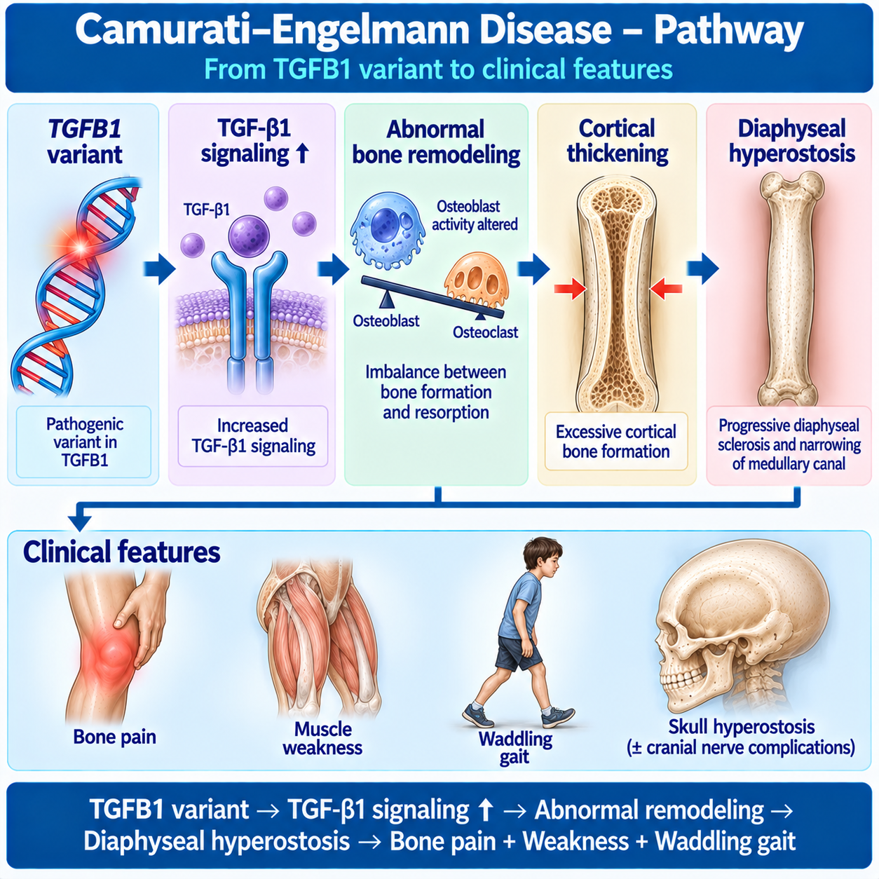
Skeletal Distribution
Which Bones Are Most Affected?
Most commonly:
- femur
- tibia
- fibula
- humerus
- radius
- ulna
Disease is often:
BILATERALand
SYMMETRICDiaphysis Versus Epiphysis
Students must learn:
Diaphysis
Bone shaft.
Metaphysis
Transition zone.
Epiphysis
Bone end.
CED mainly affects:
DIAPHYSESMetaphyses may become involved later.
Epiphyses are often relatively spared.
Clinical Presentation
Bone Pain
Most common symptom.
Pain often affects:
- legs
- thighs
- shins
Pain may worsen with:
- activity
- prolonged walking
Muscle Weakness
Many patients report:
PROXIMAL MUSCLE WEAKNESSCommon findings:
- difficulty climbing stairs
- difficulty rising from floor
- reduced exercise tolerance
Waddling Gait
A classic manifestation.
Caused by:
- pain
- weakness
- skeletal changes
The gait may resemble some neuromuscular disorders.
Fatigue
Patients often complain of:
EASY FATIGABILITYThis may become one of the earliest symptoms.
Limb Tenderness
Affected long bones may be tender.
This helps distinguish disease from purely neurological conditions.
Reduced Muscle Bulk
Some patients develop:
APPARENT MUSCLE WASTINGparticularly around the pelvic girdle.
Childhood Presentation
Many patients present in:
CHILDHOODor
EARLY ADOLESCENCESeverity varies considerably.
Adult Presentation
Some individuals remain undiagnosed until adulthood.
Milder disease can present with:
- chronic leg pain
- unexplained weakness
- incidental radiological abnormalities
Skull and Cranial-Nerve Disease
Skull Involvement
The skull may become affected.
This is important because:
SKULL HYPEROSTOSIS CAN CAUSE COMPLICATIONSCranial Nerve Compression
Progressive skull-base thickening may compress:
- optic nerve
- vestibulocochlear nerve
- facial nerve
leading to:
- visual problems
- hearing loss
- facial symptoms
Headache
Headache may occur due to:
SKULL INVOLVEMENTespecially when hyperostosis becomes extensive.
Hearing Loss
A recognized complication.
Mechanism:
NARROWING OF CRANIAL NERVE CANALSVisual Disturbance
May occur when:
OPTIC NERVE COMPRESSIONdevelops.
This requires urgent specialist evaluation.
Radiology
Radiology Overview
Radiology is central.
Most characteristic finding:
SYMMETRIC DIAPHYSEAL HYPEROSTOSISX-Ray Findings
Common findings:
- cortical thickening
- medullary narrowing
- diaphyseal sclerosis
- bilateral involvement
Femur Radiology
The femur is among the most commonly affected bones.
Students should learn to recognize:
SYMMETRIC FEMORAL SHAFT SCLEROSISTibial Radiology
Tibial involvement is common.
Look for:
CORTICAL THICKENING OF TIBIAL SHAFTSCT Imaging
Useful for:
- skull involvement
- foraminal narrowing
- cranial nerve compression
MRI
May assist in:
- complications
- differential diagnosis
- marrow evaluation
But plain radiographs remain fundamental.
Bone Scan
May demonstrate:
INCREASED ACTIVITY IN AFFECTED DIAPHYSES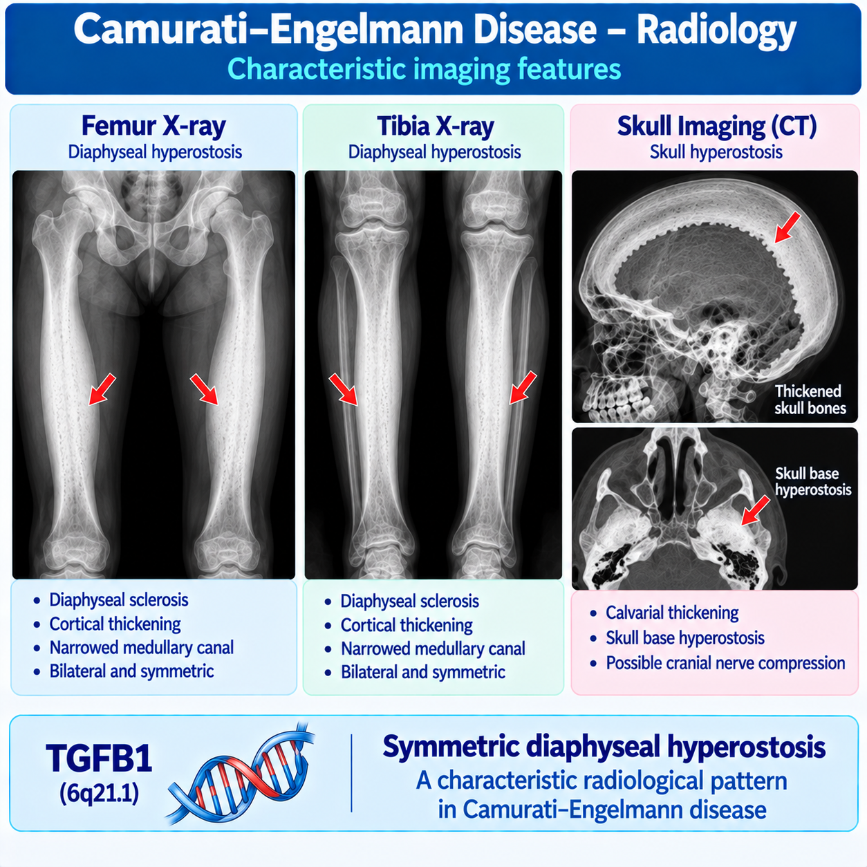
Diagnosis, Genetics and Inheritance
Laboratory Findings
There is no single diagnostic blood test.
Routine labs may be normal.
Some patients show:
- elevated alkaline phosphatase
- elevated inflammatory markers
These findings are nonspecific.
Diagnosis
Diagnosis relies upon:
CLINICAL FEATURES RADIOLOGY TGFB1 TESTINGHigh-Yield Diagnostic Pattern
BONE PAIN MUSCLE WEAKNESS WADDLING GAIT BILATERAL DIAPHYSEAL SCLEROSIS THINK CAMURATI–ENGELMANN DISEASEMolecular Diagnosis
Established by identifying:
PATHOGENIC TGFB1 VARIANTInheritance
Inheritance pattern:
AUTOSOMAL DOMINANTEach child has:
50% RISKof inheriting the variant.
Variable Expression
Severity varies greatly.
Some relatives may have:
- mild symptoms
- severe symptoms
- delayed diagnosis
Differential Diagnosis
Differential Diagnosis
Must include:
- Osteopetrosis
- Pycnodysostosis
- Van Buchem disease
- Sclerosteosis
- Ribbing disease
- Chronic osteomyelitis
Camurati–Engelmann Vs Osteopetrosis
| Feature | CED | Osteopetrosis |
|---|---|---|
| Gene | TGFB1 | Multiple |
| Main lesion | Diaphyseal hyperostosis | Generalized osteosclerosis |
| Distribution | Long-bone shafts | Whole skeleton |
| Pain | Common | Variable |
| Weakness | Common | Less characteristic |
| Marrow failure | Usually absent | Important |
Bottom memory:
DIAPHYSEAL DISEASE → CED GENERALIZED OSTEOSCLEROSIS → OSTEOPETROSISCamurati–Engelmann Vs Pycnodysostosis
| Feature | CED | Pycnodysostosis |
|---|---|---|
| Gene | TGFB1 | CTSK |
| Mechanism | Remodeling dysregulation | Osteoclast matrix degradation defect |
| Acroosteolysis | Absent | Characteristic |
| Open sutures | Not dominant feature | Common |
| Bone pain | Common | Less prominent |
| Diaphyseal sclerosis | Classic | Not classic |
Camurati–Engelmann Vs Ribbing Disease
Important because both show diaphyseal sclerosis.
CED usually:
- childhood onset
- bilateral disease
- TGFB1 mutation
Ribbing disease often:
- adult onset
- asymmetric disease
Treatment Principles
Treatment Principles
No curative therapy currently exists.
Management focuses on:
SYMPTOM CONTROLand
FUNCTION PRESERVATIONCorticosteroids
Glucocorticoids have been reported to improve:
- pain
- function
in some patients.
Losartan
Losartan has been used because of:
TGF-β PATHWAY EFFECTSResults vary.
It is not universally effective.
Pain Management
May include:
- analgesics
- rehabilitation
- activity modification
Physical Therapy
Can help maintain:
- mobility
- strength
- function
Monitoring Skull Disease
Patients with skull involvement require monitoring for:
- hearing loss
- visual symptoms
- cranial nerve dysfunction
Surgical Management
Reserved for selected complications.
Master Diagnostic Algorithm
BONE PAIN LONG-BONE TENDERNESS X-RAYBilateral Diaphyseal Hyperostosis?
If yes:
TGFB1 TESTING CAMURATI–ENGELMANN DISEASECED in One Minute
TGFB1 TGF-β1 OVERACTIVITY DIAPHYSEAL HYPEROSTOSIS BONE PAIN MUSCLE WEAKNESS WADDLING GAITDiagnosis:
RADIOLOGY + TGFB1Inheritance:
AUTOSOMAL DOMINANTMemory:
PAIN + WEAKNESS + DIAPHYSEAL SCLEROSIS = CEDMechanistic Qualification
CED should not be reduced to:
TGFB1 mutation → osteoblasts become overactive → too much bone
That is inadequate.
Use:
TGFB1 PATHOGENIC VARIANT TGF-β1 SIGNALING DYSREGULATION ABNORMAL COUPLING OF BONE FORMATION AND RESORPTION / REMODELING PROGRESSIVE CORTICAL HYPEROSTOSISTGFB1 variant → TGF-β1 signaling ↑ → abnormal remodeling → hyperostosis
but the article text must explain that bone remodeling involves coordinated osteoblast and osteoclast biology.
Sclerosing-Bone Differential Table
| Feature | Camurati–Engelmann | Osteopetrosis | Pycnodysostosis | Fibrous Dysplasia |
|---|---|---|---|---|
| Major molecular driver | TGFB1 | Multiple genes | CTSK | GNAS mosaicism |
| Main biological concept | Remodeling/signaling dysregulation | Osteoclast resorption failure | Osteoclast matrix-degradation failure | Abnormal mosaic bone formation |
| Distribution | Symmetric diaphyseal | Generalized | Generalized osteosclerosis | Usually focal/multifocal |
| Cortical thickening | Prominent | Can occur | Can occur | Different lesion architecture |
| Bone pain | Characteristic | Variable | Variable | Common in symptomatic disease |
| Muscle weakness | Characteristic clue | Not defining | Not defining | Not defining |
| Waddling gait | Characteristic clue | Not defining | Not defining | Not defining |
| Acroosteolysis | No | Not typical | Characteristic | No |
| Marrow failure | Not classic | Important in severe forms | Not typical major feature | No |
| Ground-glass lesions | No | No | No | Characteristic |
| Skull complications | Can occur | Can occur | Craniofacial phenotype | Craniofacial FD possible |
Bottom statement:
DO NOT DIAGNOSE A SCLEROSING BONE DISORDER FROM “DENSE BONE” ALONE — DISTRIBUTION + PHENOTYPE + MECHANISM MATTER.Treatment Evidence Is Limited
Treatment evidence is limited
Camurati–Engelmann disease is rare.
Much of the therapeutic literature consists of:
- case reports;
- small case series;
- observational experience.
Therefore:
REPORTED BENEFIT ≠ ESTABLISHED UNIVERSAL TREATMENTIn particular:
Glucocorticoids
May improve pain and function in some symptomatic patients.
Glucocorticoids should not be described as treatments that:
- cure CED;
- reliably reverse radiographic disease;
- are required in every patient.
Losartan
Has been used in some patients because of its relationship to TGF-β signaling.
Losartan should not be described as a treatment that:
- directly corrects TGFB1;
- reliably prevents progression;
- is universally effective;
- is standard curative treatment.
Surgery
Reserve discussion for selected structural/compressive complications.
Drug doses
DO NOT ADD DOSESunless a later dedicated evidence review specifically establishes that a dose belongs in the article.
Worked Clinical Cases
Case 1 — Child With Leg Pain and Waddling Gait
A 9-year-old child develops gradually progressive pain in both legs, easy fatigability and difficulty keeping up with classmates.
Examination shows:
- waddling gait;
- proximal lower-limb weakness;
- tenderness over the femora.
Radiographs show bilateral symmetric cortical thickening involving the femoral diaphyses.
Most important diagnostic clue
The combination of:
BONE PAIN + MUSCLE WEAKNESS + SYMMETRIC DIAPHYSEAL HYPEROSTOSISstrongly suggests:
CAMURATI–ENGELMANN DISEASEThe diagnosis should then be evaluated in the complete clinical, radiographic and genetic context.
Case 2 — “Growing Pains”
An 11-year-old has experienced bilateral leg pain for several years.
The symptoms were repeatedly attributed to:
“growing pains.”
However, the pain persists and the child develops:
- reduced exercise tolerance;
- muscle weakness;
- abnormal gait.
Important lesson
Persistent bone pain associated with:
OBJECTIVE WEAKNESS OR GAIT ABNORMALITYshould not automatically be dismissed as benign growing pains.
Radiographs may reveal the characteristic diaphyseal abnormalities of CED.
Case 3 — Symmetric Femoral and Tibial Disease
A teenager has chronic bilateral leg pain.
Radiographs demonstrate:
- cortical thickening of both femoral shafts;
- cortical thickening of both tibial shafts;
- narrowing of the medullary canals.
The epiphyses are relatively spared.
Pattern recognition
BILATERAL + SYMMETRIC + DIAPHYSEALis the key pattern.
This distribution strongly supports progressive diaphyseal dysplasia in the appropriate clinical context.
Case 4 — Apparent Neuromuscular Disease
A child is referred because of:
- proximal muscle weakness;
- difficulty climbing stairs;
- waddling gait;
- reduced muscle bulk.
A primary neuromuscular disorder is initially suspected.
However, the patient also reports:
DEEP LONG-BONE PAINand radiographs show bilateral diaphyseal hyperostosis.
Correct principle
CED can produce a phenotype that resembles neuromuscular disease.
Therefore:
WEAKNESS + WADDLING GAIT DOES NOT ALWAYS MEAN PRIMARY MUSCLE DISEASEThe skeletal symptoms and radiographs are critical.
Case 5 — Mild Adult Presentation
A 35-year-old has chronic aching pain in both legs and reduced exercise tolerance.
Radiographs obtained for another reason show symmetric diaphyseal cortical thickening.
The patient has never been diagnosed with a skeletal dysplasia.
Important lesson
Although CED often becomes apparent during childhood or adolescence:
MILD DISEASE MAY REMAIN UNRECOGNIZED UNTIL ADULTHOODAge at diagnosis does not necessarily equal age at biological disease onset.
Case 6 — Hearing Loss
A patient with established CED develops progressive hearing impairment.
Imaging demonstrates substantial skull-base hyperostosis.
Mechanism to consider
Progressive cranial hyperostosis can affect structures surrounding cranial nerves and foramina.
Therefore new:
HEARING LOSSin a patient with CED requires appropriate specialist evaluation.
Hearing problems in CED may arise from different anatomical lesions.
Case 7 — Visual Symptoms
A patient with known CED develops new visual deterioration.
Skull imaging shows extensive hyperostotic disease.
Important principle
New visual symptoms raise concern for:
CRANIAL / OPTIC PATHWAY COMPRESSIONand require prompt specialist assessment.
This requires management beyond that used for uncomplicated long-bone CED.
Case 8 — CED Versus Osteopetrosis
A child has markedly dense bones.
One clinician labels the condition:
“osteopetrosis.”
Closer review shows that the major abnormality is:
- bilateral;
- symmetric;
- predominantly diaphyseal;
- associated with bone pain, weakness and waddling gait.
There is no major marrow-failure phenotype.
Better diagnostic direction
CAMURATI–ENGELMANN DISEASEshould be considered.
Teaching distinction
CED → PREDOMINANTLY DIAPHYSEAL HYPEROSTOSISwhereas:
OSTEOPETROSIS → GENERALIZED OSTEOSCLEROSIS FROM OSTEOCLAST-RESORPTION FAILURECase 9 — CED Versus Pycnodysostosis
A patient has skeletal sclerosis and recurrent skeletal symptoms.
Which finding would strongly redirect the diagnosis toward pycnodysostosis?
ACROOSTEOLYSISespecially when accompanied by:
- short stature;
- delayed cranial-suture closure;
- characteristic jaw/dental findings.
Teaching distinction
DIAPHYSEAL HYPEROSTOSIS + PAIN/WEAKNESS → CEDversus:
OSTEOSCLEROSIS + ACROOSTEOLYSIS → PYCNODYSOSTOSISCase 10 — CED Versus Ribbing-Type Diaphyseal Dysplasia
An adult presents with painful diaphyseal sclerosis.
The disease is relatively localized and asymmetric rather than the classic bilateral symmetric long-bone pattern expected in CED.
Important principle
Not every diaphyseal sclerosing disorder is Camurati–Engelmann disease.
Consider other diaphyseal dysplasias and alternative causes according to:
- age;
- symmetry;
- distribution;
- family history;
- imaging;
- molecular findings.
CED cannot be diagnosed solely from the finding:
DIAPHYSEAL SCLEROSISCase 11 — Mildly Affected Parent
A child has classic symptomatic CED and a pathogenic TGFB1 variant.
The child's parent reports only mild intermittent leg discomfort but carries the familial pathogenic variant.
Explanation
CED shows:
VARIABLE EXPRESSIVITYand reduced penetrance has been described.
Therefore:
THE SAME FAMILY CAN CONTAIN VERY DIFFERENT PHENOTYPESA mildly affected or apparently unaffected parent does not automatically exclude autosomal-dominant transmission.
Case 12 — Genetic Counseling
A molecularly confirmed affected adult with a heterozygous pathogenic TGFB1 variant asks about transmission to future children.
Inheritance
CED is:
AUTOSOMAL DOMINANTTherefore each pregnancy has a:
50% CHANCE OF INHERITING THE PATHOGENIC VARIANTHowever:
INHERITING THE VARIANT DOES NOT PREDICT EXACT DISEASE SEVERITYbecause clinical expression can vary.
Common Mistakes
Mistake 1 — “Camurati–Engelmann disease is another name for osteopetrosis.”
Wrong.
CED is a distinct TGFB1-related sclerosing bone dysplasia characterized particularly by progressive diaphyseal hyperostosis.
Mistake 2 — “Every patient with dense bones has osteopetrosis.”
Wrong.
The distribution and mechanism matter.
CED characteristically produces:
SYMMETRIC DIAPHYSEAL HYPEROSTOSISrather than simply generalized osteosclerosis.
Mistake 3 — “TGFB1 encodes an osteoclast enzyme.”
Wrong.
TGFB1 encodes transforming growth factor beta 1, a signaling molecule involved in numerous biological processes including regulation of bone remodeling.
Mistake 4 — “CED is simply excessive osteoblast activity.”
Oversimplified.
The disease involves dysregulated TGF-β1 signaling and abnormal coupling/remodeling of bone.
Its mechanism cannot be reduced to one cell type acting independently.
Mistake 5 — “Diaphyseal sclerosis alone proves CED.”
Wrong.
Other disorders can produce diaphyseal sclerosis.
Interpret:
- distribution;
- symmetry;
- age;
- clinical phenotype;
- family history;
- molecular findings.
Mistake 6 — “The epiphyses are the primary site of disease.”
Wrong.
The characteristic abnormality predominantly involves the:
DIAPHYSESwith possible extension toward metaphyseal regions.
Mistake 7 — “The disease is usually unilateral.”
Wrong.
Classic CED typically produces:
BILATERAL SYMMETRIClong-bone abnormalities.
Mistake 8 — “Bone pain is the only important symptom.”
Wrong.
The characteristic clinical phenotype can also include:
- proximal muscle weakness;
- reduced muscle bulk;
- fatigue;
- waddling gait.
Mistake 9 — “A waddling gait always means primary muscular disease.”
Wrong.
CED can cause waddling gait through the combined effects of skeletal disease, pain and muscle weakness.
Mistake 10 — “Normal routine laboratory tests exclude CED.”
Wrong.
There is no single routine biochemical marker that excludes or confirms the disorder.
Diagnosis depends heavily on:
PHENOTYPE + RADIOLOGY + GENETICSMistake 11 — “Skull involvement must be present to diagnose CED.”
Wrong.
Skull involvement is variable and is not required in every patient.
Mistake 12 — “Skull hyperostosis is clinically unimportant.”
Wrong.
It can lead to important cranial complications, including hearing and visual problems and other cranial-nerve manifestations.
Mistake 13 — “Marrow failure is a classic major manifestation of CED.”
Wrong.
This is much more characteristic of severe osteopetrosis and is useful in distinguishing the disorders.
Mistake 14 — “Acroosteolysis is a characteristic feature of CED.”
Wrong.
Acroosteolysis is a particularly important clue to:
PYCNODYSOSTOSISnot CED.
Mistake 15 — “An unaffected-looking parent excludes inherited CED.”
Wrong.
Clinical expression can be variable, and reduced penetrance has been described.
Mistake 16 — “Every affected child must have an affected parent.”
Wrong.
De novo pathogenic variants can occur.
Mistake 17 — “Losartan cures CED.”
Wrong.
Losartan has been used in some patients based partly on its effects on TGF-β signaling, but evidence is limited and response is variable.
It is not curative therapy.
Mistake 18 — “Glucocorticoids cure the genetic disease.”
Wrong.
They may improve symptoms in selected patients but do not correct the underlying TGFB1 pathogenic variant.
Mistake 19 — “Surgery is routine treatment for long-bone disease.”
Wrong.
Management is individualized. Surgery is principally relevant to selected complications and should not be presented as universal therapy.
Mistake 20 — “A positive TGFB1 result tells us exactly how severe the disease will become.”
Wrong.
Genotype alone does not provide a precise individual severity forecast.
Clinical expression can vary substantially.
Frequently Asked Questions
What is Camurati–Engelmann disease?
Camurati–Engelmann disease is a rare inherited sclerosing bone dysplasia characterized particularly by progressive hyperostosis of the diaphyses of long bones. Common manifestations include bone pain, muscle weakness, reduced exercise tolerance and waddling gait. ---
What is another name for Camurati–Engelmann disease?
It is also known as: PROGRESSIVE DIAPHYSEAL DYSPLASIA The name reflects its characteristic progressive involvement of long-bone shafts. ---
What gene causes Camurati–Engelmann disease?
Most molecularly confirmed classic cases are caused by heterozygous pathogenic variants in: TGFB1 which encodes transforming growth factor beta 1. ---
What does TGF-β1 normally do in bone?
TGF-β1 is an important signaling molecule involved in regulation of bone remodeling and communication among cells involved in skeletal turnover. CED-associated TGFB1 variants disturb normal regulation of this pathway. ---
Why does CED cause bone pain?
Affected long bones undergo abnormal remodeling with progressive cortical thickening and hyperostosis. The exact relationship between the structural changes and pain is complex, but bone pain is one of the characteristic clinical manifestations. ---
What does “diaphyseal dysplasia” mean?
The: DIAPHYSIS is the shaft of a long bone. CED characteristically produces abnormal cortical thickening and sclerosis predominantly involving these long-bone shafts. ---
Which bones are commonly affected?
Long bones are particularly important, including the: femur; tibia; fibula; humerus; radius; ulna. The pattern is often bilateral and symmetric. ---
Why do patients develop muscle weakness and waddling gait?
CED can produce proximal muscle weakness, reduced muscle bulk, pain and impaired mobility. Together these can result in a characteristic: WADDLING GAIT The weakness should not automatically be interpreted as a primary muscular disease. ---
Can Camurati–Engelmann disease affect the skull?
Yes. Skull involvement is variable but can produce hyperostosis, particularly involving the skull base. Severe cranial disease can produce clinically important compressive complications. ---
Can CED cause hearing or visual problems?
Yes. Skull-base hyperostosis can affect cranial nerves or surrounding foramina and may contribute to hearing impairment, visual problems and other neurological manifestations. New hearing, visual or neurological symptoms require appropriate specialist assessment. ---
What does the radiograph show?
The characteristic pattern is: BILATERAL SYMMETRIC DIAPHYSEAL CORTICAL THICKENING AND SCLEROSIS with narrowing of the medullary cavity. The pattern and distribution are more informative than simply describing the bones as “dense.” ---
How is Camurati–Engelmann disease diagnosed?
Diagnosis is based on the combination of: CLINICAL PHENOTYPE CHARACTERISTIC RADIOGRAPHIC FINDINGS MOLECULAR TESTING FOR TGFB1 where appropriate. No single routine biochemical blood test establishes the diagnosis. ---
How is CED inherited?
CED is usually: AUTOSOMAL DOMINANT An affected individual carrying a heterozygous pathogenic TGFB1 variant has a 50% chance of transmitting that variant in each pregnancy. Disease severity can differ substantially among affected relatives. ---
How is Camurati–Engelmann disease treated?
There is no established therapy that corrects the underlying genetic defect. Management is individualized and may include: symptom control; pain management; rehabilitation/physical therapy; selected medical therapy such as glucocorticoids or losartan under specialist supervision; assessment and management of cranial complications. Do not present one regimen as universally effective. ---
How is CED different from osteopetrosis and pycnodysostosis?
The characteristic pattern of CED is: TGFB1 + SYMMETRIC DIAPHYSEAL HYPEROSTOSIS + BONE PAIN/MUSCLE WEAKNESS Osteopetrosis is a broader group of disorders characterized by impaired osteoclast-mediated bone resorption and generalized osteosclerosis. Pycnodysostosis is caused by CTSK-related cathepsin K deficiency and is particularly associated with: OSTEOSCLEROSIS + ACROOSTEOLYSIS + SHORT STATURE + CRANIOFACIAL/DENTAL FEATURES
Key Take-Home Messages
The article's permanent mechanistic memory should be:
TGFB1 PATHOGENIC VARIANT TGF-β1 SIGNALING DYSREGULATION ABNORMAL BONE REMODELING SYMMETRIC DIAPHYSEAL CORTICAL HYPEROSTOSIS BONE PAIN + PROXIMAL MUSCLE WEAKNESS + WADDLING GAITThe radiological memory:
BILATERAL + SYMMETRIC + DIAPHYSEAL = THINK CAMURATI–ENGELMANNThe differential memory:
GENERALIZED OSTEOSCLEROSIS → CONSIDER OSTEOPETROSIS OSTEOSCLEROSIS + ACROOSTEOLYSIS → PYCNODYSOSTOSIS FOCAL/MULTIFOCAL GROUND-GLASS BONE LESIONS → FIBROUS DYSPLASIA SYMMETRIC DIAPHYSEAL HYPEROSTOSIS + PAIN/WEAKNESS → CAMURATI–ENGELMANN