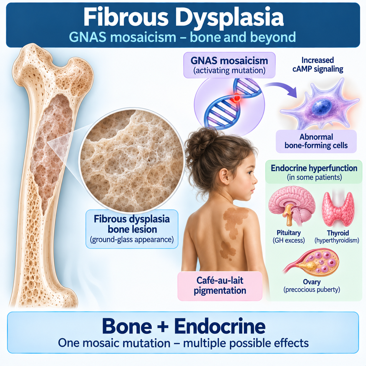
One mosaic mutation, different affected tissues
Postzygotic GNAS activation → Gsα/cAMP signaling ↑ → abnormal function of affected cells.
Bone: fibrous dysplasia · Skin: café-au-lait pigmentation · Endocrine tissue: autonomous hyperfunction
What Is Fibrous Dysplasia?
Central Concept
ONE MOSAIC MUTATION → DIFFERENT AFFECTED TISSUES → DIFFERENT MANIFESTATIONSCentral pathway:
POSTZYGOTIC ACTIVATING GNAS VARIANT Gsα SIGNALING ↑ cAMP SIGNALING ↑ ABNORMAL FUNCTION OF AFFECTED CELLSBone
FIBROUS DYSPLASIASkin
CAFÉ-AU-LAIT PIGMENTATIONEndocrine tissue
AUTONOMOUS ENDOCRINE HYPERFUNCTIONTogether, this spectrum is:
FD/MASFD/MAS is caused by somatic gain-of-function GNAS variants and therefore has a mosaic distribution. (PubMed Central (PMC))
Opening — Use This Content
A child develops a painless expansile bone lesion.
Radiography shows a characteristic:
GROUND-GLASS APPEARANCEAnother patient has multiple skeletal lesions, recurrent fractures and deformity.
Another presents with:
- café-au-lait pigmentation;
- precocious puberty;
- hyperthyroidism;
- or growth-hormone excess.
At first these findings may appear unrelated.
But they can result from the same biological event:
A POSTZYGOTIC ACTIVATING GNAS VARIANTThe mutation occurs after fertilization.
Therefore only some cells inherit it.
This creates:
SOMATIC MOSAICISMIf affected cells are mainly skeletal, the patient may present primarily with:
FIBROUS DYSPLASIAIf multiple tissues are involved, manifestations may include:
FIBROUS DYSPLASIA + SKIN PIGMENTATION + ENDOCRINE HYPERFUNCTIONforming the spectrum traditionally called:
McCUNE–ALBRIGHT SYNDROMEThe phenotype depends largely on:
WHICH TISSUES CONTAIN THE MUTATIONand:
HOW EXTENSIVELY THEY ARE INVOLVEDWhat Is Fibrous Dysplasia?
Fibrous dysplasia is a mosaic skeletal disorder in which affected bone is replaced by abnormal:
FIBRO-OSSEOUS TISSUENormal lamellar bone architecture is disrupted.
Affected bone may become:
- structurally weak;
- expanded;
- deformed;
- painful;
- susceptible to fracture.
Important:
FIBROUS DYSPLASIA IS NOT SIMPLY “FIBROSIS OF BONE”It is a developmental disorder of skeletal cells caused by mosaic GNAS activation.
Normal Bone Versus Fibrous Dysplasia
Normal bone depends on coordinated activity of:
- skeletal progenitor cells;
- osteoblasts;
- osteocytes;
- osteoclasts;
- mineralization pathways.
In fibrous dysplasia, affected skeletal progenitor cells fail to produce normal mature bone architecture.
Instead, normal bone and marrow are replaced to varying degrees by:
ABNORMAL FIBRO-OSSEOUS TISSUEThis compromises normal skeletal structure.
GNAS and cAMP Signaling
GNAS
The major molecular driver is:
GNASGNAS encodes the stimulatory G-protein alpha subunit:
GsαGsα participates in receptor-mediated signaling through:
ADENYLYL CYCLASE cAMPActivating GNAS variants cause inappropriate persistent signaling.
Therefore:
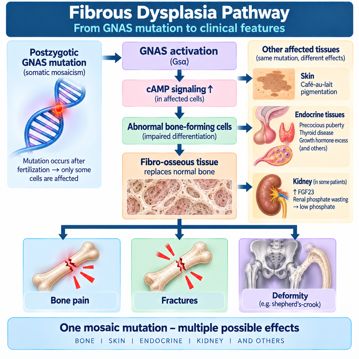
GNAS ACTIVATION Gsα ACTIVITY ↑ cAMP ↑ CELLULAR FUNCTION BECOMES AUTONOMOUS / DYSREGULATEDThis explains both skeletal and endocrine manifestations. (PubMed Central (PMC))
Why Is The Mutation Mosaic?
The pathogenic variant occurs:
AFTER FERTILIZATIONTherefore the embryo already contains multiple cells when the mutation arises.
Only descendants of the mutated cell carry the variant.
Thus:
SOME CELLS = MUTATED OTHER CELLS = NORMALThis is:
SOMATIC MOSAICISMTiming Matters
A useful conceptual principle is:
Earlier postzygotic mutation
Potentially:
MORE TISSUES INVOLVEDLater postzygotic mutation
Potentially:
MORE LIMITED DISTRIBUTIONBut do not present this as a precise rule predicting an individual patient's phenotype.
The key memory is:
MOSAIC DISTRIBUTION EXPLAINS VARIABLE DISEASEIs Fd/Mas Inherited?
This deserves a prominent callout:
FD/MAS IS NOT A CLASSIC INHERITED MENDELIAN DISORDERThe pathogenic GNAS variant is postzygotic and somatic.
GeneReviews reports no verified vertical transmission and considers recurrence risk to siblings essentially similar to the general population. (NCBI)
Therefore:
A PATIENT'S MOSAIC GNAS MUTATION IS NOT SIMPLY “PASSED DOWN” LIKE AUTOSOMAL-DOMINANT OIThis makes a useful contrast with the preceding skeletal genetics articles.
The FD/MAS Spectrum
Disease Spectrum
Use:
FD/MAS IS A SPECTRUMPotential manifestations include:
Skeleton
Fibrous dysplasia
Skin
Café-au-lait pigmentation
Gonads
Autonomous sex-steroid production
Thyroid
Thyroid lesions/hyperthyroidism
Pituitary
Growth-hormone excess ± hyperprolactinemia
Kidney/bone
FGF23-mediated renal phosphate wasting
Adrenal gland
Rare neonatal hypercortisolism
Not every patient has every feature.
Monostotic Fibrous Dysplasia
Monostotic FD means:
ONE SKELETAL SITE IS INVOLVEDIt may be:
- discovered incidentally;
- identified after pain;
- identified after fracture;
- detected because of swelling or deformity.
It does not automatically imply systemic McCune–Albright syndrome.
Polyostotic Fibrous Dysplasia
Polyostotic FD means:
MULTIPLE SKELETAL SITES ARE INVOLVEDPatients may have greater risk of:
- deformity;
- fractures;
- scoliosis;
- mobility problems;
- clinically important skeletal burden.
Disease burden varies greatly.
Mccune–Albright Syndrome
McCune–Albright syndrome represents the multisystem end of the FD/MAS spectrum.
Classic teaching emphasizes the triad:
FIBROUS DYSPLASIA CAFÉ-AU-LAIT PIGMENTATION PRECOCIOUS PUBERTYBut:
THE COMPLETE TRIAD IS NOT REQUIRED IN EVERY PATIENTModern understanding recognizes a broader mosaic GNAS disorder involving various endocrine and non-endocrine tissues. (NCBI)
Café-Au-Lait Pigmentation
Skin lesions may be present at birth or appear early.
They are typically:
- hyperpigmented;
- irregularly bordered;
- often segmental;
- related to mosaic distribution.
A classic descriptive term for the irregular border is:
“COAST OF MAINE”Café-au-lait lesions occur in other disorders.
Why Endocrine Hyperfunction Occurs
The same activating GNAS signaling that affects skeletal cells can produce autonomous activity in endocrine tissue.
Simplified:
GNAS ACTIVATION cAMP SIGNALING ↑ ENDOCRINE CELL ACTIVITY BECOMES INAPPROPRIATELY AUTONOMOUSThis may produce:
- estrogen excess;
- testosterone excess;
- thyroid hormone excess;
- growth-hormone excess;
- cortisol excess in rare neonatal disease.
Therefore:
MAS IS A MOSAIC SIGNALING DISORDER — NOT A COLLECTION OF UNRELATED ENDOCRINE DISEASESEndocrine Manifestations
Precocious Puberty In Girls
This is one of the classic manifestations.
Autonomous ovarian cyst activity can produce episodic:
ESTROGEN EXCESSleading to:
- breast development;
- vaginal bleeding;
- accelerated growth;
- advancement of bone age.
The process can be intermittent.
Therefore a single normal estradiol measurement does not necessarily exclude prior autonomous ovarian activity.
Why Precocious Puberty Matters
Repeated estrogen exposure can accelerate:
BONE AGEIf progression is substantial:
EPIPHYSEAL MATURATION ACCELERATES FINAL ADULT HEIGHT MAY BE COMPROMISEDCurrent GeneReviews emphasizes that treatment decisions focus particularly on preventing clinically important bone-age advancement rather than simply treating every isolated bleeding episode. (NCBI)
Gonadal Disease In Boys
Boys may develop:
- testicular lesions;
- macro-orchidism;
- Sertoli/Leydig cell abnormalities;
- autonomous testosterone production in some cases.
Not every structural testicular lesion produces precocious puberty.
Therefore:
STRUCTURAL GONADAL ABNORMALITY ≠ AUTOMATIC HORMONAL HYPERFUNCTIONRoutine biopsy is not appropriate simply because a typical lesion exists; atypical, enlarging or concerning lesions require specialist evaluation. (NCBI)
Thyroid Disease
Thyroid involvement may include:
- heterogeneous gland changes;
- nodules;
- hyperthyroidism.
Autonomous thyroid function results from activating GNAS signaling.
Untreated hyperthyroidism may worsen:
- bone turnover;
- skeletal morbidity;
- cardiovascular burden.
Therefore endocrine disease should not be regarded as unrelated to the skeletal phenotype.
Growth-Hormone Excess
MAS can involve:
GH EXCESSoften with hyperprolactinemia.
Clinical effects may include:
- accelerated growth;
- acromegalic features;
- worsening craniofacial FD;
- increased skull-base morbidity.
This is especially important because:
UNCONTROLLED GH EXCESS CAN WORSEN CRANIOFACIAL DISEASEThe international consensus emphasizes control of GH excess, and GeneReviews describes medical therapy as central; pituitary surgery is challenging because disease is often diffuse and skull-base FD complicates access. (PubMed Central (PMC))
Avoid Pituitary Radiotherapy When Possible
This requires careful wording.
Radiation exposure involving FD tissue has historically raised concern about malignant transformation.
Therefore radiotherapy for MAS-associated pituitary disease should generally be avoided where effective alternatives exist and reserved for exceptional specialist circumstances.
The international consensus and GeneReviews treat radiation as a last-resort approach. (PubMed Central (PMC))
Neonatal Hypercortisolism
Rare infants with extensive MAS may develop:
CORTISOL EXCESSThis can be severe and potentially life-threatening.
It is not a common manifestation.
FGF23 and Renal Phosphate Wasting
FGF23 And Fibrous Dysplasia
This is an essential connection to the existing phosphate cluster.
FD lesions can produce excessive:
FGF23FGF23 acts in the kidney to:
REDUCE PHOSPHATE REABSORPTIONTherefore:
FD BURDEN / FGF23 ↑ RENAL PHOSPHATE WASTING SERUM PHOSPHATE ↓ MINERALIZATION IMPAIRED BONE PAIN / FRACTURE RISK / OSTEOMALACIA MAY WORSENImportant Nuance: FGF23 Level Versus Phosphate Wasting
Not every patient with FD has hypophosphatemia.
Clinically important phosphate wasting is more likely with substantial disease burden, but the phenotype varies.
The student should assess:
SERUM PHOSPHATEand, when indicated:
RENAL PHOSPHATE HANDLINGrather than assuming phosphate status from the diagnosis alone.
Why Phosphate Wasting Matters
Hypophosphatemia can independently worsen:
- bone pain;
- weakness;
- fractures;
- mineralization;
- skeletal deformity.
Therefore:
DO NOT ATTRIBUTE ALL BONE PAIN TO THE FD LESION ITSELFThe patient may have a treatable superimposed:
FGF23-MEDIATED PHOSPHATE-WASTING DISORDERGeneReviews specifically emphasizes correcting inadequately treated hypophosphatemia before considering bisphosphonate treatment for FD-related pain. (NCBI)
- Renal Phosphate Wasting Explained
- FGF23 Disorders Explained
- Hypophosphatemia Explained
- Osteomalacia Explained
Pain, Fractures and Deformity
Bone Pain
Bone pain is common but variable.
Potential contributors include:
- active FD lesions;
- fractures;
- stress injury;
- deformity;
- mechanical loading;
- arthritis;
- muscle dysfunction;
- hypophosphatemia;
- other unrelated pain disorders.
Therefore:
NEW OR CHANGING PAIN REQUIRES A CAUSE — NOT JUST THE LABEL “FD”Fractures
FD bone may be mechanically weak.
Fractures may occur particularly in weight-bearing bones.
Potential consequences include:
- pain;
- deformity;
- loss of mobility;
- progressive angulation.
Repeated fractures around the proximal femur are particularly important.
Shepherd'S-Crook Deformity
A classic severe proximal femoral deformity is:
SHEPHERD'S-CROOK DEFORMITYIt reflects progressive:
COXA VARA + PROXIMAL FEMORAL BOWINGassociated with mechanically weak dysplastic bone.
This is an important radiological and orthopedic teaching point.
Scoliosis
Spinal FD can contribute to:
SCOLIOSISProgression may occur, particularly with substantial skeletal disease.
Severe scoliosis can impair:
- posture;
- function;
- respiratory mechanics.
Craniofacial Fibrous Dysplasia
Craniofacial Fibrous Dysplasia
Craniofacial FD can involve:
- skull;
- skull base;
- facial bones;
- orbit;
- paranasal sinuses.
It may cause:
- facial asymmetry;
- expansion;
- pain;
- dental abnormalities;
- hearing problems;
- visual concerns.
However:
RADIOGRAPHIC EXTENT DOES NOT ALWAYS EQUAL FUNCTIONAL IMPAIRMENTThis is especially important for optic-nerve encasement.
Optic-Nerve Encasement
Craniofacial FD may surround the:
OPTIC CANALImaging can therefore appear alarming.
But:
OPTIC-NERVE ENCASEMENT IS OFTEN ASYMPTOMATICTherefore:
ENCASEMENT ≠ OPTIC NEUROPATHYThis distinction is essential. (NCBI)
Do Not Prophylactically Decompress An Asymptomatic Optic Nerve
NO OPTIC NEUROPATHY → DO NOT DECOMPRESS SIMPLY BECAUSE CT SHOWS ENCASEMENTProphylactic optic-nerve decompression in an asymptomatic patient is contraindicated because intervention itself can worsen visual outcome.
Patients with actual optic neuropathy require expert craniofacial/neuro-ophthalmological assessment. (PubMed Central (PMC))
This is one of the article's highest-yield clinical messages.
Hearing
Craniofacial FD may affect auditory structures.
Potential problems include:
- conductive hearing loss;
- sensorineural hearing impairment;
- external auditory canal narrowing.
Symptoms should trigger appropriate audiological/ENT evaluation.
Radiology
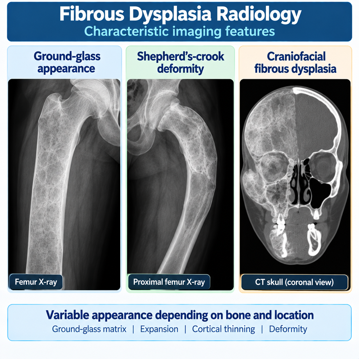
Radiology — Central Section
Radiographic appearance varies according to:
- age;
- skeletal location;
- lesion composition.
The classic descriptor is:
GROUND-GLASS MATRIXOther features can include:
- expansile lesions;
- cortical thinning;
- bone enlargement;
- deformity;
- heterogeneous density.
Why “Ground Glass”?
The abnormal mixture of:
- immature woven bone;
- fibrous stroma;
- variable mineralization;
produces a relatively homogeneous hazy radiographic density traditionally described as:
GROUND GLASSIt is a radiological descriptor, not the underlying pathology itself.
Plain Radiography
Plain radiographs are useful for:
- initial lesion characterization;
- fractures;
- long-bone deformity;
- follow-up of specific orthopedic problems.
Characteristic appearances can strongly support FD in the correct clinical context.
CT
CT is particularly useful for:
CRANIOFACIAL FDbecause it defines:
- lesion anatomy;
- skull-base involvement;
- orbital relationships;
- sinus involvement;
- cranial foramina.
Imaging should answer a clinical question.
MRI
MRI may be useful when evaluating:
- soft-tissue relationships;
- neurological complications;
- atypical lesions;
- possible cystic change;
- suspected malignant transformation.
FD signal characteristics can be variable.
Therefore:
MRI APPEARANCE ALONE MAY BE NONSPECIFICBone Scintigraphy
Bone scintigraphy can help determine:
SKELETAL DISEASE DISTRIBUTIONparticularly when evaluating polyostotic involvement.
The international consensus includes skeletal burden assessment as part of staging. (PubMed Central (PMC))
Diagnosis, Biopsy and Genetic Testing
Biopsy
Biopsy is not mandatory for every classic lesion.
It becomes more useful when:
- imaging is atypical;
- diagnosis is uncertain;
- an isolated lesion lacks convincing clinical context;
- malignancy or another pathology is suspected.
The principle is:
CLASSIC CLINICAL + RADIOLOGICAL FD MAY NOT NEED BIOPSYbut:
ATYPICAL OR CONCERNING LESIONS MAYHistology
Histology classically demonstrates irregular trabeculae of woven bone within fibrous stroma.
A traditional descriptive phrase is:
“CHINESE LETTER” TRABECULAEBut do not overemphasize this phrase as if it alone establishes diagnosis.
The complete clinicoradiological context matters.
Genetic Testing
Molecular testing may identify an activating:
GNAS VARIANTBut mosaicism creates an important limitation.
Peripheral blood may contain:
NO DETECTABLE MUTANT CELLSeven when the lesion itself carries the mutation.
Therefore:
NEGATIVE BLOOD TEST ≠ NO FD/MASAffected tissue generally has greater diagnostic yield than blood.
GeneReviews emphasizes that molecular sensitivity depends heavily on the tissue tested and assay methodology. (NCBI)
Diagnosis
Diagnosis integrates:
CLINICAL PHENOTYPE RADIOLOGY ENDOCRINE ASSESSMENT BIOCHEMISTRY GENETIC / HISTOLOGICAL CONFIRMATION WHEN NEEDEDInitial Skeletal Assessment
The clinical assessment should determine:
- monostotic versus polyostotic disease;
- pain;
- previous fractures;
- deformity;
- gait/function;
- scoliosis;
- craniofacial involvement;
- dental symptoms.
Biochemistry should particularly consider:
- calcium;
- phosphate;
- renal function;
- ALP;
- vitamin-D context;
- PTH where indicated;
- renal phosphate wasting where phosphate is low.
Endocrine Assessment
The endocrine phenotype should be assessed based on:
- age;
- symptoms;
- growth pattern;
- pubertal development;
- thyroid findings;
- craniofacial disease;
- other clinical clues.
Malignant Transformation and Red Flags
Malignant Transformation
FD lesions are overwhelmingly:
BENIGNMalignant transformation is:
RAREPossible malignancies include sarcomatous transformation.
Instead teach:
A NEW CHANGE IN A STABLE LESION REQUIRES ATTENTIONRed Flags
Concerning features include:
- new rapidly progressive pain;
- rapidly enlarging mass;
- new swelling;
- new neurological deficit;
- unexpected destructive imaging changes;
- soft-tissue mass.
These require specialist assessment and appropriate imaging/biopsy.
Radiation Exposure
Radiotherapy involving FD lesions has historically been associated with concern for malignant transformation.
Therefore:
THERAPEUTIC RADIATION SHOULD GENERALLY BE AVOIDED WHEN EFFECTIVE ALTERNATIVES EXISTThis is particularly relevant in MAS-associated pituitary disease. (PubMed Central (PMC))
Clinical Staging
Management Principles
There is currently no established routinely used medication that eradicates GNAS-mutant skeletal lesions or reliably reverses the overall FD disease course.
Management therefore focuses on:
- preserving function;
- treating fractures;
- correcting deformity;
- controlling pain;
- correcting phosphate wasting;
- controlling endocrine hyperfunction;
- protecting vision/hearing;
- dental care;
- monitoring clinically important complications.
The international consensus emphasizes multidisciplinary management. (PubMed Central (PMC))
Treat Endocrine Disease
This is a major skeletal-management principle:
ENDOCRINE DISEASE CAN WORSEN BONE DISEASETherefore identify and appropriately manage:
- precocious puberty;
- hyperthyroidism;
- GH excess;
- clinically important phosphate wasting;
- other relevant endocrinopathies.
Treat Phosphate Wasting
Clinically significant renal phosphate wasting should be addressed because untreated hypophosphatemia can worsen:
- pain;
- weakness;
- mineralization;
- fracture risk.
The exact phosphate/active-vitamin-D regimen belongs under specialist metabolic-bone management and should not be invented here.
Internal link prominently to:
and:
Treatment Principles
Bisphosphonates
Bisphosphonates are antiresorptive drugs.
In FD:
THE MAIN SUPPORTED ROLE IS MANAGEMENT OF PERSISTENT FD-RELATED BONE PAIN IN SELECTED PATIENTSThey have not been shown to:
ERADICATE FD LESIONSor:
RELIABLY STOP DISEASE PROGRESSIONGeneReviews specifically states that bisphosphonates have not been shown to affect progression and limits their role primarily to FD-related bone pain. (NCBI)
Before Bisphosphonate Treatment
Before attributing pain to FD and moving toward antiresorptive therapy, evaluate potentially correctable contributors such as:
HYPOPHOSPHATEMIAInadequately treated phosphate wasting can substantially worsen bone pain. (NCBI)
Also assess:
- fracture/stress fracture;
- mechanical deformity;
- vitamin-D status where clinically appropriate;
- other causes of pain.
Oral Versus IV Bisphosphonate Evidence
The educational message is:
- IV bisphosphonates have been used for significant FD-related pain;
- oral bisphosphonate trials have not demonstrated convincing benefit for FD pain;
- BMD changes should not be mistaken for evidence that the underlying lesion has been cured.
Denosumab
Denosumab is:
NOT ROUTINE STANDARD THERAPY FOR FD/MASIt inhibits RANKL and can markedly suppress lesion activity and bone turnover.
Research has shown potentially substantial effects on lesion activity and pain, but discontinuation can produce:
REBOUND BONE TURNOVERand:
SEVERE HYPERCALCEMIAGeneReviews describes promising adult data but emphasizes important safety concerns and ongoing study. A small 2026 pediatric open-label study also reported improvements, but only five patients were studied, so it does not establish routine pediatric therapy. (NCBI)
Therefore:
PROMISING ≠ STANDARD OF CARESurgery
Surgery may be needed for:
- fractures;
- progressive deformity;
- mechanical dysfunction;
- selected craniofacial problems;
- severe scoliosis.
Surgery should focus on:
FUNCTION + MECHANICAL STABILITYnot simply removing every radiographically abnormal area.
Curettage And Bone Grafting
Simple curettage of extensive FD lesions often has limited long-term effectiveness because abnormal tissue can persist or recur.
In craniofacial disease, the international consensus specifically advises against simple curettage as a routine strategy because it is ineffective and may increase complications. (PubMed Central (PMC))
Proximal Femoral Disease
Proximal femoral FD deserves special attention because repeated loading can drive:
- fracture;
- progressive varus;
- bowing;
- shepherd's-crook deformity.
Orthopedic management aims to preserve:
ALIGNMENT + STABILITY + FUNCTIONCraniofacial Surgery
Surgery may be appropriate for:
- functional impairment;
- significant deformity;
- progressive symptomatic disease;
- selected compressive complications.
But:
DO NOT OPERATE ON CT APPEARANCE ALONEThe goal is preservation/restoration of function and appropriate correction of deformity.
Pain Management
Pain management should begin by asking:
WHY DOES THIS PATIENT HURT?Possible causes:
- FD lesion activity;
- fracture;
- stress fracture;
- deformity;
- arthritis;
- muscular/mechanical pain;
- hypophosphatemia;
- neuropathic pain;
- unrelated pathology.
Management can include:
- treating the cause;
- rehabilitation;
- appropriate analgesia;
- selected bisphosphonate therapy for persistent FD-related pain;
- multidisciplinary pain care where necessary.
Rehabilitation
Physical therapy and rehabilitation can support:
- muscle strength;
- gait;
- balance;
- mobility;
- function;
- recovery after fractures/surgery;
- adaptation to deformity.
The goal is:
SAFE FUNCTION, NOT FEAR-BASED IMMOBILITYPregnancy
Pregnancy can occur in women with FD/MAS.
Clinical management should consider:
- endocrine status;
- skeletal burden;
- pain;
- mobility;
- previous fractures;
- obstetric factors.
Management should be individualized.
Surveillance
Surveillance should be:
MANIFESTATION-DEPENDENTnot identical for every patient.
Important domains include:
- skeletal pain/function;
- deformity;
- fracture;
- scoliosis;
- phosphate status;
- growth/puberty in children;
- thyroid disease;
- GH excess where relevant;
- vision/hearing in craniofacial disease.
For craniofacial FD, GeneReviews recommends neuro-ophthalmologic assessment and notes that CT frequency should be individualized, with approximately five-year intervals suggested in stable contexts and earlier imaging when clinically indicated. (NCBI)
Master Pathophysiology Algorithm
POSTZYGOTIC GNAS ACTIVATION Gsα SIGNALING ↑ cAMP ↑Three branches:
BONE
ABNORMAL SKELETAL PROGENITOR FUNCTION FIBRO-OSSEOUS LESIONPain / fracture / deformity
ENDOCRINE
AUTONOMOUS HORMONE PRODUCTIONPrecocious puberty / hyperthyroidism / GH excess / others
SKIN
MOSAIC PIGMENTATIONCafé-au-lait lesions
Bottom:
ONE MOSAIC SIGNALING DEFECT — MANY TISSUESMaster Diagnostic Algorithm
SUSPECTED FIBROUS DYSPLASIA CLINICAL HISTORY + EXAMINATIONPain?
Fracture?
Deformity?
Café-au-lait?
Early puberty?
Thyroid/GH clues?
RADIOGRAPHYGround-glass?
Expansion?
Deformity?
DEFINE SKELETAL EXTENTMonostotic?
Polyostotic?
Craniofacial?
BONE / MINERAL BIOCHEMISTRYEspecially:
Phosphate
Phosphate low?
ASSESS RENAL PHOSPHATE WASTING / FGF23 CONTEXT ASSESS ENDOCRINE MANIFESTATIONSbased on phenotype.
Classic phenotype?
Clinical + radiological diagnosis may be sufficient.
Atypical / uncertain?
BIOPSY ± GNAS TESTING DEFINE FD/MAS PHENOTYPE MANAGE SKELETAL + ENDOCRINE + FUNCTIONAL COMPLICATIONSCraniofacial Decision Pathway
CRANIOFACIAL FD VISION NORMAL?Yes
Even if optic canal is encased:
OBSERVE + CLINICAL / NEURO-OPHTHALMIC FOLLOW-UP NO PROPHYLACTIC OPTIC-NERVE DECOMPRESSIONNo — optic neuropathy suspected
URGENT EXPERT EVALUATIONDetermine cause and appropriate intervention.
Bottom:
TREAT FUNCTION — NOT THE CT IMAGE(NCBI)
Differential Comparisons
Fd Versus Osteogenesis Imperfecta
| Feature | Fibrous dysplasia | Osteogenesis imperfecta |
|---|---|---|
| Molecular pattern | Somatic mosaic GNAS activation | Usually germline genetic disorder |
| Main skeletal problem | Focal/multifocal fibro-osseous lesions | Generalized collagen-matrix fragility |
| Distribution | Mosaic | Systemic |
| Ground-glass lesions | Characteristic | Not typical |
| Blue sclerae | Not typical | Important clue |
| DI | Not defining | May occur |
| Endocrine hyperfunction | MAS spectrum | Not defining |
| Inheritance | Not classically inherited | Often Mendelian |
| Major pathway | Gsα/cAMP | Type I collagen |
Fd Versus Hypophosphatasia
| Feature | Fibrous dysplasia | Hypophosphatasia |
|---|---|---|
| Main defect | Mosaic skeletal-cell dysregulation | TNSALP deficiency |
| Gene | GNAS | ALPL |
| ALP | May reflect skeletal activity; not persistently low defining feature | Persistently low is key |
| Lesions | Focal/multifocal | Generalized metabolic phenotype |
| Ground glass | Characteristic FD clue | Not defining |
| FGF23 | Can be excessive | Not primary mechanism |
| Hypophosphatemia | Can occur from renal wasting | Not defining mechanism |
| Targeted therapy | No established lesion-eradicating therapy | Asfotase alfa in appropriate disease |
Fd Versus Osteopetrosis
| Feature | Fibrous dysplasia | Osteopetrosis |
|---|---|---|
| Main mechanism | Abnormal mosaic bone-forming cells | Osteoclast resorption failure |
| Distribution | Focal/multifocal | Usually generalized skeletal phenotype |
| Density | Ground-glass/variable | Diffuse osteosclerosis |
| Marrow failure | Not classic | Severe forms |
| Optic issue | Craniofacial FD/optic canal | Skull-base sclerosis/nerve compression |
| Endocrine disease | MAS | Not defining |
| HSCT | No | Selected severe forms |
Fd Versus Paget Disease
| Feature | Fibrous dysplasia | Paget disease |
|---|---|---|
| Typical onset | Childhood/young life lesion development | Usually older adults |
| Molecular context | Mosaic GNAS | Different remodeling disorder |
| Distribution | Monostotic/polyostotic | Monostotic/polyostotic |
| Ground-glass lesion | Classic clue | Not classic pattern |
| Endocrine hyperfunction | MAS possible | Not defining |
| Café-au-lait | May occur | No |
Internal link to the existing Paget article.
Worked Clinical Cases
Case 1 — Ground-glass lesion
A teenager has an incidental expansile femoral lesion with ground-glass matrix.
Key thought: fibrous dysplasia.
Determine skeletal extent and clinical context before assuming MAS.
Case 2 — One bone only
Patient has a classic FD lesion confined to one rib and no extraskeletal manifestations.
Classification
MONOSTOTIC FIBROUS DYSPLASIANot automatically McCune–Albright syndrome.
Case 3 — Multiple bones
Child has lesions involving femur, pelvis and skull.
Classification
POLYOSTOTIC FIBROUS DYSPLASIAAssess endocrine and phosphate abnormalities.
Case 4 — Early vaginal bleeding
Young girl has café-au-lait pigmentation and episodic vaginal bleeding with ovarian cyst activity.
Mechanism
AUTONOMOUS OVARIAN ESTROGEN PRODUCTIONConsider MAS.
Case 5 — Low phosphate
Patient with extensive polyostotic FD develops bone pain and hypophosphatemia.
Wrong approach
“Pain is simply from the lesions.”
Better approach
ASSESS FGF23-MEDIATED RENAL PHOSPHATE WASTINGCase 6 — Optic canal encasement
CT shows craniofacial FD completely surrounding an optic canal, but visual examination is normal.
Wrong action
Automatic prophylactic decompression.
Correct principle
ENCASEMENT ≠ OPTIC NEUROPATHYObserve with appropriate specialist follow-up. (NCBI)
Case 7 — Progressive visual loss
Patient with craniofacial FD develops objective deterioration in vision.
Action
URGENT EXPERT CRANIOFACIAL / NEURO-OPHTHALMOLOGICAL ASSESSMENTThis is different from asymptomatic radiological encasement.
Case 8 — Shepherd's crook
Patient with polyostotic FD develops progressive proximal femoral varus and bowing.
Diagnosis
SHEPHERD'S-CROOK DEFORMITYOrthopedic mechanical assessment is required.
Case 9 — Bone pain and bisphosphonate request
Adult with FD requests bisphosphonates to “remove the lesion.”
Answer
Bisphosphonates may have a role for selected persistent FD-related bone pain but:
THEY DO NOT ERADICATE FD OR RELIABLY STOP LESION PROGRESSION(NCBI)
Case 10 — Denosumab request
Patient asks whether denosumab is now established treatment.
Answer
NOResearch is promising, but important rebound mineral-metabolism risks remain and evidence is insufficient for routine standard use. (NCBI)
Case 11 — Negative blood GNAS test
Patient has convincing polyostotic FD but peripheral-blood testing is negative.
Wrong conclusion
“No GNAS mutation, therefore no FD.”
Correct principle
MOSAICISM CAN MAKE BLOOD TESTING NEGATIVEAffected tissue may have higher diagnostic yield.
Case 12 — Skull FD + GH excess
Patient has progressive craniofacial FD and elevated GH/IGF-1.
Lesson
ENDOCRINE HYPERFUNCTION CAN DRIVE SKELETAL MORBIDITYControl of GH excess is an important component of skeletal management.
Common Mistakes
Mistake 1
Fibrous dysplasia is simply fibrosis of bone. This is incorrect.
Mistake 2
FD is a germline inherited GNAS disorder. This is incorrect.
Mistake 3
Every child of a patient with FD/MAS has a high inheritance risk. This is incorrect.
Mistake 4
Every FD patient has McCune–Albright syndrome. This is incorrect.
Mistake 5
The complete classic MAS triad is required for diagnosis. This is incorrect.
Mistake 6
Monostotic FD means mild MAS. This is incorrect.
Mistake 7
Every FD lesion has an identical ground-glass appearance. This is incorrect.
Mistake 8
Ground-glass appearance alone proves FD in every setting. This is incorrect.
Mistake 9
Every classic FD lesion requires biopsy. This is incorrect.
Mistake 10
A negative blood GNAS test excludes FD/MAS. This is incorrect.
Mistake 11
All FD-related pain comes directly from the lesion. This is incorrect.
Mistake 12
Hypophosphatemia is unrelated to FD. This is incorrect.
Mistake 13
Every patient with FD has phosphate wasting. This is incorrect.
Mistake 14
Optic-canal encasement means the patient needs decompression. This is incorrect.
Mistake 15
Prophylactic optic-nerve decompression protects vision. This is incorrect.
Mistake 16
Bisphosphonates remove FD lesions. This is incorrect.
Mistake 17
Bisphosphonates are proven to stop FD progression. This is incorrect.
Mistake 18
Denosumab is now routine first-line FD therapy. This is incorrect.
Mistake 19
Endocrine abnormalities are unrelated to skeletal disease. This is incorrect.
Mistake 20
FD/MAS is only a bone disorder. This is incorrect.
FD/MAS in One Minute
FD/MAS IN ONE MINUTE POSTZYGOTIC GNAS ACTIVATION Gsα ↑ cAMP ↑Three major tissues:
Bone
FIBROUS DYSPLASIAGround glass
Pain
Fracture
Deformity
Skin
CAFÉ-AU-LAITEndocrine
AUTONOMOUS HYPERFUNCTIONPrecocious puberty
Hyperthyroidism
GH excess
Also remember:
FD → FGF23 ↑ → RENAL PHOSPHATE WASTINGCraniofacial rule:
OPTIC CANAL ENCASEMENT ≠ OPTIC NEUROPATHYTreatment:
TREAT COMPLICATIONS + ENDOCRINE DRIVERS + PHOSPHATE WASTINGNot:
“REMOVE EVERY LESION”Final:
ONE MOSAIC MUTATION — MANY TISSUESFrequently Asked Questions
What is fibrous dysplasia?
Fibrous dysplasia is a mosaic skeletal disorder in which normal bone is replaced by abnormal fibro-osseous tissue due to postzygotic activating GNAS variants.
What causes fibrous dysplasia?
It is caused by somatic activating variants in GNAS that increase Gsα/cAMP signaling in affected cells.
Is fibrous dysplasia inherited?
FD/MAS is generally not inherited because the GNAS mutation arises postzygotically and produces somatic mosaicism.
What is monostotic fibrous dysplasia?
It is fibrous dysplasia involving a single skeletal site.
What is polyostotic fibrous dysplasia?
It is fibrous dysplasia involving multiple skeletal sites.
What is McCune–Albright syndrome?
It is the multisystem FD/MAS phenotype in which mosaic GNAS activation can produce fibrous dysplasia together with skin pigmentation and/or autonomous endocrine abnormalities.
What is the classic radiological appearance?
A ground-glass matrix is a characteristic imaging feature, although appearance varies with age, site and lesion composition.
What is shepherd's-crook deformity?
It is progressive proximal femoral varus and bowing associated with significant FD involvement.
Can FD cause low phosphate?
Yes. FD lesions can overproduce FGF23, leading to renal phosphate wasting and hypophosphatemia in some patients.
Why does phosphate wasting matter?
Hypophosphatemia can worsen mineralization, bone pain, weakness, fractures and deformity.
Does optic-nerve encasement require surgery?
No. Asymptomatic optic-nerve encasement is common, and prophylactic decompression without optic neuropathy is contraindicated.
Do bisphosphonates cure fibrous dysplasia?
No. They may be used for selected FD-related bone pain, but they have not been shown to eradicate lesions or stop disease progression.
Is denosumab standard treatment?
No. It remains an investigational/specialist approach. Potential benefits have been reported, but rebound bone turnover and severe hypercalcemia are important concerns.
Does a negative blood GNAS test exclude FD/MAS?
No. Because the disease is mosaic, the pathogenic variant may not be detectable in peripheral blood.
Can fibrous dysplasia become malignant?
Malignant transformation is rare. New rapidly progressive pain, swelling, mass formation, neurological deficit or destructive imaging changes warrant specialist investigation.
Key Take-Home Messages
Fibrous dysplasia/McCune–Albright syndrome begins with:
POSTZYGOTIC GNAS ACTIVATIONTherefore:
THE DISEASE IS MOSAICAffected skeletal cells produce:
ABNORMAL FIBRO-OSSEOUS TISSUEleading to:
GROUND-GLASS LESIONS FRACTURES DEFORMITY PAINThe same mutation in endocrine tissues can cause:
AUTONOMOUS HORMONE PRODUCTIONTherefore always think beyond the skeleton.
The same skeletal lesions can also increase:
FGF23leading to:
RENAL PHOSPHATE WASTINGwhich may independently worsen skeletal disease.
For craniofacial FD:
OPTIC-NERVE ENCASEMENT ≠ OPTIC NEUROPATHYand:
DO NOT OPERATE ON AN IMAGE ALONEFor treatment:
CONTROL ENDOCRINE DISEASE CORRECT IMPORTANT PHOSPHATE WASTING TREAT FRACTURES AND DEFORMITY PRESERVE FUNCTION MANAGE PAINBisphosphonates:
MAY HELP SELECTED BONE PAINbut:
DO NOT CURE FDDenosumab:
PROMISING ≠ ESTABLISHED ROUTINE THERAPYAnd the final memory statement:
FD/MAS IS ONE MOSAIC GNAS SIGNALING DISORDER EXPRESSED DIFFERENTLY IN BONE, SKIN AND ENDOCRINE TISSUES.