Dense bone does not necessarily mean strong bone
Osteoclast function ↓ → bone resorption ↓ → abnormal bone accumulation.
FRACTURES + MARROW-SPACE LOSS + NERVE COMPRESSION
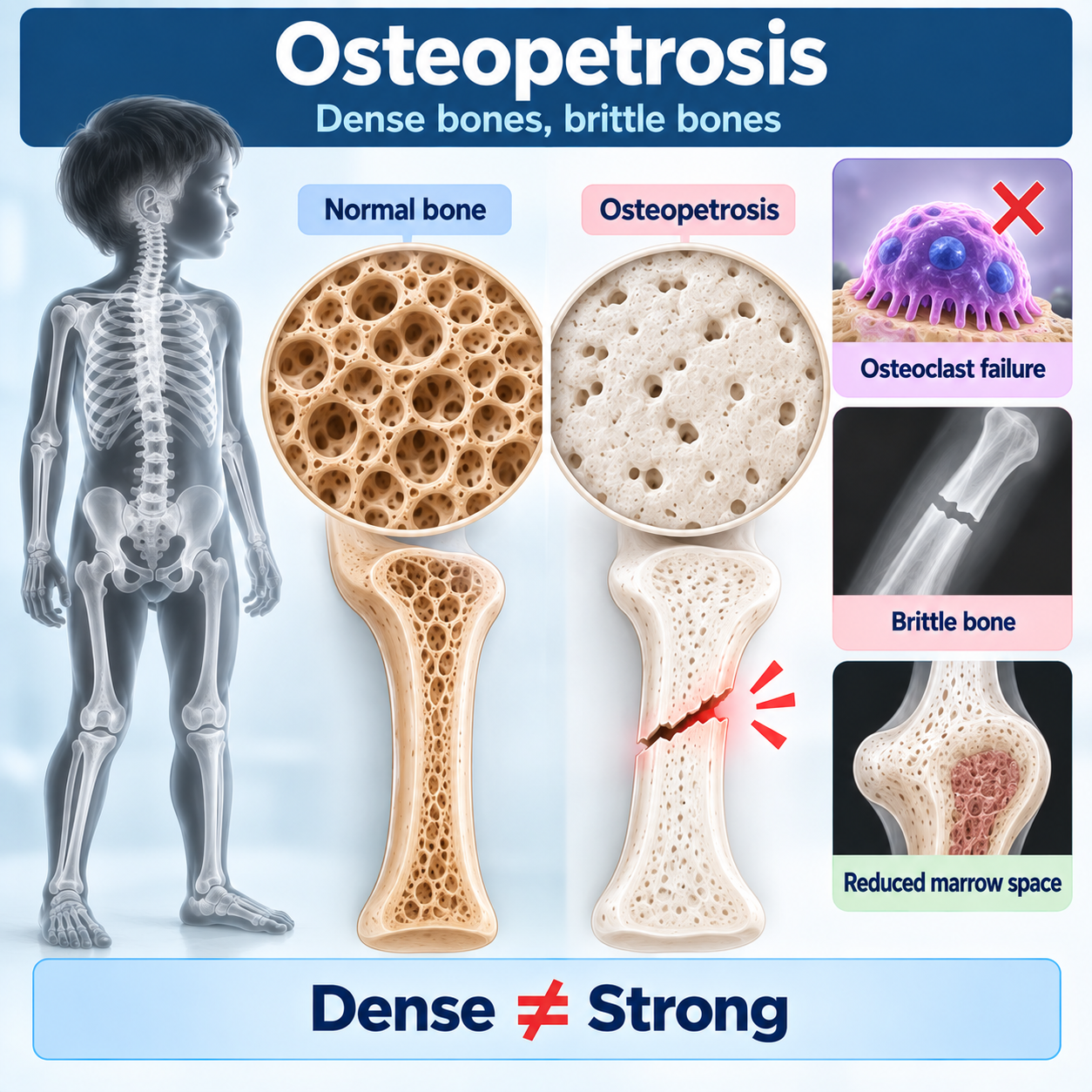
What Is Osteopetrosis?
Central Teaching Concept
The entire article should revolve around one apparent paradox:
DENSE BONE ≠ STRONG BONEOsteopetrosis is fundamentally a disorder in which:
BONE RESORPTION FAILSThe central pathway is:
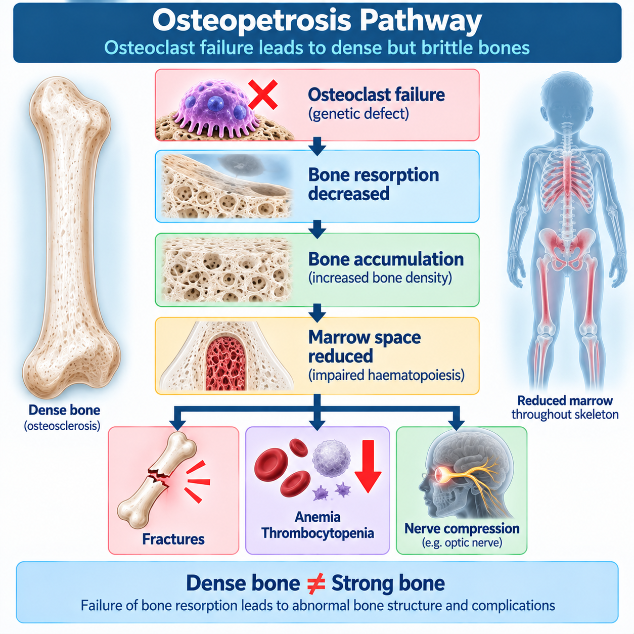
OSTEOCLAST FUNCTION ↓ BONE RESORPTION ↓ OLD BONE IS NOT REMOVED NORMALLY BONE REMODELING FAILS SKELETAL DENSITY ↑but:
BONE QUALITY AND ARCHITECTURE ARE ABNORMAL FRACTURES + MARROW-SPACE LOSS + NERVE COMPRESSIONThis is the article's central memory.
Opening — Use This Content
A radiograph shows extraordinarily dense bones.
At first glance, this might appear protective.
More bone should mean stronger bone.
But the patient has:
- recurrent fractures;
- anemia;
- thrombocytopenia;
- hepatosplenomegaly;
- visual impairment;
- or cranial-nerve abnormalities.
This apparent contradiction is one of the most memorable concepts in metabolic bone disease:
OSTEOPETROSISOsteopetrosis is a genetically heterogeneous group of disorders characterized by abnormally increased skeletal density caused predominantly by impaired:
OSTEOCLAST-MEDIATED BONE RESORPTIONNormal bone requires continuous remodeling.
Osteoblasts form bone.
Osteoclasts remove bone.
If osteoclast resorption fails, bone accumulates—but the accumulated bone is structurally abnormal.
Therefore:
MORE BONE DOES NOT NECESSARILY MEAN BETTER BONESevere disease can also prevent normal expansion of the marrow cavity and narrow cranial foramina.
This produces the distinctive combination:
DENSE BONES FRACTURES BONE MARROW FAILURE CRANIAL-NERVE COMPRESSIONNormal Bone Remodeling
Normal Bone Remodeling
Normal adult bone is not static.
It undergoes continuous:
REMODELINGTwo major cell types cooperate.
Osteoclasts
RESORB BONEOsteoblasts
FORM BONEThe simplified remodeling cycle is:
ACTIVATION OSTEOCLAST RESORPTION REVERSAL OSTEOBLAST FORMATION MINERALIZATIONNormal skeletal architecture depends on balance between these processes.
Why Bone Must Be Resorbed
Bone resorption is not inherently harmful.
Osteoclast activity is necessary for:
- skeletal modeling during growth;
- remodeling of old bone;
- repair of microscopic damage;
- shaping of long bones;
- formation and maintenance of marrow spaces;
- adaptation of skeletal architecture.
Therefore:
TO BUILD NORMAL BONE, THE BODY MUST ALSO REMOVE BONEThis is a central physiological lesson.
What Osteoclasts Do
What Is An Osteoclast?
Osteoclasts are large multinucleated cells derived from:
HEMATOPOIETIC / MONOCYTE-MACROPHAGE LINEAGE PRECURSORSTheir function requires:
- attachment to the bone surface;
- formation of a sealed resorption compartment;
- acidification;
- dissolution of mineral;
- degradation of organic matrix.
This hematopoietic origin later explains why:
HSCT CAN TREAT SOME FORMS OF OSTEOPETROSISHow Osteoclasts Resorb Bone
The osteoclast creates a specialized:
RUFFLED BORDERagainst the bone surface.
Hydrogen ions are transported into the resorption compartment.
This creates an acidic environment that helps dissolve:
HYDROXYAPATITEThe organic matrix is then degraded by proteolytic mechanisms.
Therefore normal osteoclast function requires:
ACID GENERATION PROTON TRANSPORT ION BALANCE MATRIX DEGRADATIONDefects in these processes can produce osteopetrosis.
How Osteopetrosis Develops
TCIRG1 Mechanism
One of the most important genes in severe autosomal-recessive osteopetrosis is:
TCIRG1TCIRG1 encodes the a3 subunit of the vacuolar proton pump involved in osteoclast acidification.
Loss of normal function impairs acidification of the osteoclast resorption compartment.
Therefore:
TCIRG1 DEFECT OSTEOCLAST ACIDIFICATION ↓ BONE RESORPTION ↓ OSTEOPETROSISThe 2025 GeneReview describes TCIRG1-related disease as ranging from severe infantile osteopetrosis to milder phenotypes. (NCBI)
CLCN7 Mechanism
Another major gene is:
CLCN7It encodes a chloride/proton exchanger important for lysosomal and osteoclast function.
CLCN7-related disease spans:
- severe autosomal-recessive osteopetrosis;
- intermediate osteopetrosis;
- autosomal-dominant osteopetrosis type II.
Therefore:
ONE GENE CAN PRODUCE DIFFERENT CLINICAL SEVERITIESCLCN7-related osteopetrosis is an important example of why classification cannot rely only on the gene name. (NCBI)
Do Not Turn The Article Into A Gene Catalogue
Mention that additional genes can cause osteopetrosis and related osteoclast disorders.
The important principle is:
OSTEOPETROSIS IS GENETICALLY HETEROGENEOUSDifferent genetic defects can affect:
- osteoclast development;
- osteoclast acidification;
- ion transport;
- vesicular function;
- osteoclast-bone interaction;
- signaling pathways.
The Central Paradox
Create a prominent callout:
WHY ARE DENSE BONES BRITTLE?Bone strength is not determined simply by:
HOW MUCH BONE IS PRESENTIt also depends on:
- architecture;
- remodeling;
- microdamage repair;
- material quality;
- geometry.
In osteopetrosis, old and abnormal bone is retained.
Normal modeling and remodeling are impaired.
Therefore the skeleton can become:
RADIOGRAPHICALLY DENSEyet:
MECHANICALLY FRAGILEThis explains pathological fractures despite osteosclerosis.
Genetics of Osteopetrosis
Mention that additional genes can cause osteopetrosis and related osteoclast disorders.
The important principle is:
OSTEOPETROSIS IS GENETICALLY HETEROGENEOUSDifferent genetic defects can affect:
- osteoclast development;
- osteoclast acidification;
- ion transport;
- vesicular function;
- osteoclast-bone interaction;
- signaling pathways.
Clinical Spectrum
Osteopetrosis Is Not Osteoporosis
The names sound similar but describe almost opposite radiographic phenotypes.
Osteoporosis
BONE MASS ↓Osteopetrosis
BONE DENSITY ↑But both may cause:
FRACTURESTherefore:
BONE DENSITY ALONE DOES NOT EQUAL BONE STRENGTHClinical Spectrum
Osteopetrosis should be taught as:
A SPECTRUMAt one extreme:
SEVERE INFANTILE AUTOSOMAL-RECESSIVE OSTEOPETROSISAt the other:
MILD AUTOSOMAL-DOMINANT ADULT DISEASEBetween them:
INTERMEDIATE FORMSSeverity depends substantially on the molecular cause and residual osteoclast function.
Major Clinical Categories
| Form | Typical onset | General severity | Common inheritance pattern |
|---|---|---|---|
| Severe infantile osteopetrosis | Infancy | Severe / potentially life-threatening | Usually autosomal recessive |
| Intermediate osteopetrosis | Childhood | Variable | AR or AD depending on cause |
| Autosomal dominant osteopetrosis | Later childhood/adolescence/adulthood | Usually milder | Autosomal dominant |
Immediately state:
THIS IS A CLINICAL FRAMEWORK, NOT A COMPLETE GENETIC CLASSIFICATIONSevere Infantile Osteopetrosis
Severe infantile disease may present early with:
- failure to thrive or growth deficiency;
- pathological fractures;
- macrocephaly;
- abnormal craniofacial appearance;
- anemia;
- thrombocytopenia;
- hepatosplenomegaly;
- recurrent infection;
- hypocalcemia;
- secondary hyperparathyroidism;
- visual impairment;
- hearing abnormalities;
- delayed tooth eruption;
- neurological complications.
Severe TCIRG1-related disease frequently presents in infancy and can cause early death without successful treatment. (NCBI)
Bone Marrow Failure
Bone Marrow Failure
This is a major teaching section.
Normally the interior of many bones contains:
HEMATOPOIETIC MARROWIn severe osteopetrosis, failure of normal bone resorption prevents adequate development/maintenance of:
MARROW SPACEThe sequence is:
OSTEOCLAST FAILURE MEDULLARY CAVITY NARROWS HEMATOPOIETIC SPACE ↓ BLOOD-CELL PRODUCTION ↓ ANEMIA + THROMBOCYTOPENIA ± OTHER CYTOPENIASThis is why a skeletal disease can become:
A HEMATOLOGICAL EMERGENCYExtramedullary Hematopoiesis
When bone marrow cannot adequately support hematopoiesis, the body may increase blood-cell production outside normal marrow.
This is:
EXTRAMEDULLARY HEMATOPOIESISImportant sites include:
- liver;
- spleen.
Therefore:
MARROW FAILURE EXTRAMEDULLARY HEMATOPOIESIS HEPATOSPLENOMEGALYThis is a classic severe-disease pathway. (NCBI)
Anemia
Anemia may result from reduced effective marrow space.
Clinical consequences can include:
- pallor;
- fatigue;
- poor growth;
- cardiorespiratory stress in severe disease.
Thrombocytopenia
Reduced marrow function may also produce:
THROMBOCYTOPENIAThis increases bleeding risk.
Cranial-Nerve Compression
Why Cranial Nerves Are Affected
Bone sclerosis also affects:
THE SKULL BASEAs bone accumulates, foramina through which nerves pass may become narrowed.
Therefore:
SKULL-BASE SCLEROSIS NEURAL FORAMINA NARROW CRANIAL-NERVE COMPRESSIONPotential consequences include:
- visual impairment;
- hearing impairment;
- facial nerve dysfunction.
Visual Impairment
Visual loss is one of the most serious complications of severe infantile osteopetrosis.
Mechanisms can include:
OPTIC NERVE COMPRESSIONand, in some molecular forms:
PRIMARY RETINAL / NEUROLOGICAL DISEASEThis distinction matters.
If vision loss is due to irreversible optic atrophy or primary neurological disease:
HSCT MAY NOT RESTORE ITCLCN7 GeneReviews specifically notes that established cranial-nerve dysfunction is usually irreversible and that primary neurological disease in neuronopathic forms is not corrected by HSCT. (NCBI)
Why Early Treatment Matters
Create this callout:
PREVENTION OF NERVE DAMAGE IS EASIER THAN REVERSALSuccessful correction of osteoclast function may improve skeletal and marrow abnormalities.
But once:
OPTIC NERVE ATROPHYhas developed, vision may not recover.
This is one reason severe infantile disease requires:
EARLY SPECIALIST EVALUATIONHearing Loss
Hearing impairment can result from:
- skull-base sclerosis;
- narrowing of auditory pathways;
- cranial-nerve involvement.
Some genetic forms can have additional mechanisms.
Hypocalcemia and Secondary Hyperparathyroidism
Hypocalcemia
A patient with extraordinarily dense bones may paradoxically develop:
HYPOCALCEMIAWhy?
Because impaired osteoclastic resorption reduces normal release of mineral from bone.
In severe disease this can contribute to:
- hypocalcemia;
- neuromuscular symptoms;
- seizures.
TCIRG1-related disease can present with hypocalcemia and seizures. (NCBI)
Secondary Hyperparathyroidism
The physiological response to hypocalcemia is:
PTH ↑Therefore:
OSTEOCLAST DYSFUNCTION CALCIUM AVAILABILITY ↓ HYPOCALCEMIA PTH ↑ SECONDARY HYPERPARATHYROIDISMBut because osteoclast function is intrinsically impaired, simply increasing PTH cannot fully normalize bone resorption.
Osteopetrorickets
This is an important paradox.
Some children with osteopetrosis can develop:
RICKETS DESPITE VERY DENSE BONESThis is sometimes termed:
OSTEOPETRORICKETSThe concept reflects disturbed calcium/mineral physiology superimposed on defective osteoclast resorption.
Therefore:
RADIOGRAPHIC DENSITY DOES NOT GUARANTEE NORMAL MINERAL HOMEOSTASISCalcium and calcitriol management can be difficult and must be individualized. CLCN7 and TCIRG1 references both emphasize the complexity of calcium management. (NCBI)
Fractures and Orthopedic Problems
Fractures
Despite increased skeletal density:
FRACTURES ARE COMMONThey may occur after relatively minor trauma.
The reason is:
ABNORMAL REMODELING + ABNORMAL ARCHITECTURErather than insufficient radiographic density.
This should repeatedly reinforce:
DENSE ≠ STRONGOrthopedic Challenges
Osteopetrotic bone presents special surgical challenges.
It may be:
- very hard to drill;
- brittle;
- prone to iatrogenic fracture;
- slow to heal in some settings;
- associated with increased infection risk.
Therefore orthopedic procedures should involve clinicians familiar with osteopetrosis.
The 2025 TCIRG1 GeneReview specifically notes resistance to drilling and increased iatrogenic-fracture risk during operative repair. (NCBI)
Dental and Neurological Disease
Dental Manifestations
Possible dental problems include:
- delayed tooth eruption;
- abnormal tooth development;
- caries;
- infection;
- jaw osteomyelitis;
- impaired healing after dental procedures.
Dental care is particularly important because sclerotic poorly remodeled jaw bone may be vulnerable to difficult infection.
CLCN7-related ADOII is particularly associated with mandibular osteomyelitis risk. (NCBI)
Neurological Disease
Not all neurological abnormalities in osteopetrosis are simply due to:
NERVE COMPRESSIONSome molecular forms may include:
PRIMARY NEURODEGENERATIONThis is extremely important when considering HSCT.
Correcting donor-derived osteoclasts cannot necessarily correct an intrinsic neurological defect in other cell populations.
Therefore:
GENOTYPE MATTERS BEFORE TRANSPLANTATIONAutosomal Dominant Osteopetrosis
Autosomal Dominant Osteopetrosis
Milder autosomal-dominant disease often presents later.
Patients may have:
- incidental osteosclerosis;
- fractures;
- bone pain;
- scoliosis;
- degenerative joint disease;
- mandibular osteomyelitis;
- characteristic radiographic findings.
Bone marrow failure and severe cranial-nerve compression are much less typical than in severe infantile ARO.
Albers-Schönberg Disease
Autosomal dominant osteopetrosis type II is traditionally called:
ALBERS-SCHÖNBERG DISEASEIt is commonly associated with heterozygous:
CLCN7pathogenic variants.
Typical onset is later childhood or adolescence, and manifestations may include fractures, scoliosis, hip osteoarthritis and mandibular osteomyelitis. (NCBI)
Osteopetrosis Radiology
Radiology — Major Section
Radiology is central to recognizing osteopetrosis.
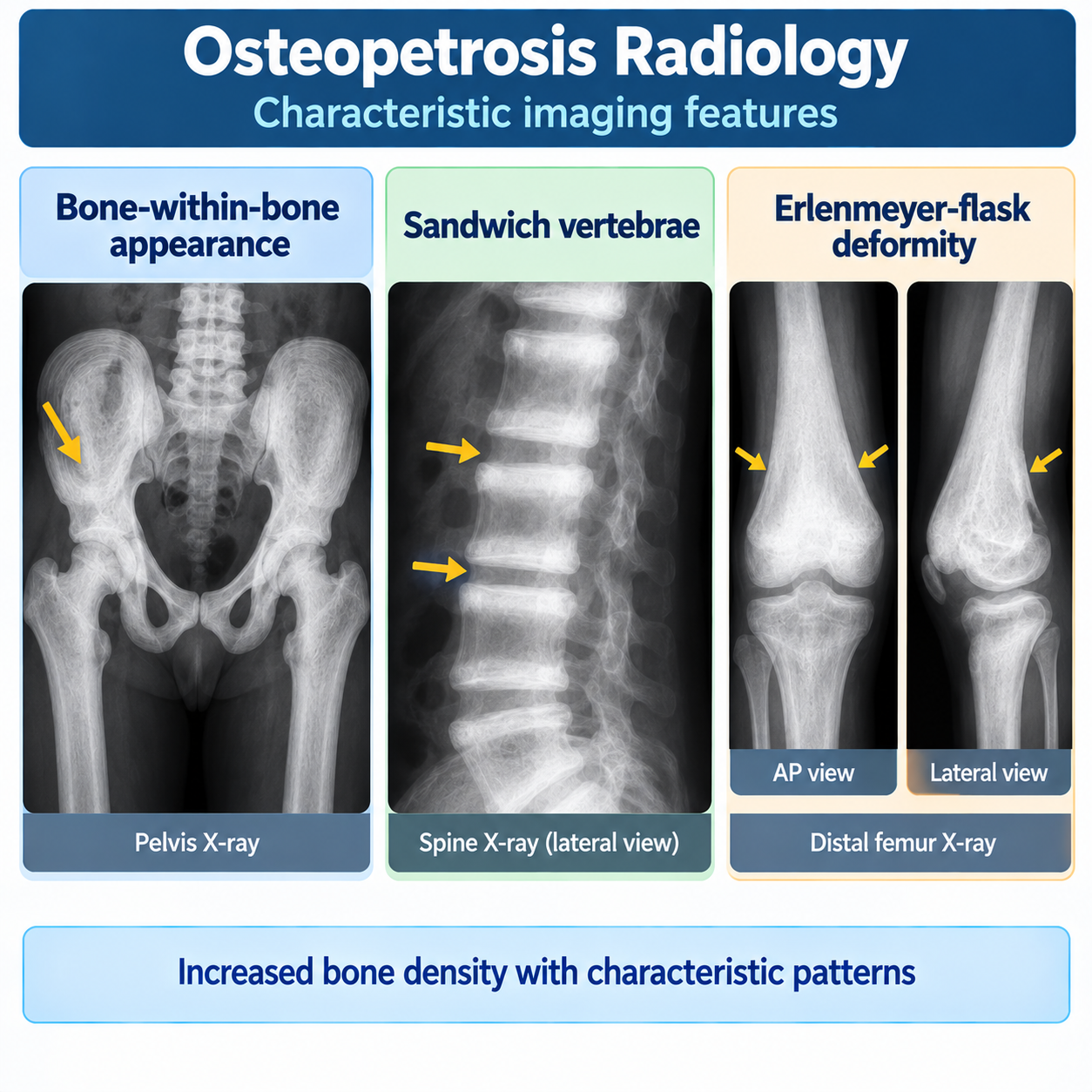
Potential findings include:
GENERALIZED OSTEOSCLEROSISplus characteristic patterns such as:
- bone-within-bone appearance;
- sandwich vertebrae;
- Erlenmeyer-flask metaphyseal modeling;
- skull-base sclerosis;
- transverse sclerotic bands.
These signs should prompt consideration of osteopetrosis and related sclerosing bone disorders. (NCBI)
Bone-Within-Bone Appearance
This classic sign appears as:
A DENSER INTERNAL BONE CONTOUR WITHIN THE OUTER BONEIt can be particularly apparent in:
- iliac wings;
- vertebrae;
- other bones.
The sign reflects abnormal cycles of bone modeling/remodeling.
Important:
BONE-WITHIN-BONE IS HIGHLY SUGGESTIVE IN CONTEXT BUT NOT PATHOGNOMONIC BY ITSELFSandwich Vertebrae
The vertebral endplates may become disproportionately sclerotic.
This creates:
DENSE SUPERIOR AND INFERIOR ENDPLATESwith a relatively different central vertebral appearance.
The result resembles:
A SANDWICHHence:
SANDWICH VERTEBRAEThis is a classic feature of autosomal dominant osteopetrosis type II. (NCBI)
Erlenmeyer-Flask Deformity
Failure of normal metaphyseal remodeling can produce widening of the metaphysis.
The distal femur may resemble:
AN ERLENMEYER FLASKThis is fundamentally a:
MODELING DEFECTbecause osteoclasts are required to sculpt the growing metaphysis.
This finding is not unique to osteopetrosis.
Skull-Base Sclerosis
Dense skull-base bone may:
- narrow neural foramina;
- compress cranial nerves;
- contribute to visual/hearing abnormalities.
Therefore skull imaging is not merely diagnostic—it may also reveal the anatomical basis for major complications.
Laboratory Evaluation
Laboratory Evaluation
There is no single universal serum biomarker that diagnoses all osteopetrosis.
Depending on severity, investigations may reveal:
- anemia;
- thrombocytopenia;
- other cytopenias;
- hypocalcemia;
- altered phosphate physiology;
- elevated PTH;
- biochemical evidence of disturbed bone remodeling.
But:
RADIOGRAPHIC OSTEOSCLEROSIS + CLINICAL PHENOTYPE + GENETICSare central.
CBC
In severe disease:
CBC IS CRITICALbecause marrow-space loss can produce:
- anemia;
- thrombocytopenia;
- leukocyte abnormalities.
A CBC can therefore reveal one of the most dangerous consequences of osteopetrosis.
Genetic Testing
Genetic Testing
Genetic testing is important because it can:
- establish molecular diagnosis;
- define inheritance;
- help predict phenotype;
- identify neurological forms;
- influence treatment decisions;
- guide transplantation considerations;
- support family counseling.
Because osteopetrosis is genetically heterogeneous, a:
MULTIGENE PANELor broader genomic approach may be appropriate rather than repeatedly testing single genes.
CLCN7 GeneReviews specifically favors multigene-panel or comprehensive genomic testing because phenotypes overlap. (NCBI)
Do Not Diagnose By Radiology Alone
DENSE BONES ≠ AUTOMATIC OSTEOPETROSISOther disorders can cause osteosclerosis.
The diagnosis should integrate:
CLINICAL PHENOTYPE RADIOLOGY LABORATORY FINDINGS MOLECULAR GENETICSDifferential Diagnosis
- other sclerosing bone dysplasias;
- pyknodysostosis;
- high-bone-mass syndromes;
- fluorosis where clinically relevant;
- heavy-metal-associated osteosclerosis in appropriate exposure contexts;
- secondary osteosclerotic disorders.
Osteopetrosis vs Osteogenesis Imperfecta
| Feature | Osteopetrosis | Osteogenesis imperfecta |
|---|---|---|
| Primary problem | Osteoclast resorption failure | Collagen matrix defect |
| Bone density | Increased | Often reduced/variable |
| Bone strength | Poor despite density | Poor |
| Fractures | Common | Common |
| Marrow failure | Severe forms | Not typical |
| Cranial nerve compression | Important severe feature | Not defining |
| Blue sclerae | Not typical | Classic clue in some forms |
| DI | Not defining | Important in some forms |
| Major genes | TCIRG1, CLCN7, others | COL1A1/COL1A2, others |
| HSCT | Disease-modifying/curative in selected forms | Not standard mechanism-directed treatment |
Bottom:
OSTEOPETROSIS = RESORPTION FAILURE OI = MATRIX FAILUREOsteopetrosis vs Hypophosphatasia
Osteopetrosis Versus Hypophosphatasia
| Feature | Osteopetrosis | Hypophosphatasia |
|---|---|---|
| Main defect | Osteoclast resorption | TNSALP/mineralization |
| Bone density | Usually increased | Variable |
| ALP | Not defining | Persistently low |
| Marrow failure | Severe forms | Not typical |
| Optic compression | Important | Not typical |
| Premature root-intact tooth loss | Not defining | Characteristic clue |
| Major genes | TCIRG1, CLCN7, others | ALPL |
| Targeted treatment | HSCT in selected forms | Asfotase alfa in appropriate patients |
Bottom:
OSTEOPETROSIS = CAN'T REMOVE BONE HPP = CAN'T MINERALIZE BONE NORMALLYThree-Way Fragility Comparison
This should be a major integration table.
| Disorder | Fundamental defect | Simplified memory |
|---|---|---|
| Osteogenesis imperfecta | Collagen matrix | Bad framework |
| Hypophosphatasia | Mineralization | Can't mineralize properly |
| Osteopetrosis | Osteoclast resorption | Can't remove/remodel bone |
Then:
ALL THREE CAN FRACTUREfor completely different biological reasons.
This is the major cluster-learning payoff.
Treatment Principles
Treatment depends strongly on:
- molecular cause;
- severity;
- age;
- marrow involvement;
- neurological phenotype;
- visual risk;
- fracture burden.
Therefore:
GENOTYPE + PHENOTYPE → TREATMENTThere is no single treatment appropriate for every form of osteopetrosis.
Why HSCT Can Treat Osteopetrosis
Why HSCT Can Work
This should be one of the strongest teaching sections.
Ask:
WHY CAN A BONE DISEASE BE TREATED WITH A STEM-CELL TRANSPLANT?Because:
OSTEOCLASTS COME FROM HEMATOPOIETIC PRECURSORSTherefore:
DONOR HEMATOPOIETIC STEM CELLS DONOR-DERIVED OSTEOCLAST PRECURSORS FUNCTIONAL OSTEOCLASTS BONE RESORPTION RESTORED SKELETAL REMODELING IMPROVESThis is an elegant example of treatment derived directly from pathophysiology.
HSCT In Severe Osteopetrosis
For suitable severe osteoclast-intrinsic forms:
ALLOGENEIC HSCT CAN BE CURATIVE / DISEASE-MODIFYINGIt can improve or reverse:
- abnormal sclerosis;
- marrow failure;
- extramedullary hematopoiesis.
However:
HSCT IS A HIGH-RISK TREATMENTand suitability depends on:
- genotype;
- phenotype;
- neurological involvement;
- age;
- transplant expertise;
- donor and patient factors.
CLCN7 GeneReviews describes HSCT as potentially curative for appropriate ARO because osteoclasts are hematopoietic in origin, while the 2025 TCIRG1 GeneReview describes HSCT as historically the most effective treatment for severe TCIRG1 disease. (NCBI)
TCIRG1 And HSCT
The 2025 TCIRG1 GeneReview identifies HSCT particularly in severe disease such as:
- severe osteopetrosis with bone-marrow failure;
- hematologic failure with threatened vision;
- severe disease in infancy.
Early treatment is emphasized because some complications become irreversible. (NCBI)
HSCT Does Not Treat Every Osteopetrosis
OSTEOPETROSIS ≠ AUTOMATIC HSCTSome molecular forms involve defects outside donor-correctable osteoclast function.
Particularly important are forms with:
PRIMARY NEUROLOGICAL DISEASEHSCT may correct osteoclast dysfunction but not an intrinsic CNS disorder.
Therefore:
MOLECULAR DIAGNOSIS BEFORE HSCT IS CRITICALEstablished Nerve Damage
Even when HSCT successfully corrects skeletal disease:
ESTABLISHED OPTIC ATROPHY MAY BE IRREVERSIBLETherefore:
EARLY DIAGNOSIS MATTERSThis is a major reason severe infantile osteopetrosis should be recognized promptly. (NCBI)
Interferon Gamma-1b
Interferon gamma-1b has been used in severe infantile osteopetrosis.
NIAMS states that it is the FDA-approved drug specifically for severe infantile osteopetrosis and is not approved for noninfantile disease. (NIAMS)
The 2025 TCIRG1 GeneReview is more cautious:
- it may be considered in infantile TCIRG1 disease;
- it has been used as a bridge to HSCT;
- evidence is insufficient to recommend it as universal monotherapy. (NCBI)
Therefore teach:
IFN-γ1b IS NOT A UNIVERSAL TREATMENT FOR ALL OSTEOPETROSISSupportive Management
Supportive Hematological Management
Severe marrow failure may require supportive care such as:
- red-cell transfusion;
- platelet transfusion;
- infection management;
- specialist hematological care.
These measures:
SUPPORT THE PATIENTbut:
DO NOT CORRECT THE UNDERLYING OSTEOCLAST DEFECTCalcium And Mineral Management
Calcium/mineral management can be complex.
Patients may develop:
- hypocalcemia;
- secondary hyperparathyroidism;
- osteopetrorickets.
Treatment must be individualized.
Importantly, excessive attempts to increase mineralization can potentially worsen skeletal sclerosis/foraminal narrowing in some settings.
The 2025 TCIRG1 GeneReview specifically advises judicious management rather than indiscriminate supplementation. (NCBI)
Therefore:
DO NOT CREATE A UNIVERSAL CALCIUM/VITAMIN-D REGIMENFracture Management
Fractures should be managed by clinicians familiar with the abnormal mechanical properties of osteopetrotic bone.
Potential challenges include:
- hard sclerotic bone;
- difficult drilling;
- iatrogenic fracture;
- delayed union/nonunion;
- infection.
Treatment should be individualized.
Dental Management
Dental management should emphasize:
- preventive oral hygiene;
- early treatment of infection;
- monitoring tooth eruption;
- cautious surgical planning;
- awareness of jaw osteomyelitis risk.
Vision Management
Visual function should be evaluated promptly in severe disease.
Potential approaches depend on:
- mechanism;
- degree of compression;
- optic-nerve viability;
- genotype;
- transplantation plan.
Optic-nerve decompression has been attempted in selected patients but is difficult and is not a universal solution. (NCBI)
Multidisciplinary Care
Depending on severity, the team may include:
- metabolic bone/endocrinology;
- pediatrics;
- genetics;
- hematology;
- transplant specialists;
- orthopedics;
- ophthalmology;
- neurology;
- ENT/audiology;
- dentistry;
- rehabilitation.
Severe osteopetrosis is:
A MULTISYSTEM GENETIC DISEASEnot simply an abnormal radiograph.
Genetic Counseling
Inheritance depends on the molecular subtype.
Severe infantile disease
Often:
AUTOSOMAL RECESSIVEADOII
Typically:
AUTOSOMAL DOMINANTCLCN7 is particularly illustrative because it can produce both recessive and dominant phenotypes. (NCBI)
Genetic counseling should use the identified molecular diagnosis.
Diagnostic Algorithm
Implement prominently.
OSTEOSCLEROSIS / UNUSUALLY DENSE BONES IS THE PHENOTYPE COMPATIBLE WITH OSTEOPETROSIS?Look for:
Fractures
Growth problems
Anemia / thrombocytopenia
Hepatosplenomegaly
Visual impairment
Hearing impairment
Dental abnormalities
RADIOGRAPHIC PATTERNGeneralized osteosclerosis
Bone-within-bone
Sandwich vertebrae
Erlenmeyer-flask modeling
Skull-base sclerosis
ASSESS SEVERITYCBC
Ca / PO₄ / PTH context
Vision
Hearing
Neurological phenotype
MOLECULAR GENETIC TESTING DEFINE MOLECULAR SUBTYPE SEVERE OSTEOCLAST-INTRINSIC / TRANSPLANT-CORRECTABLE FORM?Yes
→ urgent specialist HSCT assessment.
No / uncertain
→ subtype-specific supportive/targeted management.
Bottom:
GENOTYPE + PHENOTYPE DETERMINE TREATMENTPathophysiology Algorithm
OSTEOCLAST FAILURE RESORPTION ↓ REMODELING ↓ BONE ACCUMULATES OSTEOSCLEROSISbut simultaneously:
Branch A
ABNORMAL ARCHITECTURE FRACTURESBranch B
MARROW SPACE ↓ ANEMIA / THROMBOCYTOPENIA EXTRAMEDULLARY HEMATOPOIESIS HEPATOSPLENOMEGALYBranch C
SKULL FORAMINA NARROW OPTIC / AUDITORY / OTHER NERVE COMPRESSIONThis is the central article diagram in text form.
Worked Clinical Cases
Case 1 — Dense bones and fractures
A child has generalized osteosclerosis but recurrent low-trauma fractures.
Wrong conclusion
“Dense bone means the skeleton is strong.”
Correct thought
OSTEOPETROSISDense but abnormally remodeled bone can be brittle.
Case 2 — Infant with anemia
Infant has:
- osteosclerosis;
- anemia;
- thrombocytopenia;
- hepatosplenomegaly.
Mechanism
MARROW-SPACE LOSS MARROW FAILURE EXTRAMEDULLARY HEMATOPOIESISSevere infantile osteopetrosis should be urgently considered.
Case 3 — Visual loss
Infant with osteopetrosis develops visual impairment.
Key concern
OPTIC-NERVE COMPRESSION / ATROPHYPrompt specialist assessment is important because established damage may be irreversible.
Case 4 — Hypocalcemic seizure
Infant with severe osteopetrosis develops hypocalcemia and seizure.
Lesson
DENSE BONE DOES NOT GUARANTEE NORMAL CALCIUM HOMEOSTASISCase 5 — Adult incidental finding
A 30-year-old has very dense bones on radiographs and a history of several fractures but no marrow failure.
Consider
AUTOSOMAL-DOMINANT OSTEOPETROSISespecially with characteristic imaging.
Case 6 — Sandwich vertebrae
Radiograph shows dense vertebral endplates producing a sandwich appearance.
Association
ADOII / ALBERS-SCHÖNBERG DISEASEin the appropriate clinical context.
Case 7 — Bone within bone
Pelvic radiograph shows a bone-within-bone appearance.
Lesson
This is a classic osteopetrosis clue but must be interpreted with the complete phenotype.
Case 8 — Erlenmeyer flask
A child has widened, poorly modeled distal femoral metaphyses.
Mechanism
FAILURE OF NORMAL METAPHYSEAL MODELINGdue to impaired osteoclast activity.
Case 9 — Why transplant?
Parent asks why a bone disorder is being considered for stem-cell transplantation.
Answer
OSTEOCLASTS ARE HEMATOPOIETIC-LINEAGE CELLSDonor stem cells can generate functional donor-derived osteoclasts in appropriate molecular forms.
Case 10 — Neurological subtype
Child has severe osteopetrosis plus evidence of primary neurodegeneration.
Lesson
HSCT MAY CORRECT OSTEOCLAST DISEASE WITHOUT CORRECTING PRIMARY CNS DISEASEMolecular diagnosis matters.
Case 11 — Adult fracture surgery
Adult with ADOII requires orthopedic surgery.
Important issue
Osteopetrotic bone can be unusually hard yet brittle, making surgery technically difficult and increasing complication risk.
Case 12 — Low ALP
Patient with fractures has persistently low ALP but does not have generalized osteosclerosis.
Better differential
THINK HYPOPHOSPHATASIAnot osteopetrosis merely because fractures are present.
Common Mistakes
Mistake 1
Dense bone always means strong bone.
Wrong.
Mistake 2
Osteopetrosis is the same as osteoporosis.
Wrong.
Mistake 3
Osteopetrosis is primarily excessive osteoblast activity in every case.
Wrong.
Mistake 4
Bone resorption is unnecessary.
Wrong.
Mistake 5
Osteoclast failure affects only bone density.
Wrong.
Mistake 6
Fractures cannot occur in osteopetrosis.
Wrong.
Mistake 7
Bone marrow failure is unrelated to the skeletal disease.
Wrong.
Mistake 8
Hepatosplenomegaly is unrelated.
Wrong.
Mistake 9
Visual loss is always primary eye disease.
Wrong.
Mistake 10
All neurological problems are caused by nerve compression.
Wrong.
Mistake 11
All osteopetrosis is autosomal recessive.
Wrong.
Mistake 12
All osteopetrosis is autosomal dominant.
Wrong.
Mistake 13
TCIRG1 is the only osteopetrosis gene.
Wrong.
Mistake 14
CLCN7 produces only dominant disease.
Wrong.
Mistake 15
Bone-within-bone appearance alone proves the diagnosis.
Wrong.
Mistake 16
HSCT treats every form of osteopetrosis.
Wrong.
Mistake 17
HSCT reliably reverses established optic atrophy.
Wrong.
Mistake 18
Interferon gamma-1b is universal therapy for adult osteopetrosis.
Wrong.
Mistake 19
Calcium and vitamin D should automatically be given in high doses.
Wrong.
Mistake 20
Osteopetrosis is simply a radiological disorder.
Wrong.
Osteopetrosis in One Minute
OSTEOPETROSIS IN ONE MINUTE OSTEOCLAST FAILURE BONE RESORPTION ↓ REMODELING ↓ BONE DENSITY ↑but:
BONE QUALITY ABNORMALTherefore:
DENSE + BRITTLEThen remember three consequences:
Bone
FRACTURESMarrow
MARROW SPACE ↓ ANEMIA + THROMBOCYTOPENIASkull
FORAMINA NARROW VISION / HEARING / CRANIAL-NERVE PROBLEMSClassic imaging:
BONE WITHIN BONE SANDWICH VERTEBRAE ERLENMEYER FLASKSevere appropriate subtype:
HSCTWhy?
OSTEOCLASTS COME FROM HEMATOPOIETIC PRECURSORSFinal:
DENSE BONE ≠ STRONG BONEFrequently Asked Questions
What is osteopetrosis?
Osteopetrosis is a genetically heterogeneous group of disorders characterized by increased skeletal density, usually because osteoclast-mediated bone resorption is impaired.
Why are bones dense in osteopetrosis?
Bone that should normally be removed during modeling and remodeling is retained because osteoclast resorption is impaired.
Why do dense bones fracture?
Bone strength depends on architecture, remodeling and material quality as well as density. Osteopetrotic bone is abnormally remodeled and can therefore be brittle despite high density.
What cells are defective in osteopetrosis?
Most classic forms involve impaired osteoclast formation or function.
What genes commonly cause osteopetrosis?
Many genes can cause osteopetrosis. TCIRG1 is a major cause of severe autosomal-recessive disease, while CLCN7 can cause recessive, intermediate and autosomal-dominant forms.
Why does severe osteopetrosis cause anemia?
Excess abnormal bone reduces available marrow space, impairing hematopoiesis.
Why does hepatosplenomegaly occur?
Marrow failure can drive extramedullary hematopoiesis in organs such as the liver and spleen.
Why can osteopetrosis cause blindness?
Skull-base sclerosis can narrow the optic canal and compress the optic nerve. Some genetic forms can also cause primary retinal or neurological disease.
Can osteopetrosis cause hypocalcemia?
Yes. Severe forms may impair normal mineral mobilization and can be associated with hypocalcemia and secondary hyperparathyroidism.
What is a bone-within-bone appearance?
It is a radiographic pattern in which a dense internal contour appears within the outer bone and is a classic clue to osteopetrosis in the appropriate clinical setting.
What are sandwich vertebrae?
They are vertebrae with prominent sclerosis of the endplates, producing a sandwich-like radiographic appearance, classically seen in ADOII.
What is Albers-Schönberg disease?
It is the traditional name for autosomal dominant osteopetrosis type II, commonly associated with heterozygous CLCN7 pathogenic variants.
Why can HSCT treat osteopetrosis?
Osteoclasts arise from hematopoietic precursors. In appropriate osteoclast-intrinsic genetic forms, donor stem cells can generate functional osteoclasts and correct the underlying resorption defect.
Does every patient with osteopetrosis need HSCT?
No. HSCT is primarily considered for appropriate severe molecular forms. Molecular diagnosis and specialist assessment are essential.
What is the most important concept to remember?
DENSE BONE DOES NOT NECESSARILY MEAN STRONG BONE.
Key Take-Home Messages
Osteopetrosis is fundamentally a disorder of:
BONE RESORPTION AND REMODELINGThe central mechanism is:
OSTEOCLAST FAILURE RESORPTION ↓ ABNORMAL BONE ACCUMULATION OSTEOSCLEROSISBut:
DENSITY IS NOT THE SAME AS QUALITYTherefore osteopetrotic bone can be:
DENSEand simultaneously:
BRITTLEThis explains:
PATHOLOGICAL FRACTURESSevere disease also causes:
MARROW SPACE ↓ ANEMIA + THROMBOCYTOPENIA EXTRAMEDULLARY HEMATOPOIESIS HEPATOSPLENOMEGALYSkull-base sclerosis can produce:
CRANIAL-NERVE COMPRESSIONespecially:
OPTIC-NERVE DAMAGEThe classic radiographic clues are:
BONE-WITHIN-BONE SANDWICH VERTEBRAE ERLENMEYER-FLASK MODELINGGenetic testing matters because:
NOT ALL OSTEOPETROSIS IS THE SAMEand:
NOT ALL OSTEOPETROSIS SHOULD RECEIVE HSCTFor appropriate severe osteoclast-intrinsic disease:
HSCT CAN CORRECT THE UNDERLYING CELLULAR DEFECTbecause:
OSTEOCLASTS ARE DERIVED FROM HEMATOPOIETIC PRECURSORSBut established neurological or optic damage may not reverse.
Therefore:
EARLY DIAGNOSIS + MOLECULAR DIAGNOSIS MATTERFinal cluster memory:
OI = MATRIX FAILURE HPP = MINERALIZATION FAILURE OSTEOPETROSIS = RESORPTION FAILUREAnd the final article statement:
BONE NEEDS TO BE REMOVED AS WELL AS FORMED — WITHOUT NORMAL REMODELING, MORE BONE CAN BECOME WORSE BONE.