Osteomalacia at a Glance
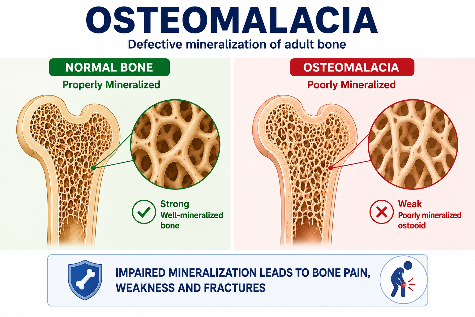
Osteomalacia is defective mineralization of newly formed osteoid in adult bone. Bone matrix is formed, but mineral deposition is inadequate or delayed.
The key question is not simply whether bone density is low. The key question is whether newly formed osteoid is mineralizing properly.
What Is Osteoid?
Bone formation occurs in stages. Osteoblasts first produce an organic bone matrix called osteoid. This matrix subsequently undergoes mineralization.
Calcium and phosphate are incorporated into mineral crystals within the bone matrix, producing normally mineralized bone.
What Happens in Osteomalacia?
In osteomalacia, osteoid is produced but mineralization is inadequate or delayed. Excessive unmineralized osteoid can accumulate.
Osteomalacia is defective mineralization of newly formed osteoid in adult bone.
The fundamental defect is mineralization failure rather than simply reduced production of bone.
Osteomalacia, Osteoporosis and Rickets
| Condition | Core Defect | Teaching Point |
|---|---|---|
| Osteomalacia | Defective mineralization of adult bone. | Osteoid is formed but inadequately mineralized. |
| Osteoporosis | Reduced bone strength associated with reduced bone mass and microarchitectural deterioration. | Low bone mass is central; osteomalacia is not the defining defect. |
| Rickets | Defective mineralization in the growing skeleton, including the growth plate. | Pediatric growth plate disease, not adult osteomalacia. |
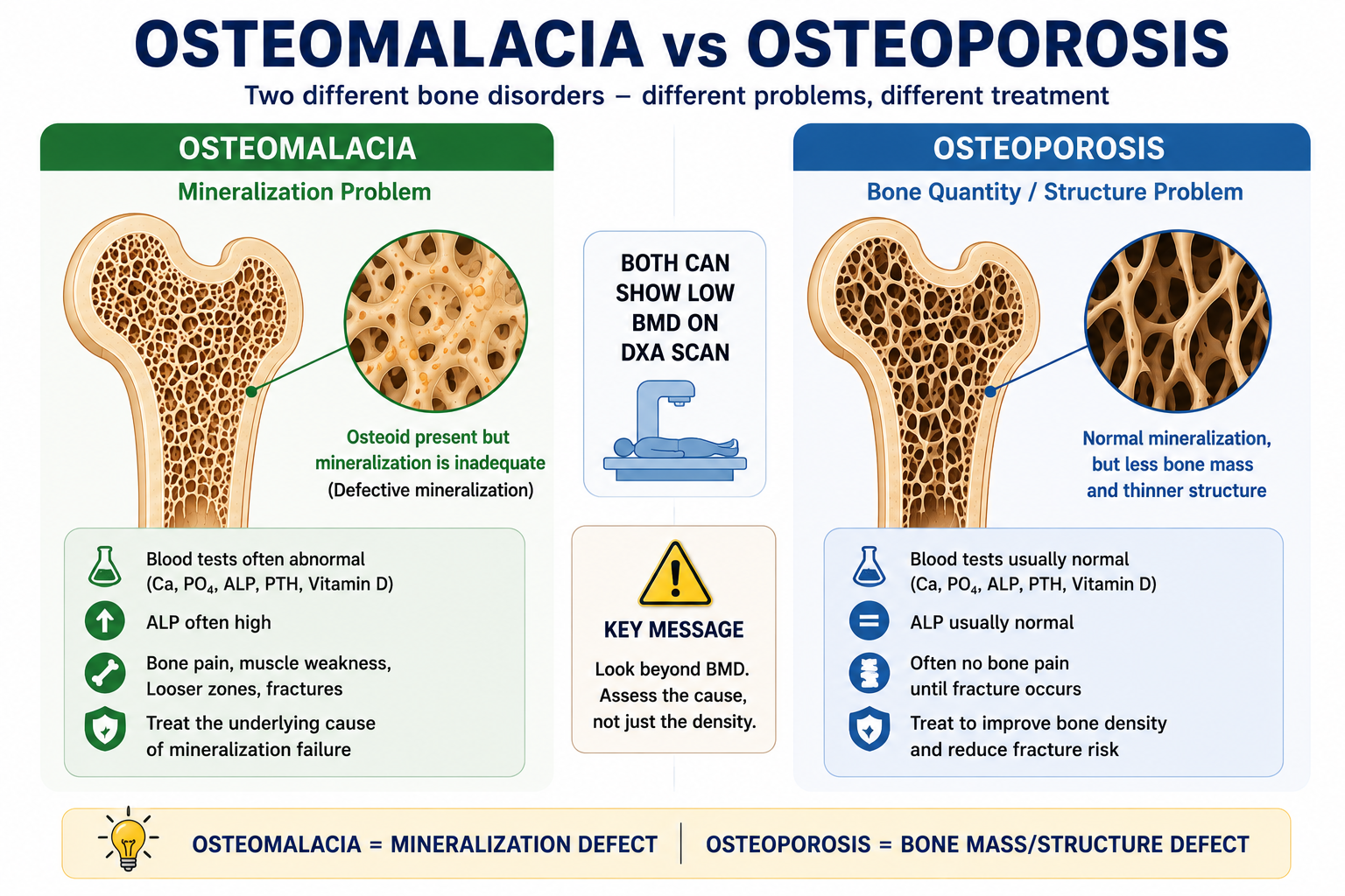
Osteomalacia is also not synonymous with vitamin D deficiency. Severe or prolonged vitamin D deficiency can cause osteomalacia, but deficiency and osteomalacia are not identical diagnoses.
Why Mineralization Fails
Normal mineralization requires an appropriate extracellular environment, especially adequate calcium and phosphate availability, as well as normal regulatory and cellular mechanisms.
Causes are best classified by mechanism.
| Mechanism | Examples | Diagnostic Direction |
|---|---|---|
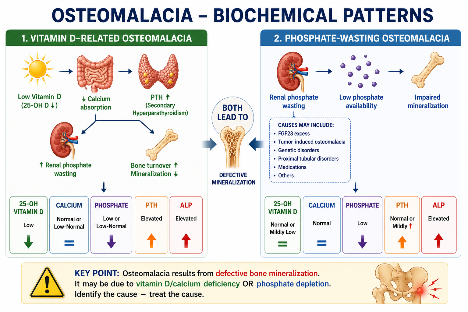
| Vitamin D / calcium-related mineralization failure | Vitamin D deficiency, malabsorption, inadequate calcium availability, impaired activation or action. | Review 25-OH D, calcium, PTH, ALP and clinical context. |
| Phosphate-depletion mineralization failure | Low phosphate intake/absorption, renal phosphate wasting, FGF23-mediated disorders. | Ask whether the kidney is conserving phosphate appropriately. |
| Renal/metabolic disorders | CKD-MBD, renal tubular disorders, metabolic acidosis and related mineral disturbances. | Interpret kidney function, phosphate handling and mineral markers together. |
| Primary or unusual mineralization defects | Selected intrinsic mineralization disorders or medication-related problems. | Consider when common calcium, vitamin D and phosphate explanations do not fit. |
Vitamin D-Related Osteomalacia
Vitamin D deficiency can cause osteomalacia when deficiency is severe or prolonged enough to impair calcium-phosphate mineralization of osteoid.
Do not assume vitamin D deficiency automatically equals osteomalacia. For the deficiency framework, see Vitamin D Deficiency Explained. For activation physiology, see Vitamin D Metabolism Explained.
Phosphate-Dependent Osteomalacia
Phosphate is a mineralization substrate. Osteomalacia can occur when phosphate availability is persistently inadequate, even if vitamin D status is not the primary problem.
Hypophosphatemic osteomalacia should prompt the question: if serum phosphate is low, is the kidney appropriately conserving phosphate?
For background, review Phosphate Homeostasis Explained and Hypophosphatemia Explained.
FGF23-Mediated and Tumor-Induced Osteomalacia
FGF23 promotes renal phosphate wasting and suppresses calcitriol production. Excess FGF23 activity can therefore produce hypophosphatemic osteomalacia.
In tumor-induced osteomalacia, evaluation depends on the biochemical context. Renal phosphate handling, such as TRP, fractional excretion of phosphate or TmP/GFR, helps decide whether phosphate wasting is present before FGF23 is interpreted.
This article introduces the logic but does not provide phosphate/FGF23 cutoff values.
Symptoms and Clinical Features
Osteomalacia can develop gradually and may be mistaken for musculoskeletal, rheumatological or neurological disease.
- Diffuse bone pain or tenderness.
- Proximal muscle weakness.
- Difficulty rising from a chair or climbing stairs.
- Waddling gait in more advanced disease.
- Fragility fractures or pseudofractures.
- Clinical features of the underlying cause, such as malabsorption or CKD.
These symptoms are not specific. They become more meaningful when combined with compatible biochemical and imaging findings.
Laboratory Assessment
Assessment usually includes calcium, phosphate, ALP, PTH, 25-OH vitamin D and renal function. Use laboratory-specific reference ranges.
| Test | Why It Matters | Interpretation Trap |
|---|---|---|
| Calcium | Assesses calcium availability and PTH stimulus. | Hypocalcemia is not required. |
| Phosphate | Critical substrate for mineralization. | Low phosphate may point toward renal wasting or FGF23 activity. |
| ALP | Useful clue to active mineralization disorder. | ALP is not diagnostic alone; normal ALP does not absolutely exclude early disease. |
| PTH | Shows secondary hyperparathyroid response or other mineral feedback. | PTH must be interpreted with calcium, phosphate and kidney function. |
| 25-OH vitamin D | Main marker of nutritional vitamin D status. | Low vitamin D alone does not prove osteomalacia. |
| Renal function | CKD and tubular disorders alter mineral handling. | CKD-MBD should not be equated automatically with osteomalacia. |
Imaging and Looser Zones
Imaging may show fractures, pseudofractures or Looser zones in established osteomalacia. However, early disease may lack characteristic radiographic abnormalities.
Looser zones are pseudofractures that reflect impaired mineralization and mechanical stress. They are supportive when present but are not required in every case.
DXA can show low bone mineral density, but DXA alone cannot reliably distinguish osteomalacia from osteoporosis because it does not directly assess osteoid mineralization.
How Osteomalacia Is Confirmed
Diagnosis usually integrates clinical features, biochemical abnormalities and imaging. The more the pattern fits a cause-specific mineralization defect, the stronger the clinical diagnosis becomes.
Undecalcified bone biopsy with histomorphometric assessment remains the reference/gold-standard method for directly demonstrating the mineralization defect of osteomalacia.
However, biopsy is invasive and is not used routinely for every suspected case. It is generally reserved for selected uncertain or complex cases where definitive confirmation is necessary.
Common Laboratory Patterns
| Pattern | Possible Direction | Next Question |
|---|---|---|
| Low 25-OH D, high ALP, high PTH | Vitamin D/calcium-related osteomalacia pattern may fit. | Is deficiency severe/prolonged and clinically compatible? |
| Low phosphate, high ALP, normal vitamin D | Phosphate-dependent osteomalacia becomes important. | Is phosphate loss renal? |
| Low phosphate with inappropriate urinary phosphate loss | Renal phosphate wasting. | Is FGF23 involved? |
| CKD with abnormal Ca2+, PO4, PTH and ALP | CKD-MBD physiology. | Is this renal osteodystrophy, osteomalacia, mixed disease or another pattern? |
Treatment Principles
Treatment is cause-specific. Do not treat osteomalacia as simply low bone mineral density, and do not treat every cause with the same regimen.
- Vitamin D-related osteomalacia requires correction of vitamin D status and relevant calcium availability.
- Phosphate-depletion osteomalacia requires correction of the phosphate problem and its cause.
- Renal phosphate wasting requires identification of the wasting mechanism.
- FGF23-mediated disease requires cause-specific management rather than generic supplementation alone.
- CKD-MBD requires CKD-context interpretation of calcium, phosphate, PTH, ALP and vitamin D physiology.
This page does not provide drug doses, phosphate regimens, FGF23 cutoffs or CKD treatment protocols.
Worked Clinical Cases
Case 1: Low vitamin D with bone pain and high ALP
This can fit vitamin D-related osteomalacia if the clinical, biochemical and imaging pattern is coherent. Low vitamin D alone is not enough; the mineralization story must fit.
Case 2: Low phosphate with normal 25-OH vitamin D
Consider phosphate-dependent osteomalacia. The next step is to ask whether the kidney is conserving phosphate appropriately or wasting it.
Case 3: Low BMD on DXA
DXA cannot by itself distinguish osteoporosis from osteomalacia. Review symptoms, calcium, phosphate, ALP, PTH, 25-OH D, renal function and imaging clues.
Common Mistakes
- Diagnosing osteomalacia from vitamin D alone.
- Diagnosing osteomalacia from ALP alone.
- Excluding osteomalacia because calcium is normal.
- Using DXA alone to distinguish osteomalacia from osteoporosis.
- Calling every CKD-MBD bone problem osteomalacia.
- Forgetting phosphate-wasting and FGF23-mediated causes.
- Assuming biopsy is required routinely for every suspected case.
- Treating osteomalacia as low BMD rather than a cause-specific mineralization disorder.
Osteomalacia in One Minute
Osteomalacia is defective mineralization of newly formed osteoid in adult bone. The problem is that bone matrix is formed but inadequately mineralized.
It is not osteoporosis, not vitamin D deficiency itself and not adult rickets. Vitamin D deficiency can cause osteomalacia, but phosphate depletion, renal phosphate wasting, FGF23-mediated disorders, CKD-related mineral disease and other mineralization defects can also matter.
Diagnosis integrates symptoms, calcium, phosphate, ALP, PTH, 25-OH vitamin D, renal function and imaging. DXA alone cannot reliably distinguish osteomalacia from osteoporosis. Bone histomorphometry remains the reference standard when definitive confirmation is required.
Frequently Asked Questions
Key Take-Home Messages
- Osteomalacia is defective mineralization of newly formed osteoid in adult bone.
- It is conceptually different from osteoporosis, vitamin D deficiency and rickets.
- Calcium, phosphate, ALP, PTH, 25-OH D and renal function should be interpreted together.
- Normal calcium does not exclude osteomalacia.
- DXA alone cannot reliably distinguish osteomalacia from osteoporosis.
- Bone histomorphometry is the reference standard but is reserved for selected cases.
- Treatment must correct the underlying mineralization defect.
Next in the Cluster
Next, review Renal Phosphate Wasting Explained to learn how TRP, FEPO4 and TmP/GFR distinguish appropriate phosphate conservation from inappropriate renal phosphate loss.
This article is intended for medical education only. It explains osteomalacia diagnostic reasoning and treatment principles, not patient-specific medical advice, drug doses, phosphate regimens, FGF23 cutoffs, biopsy indications or CKD protocols.