Vitamin D Deficiency at a Glance
Vitamin D deficiency is inadequate vitamin D status that may disturb calcium, phosphate, PTH and skeletal mineral physiology. Serum 25-OH vitamin D is the principal laboratory marker used to assess vitamin D status.
Do not reduce the topic to a single number. Ask why vitamin D is low, what the mineral system is doing, and whether skeletal mineralization has been affected.
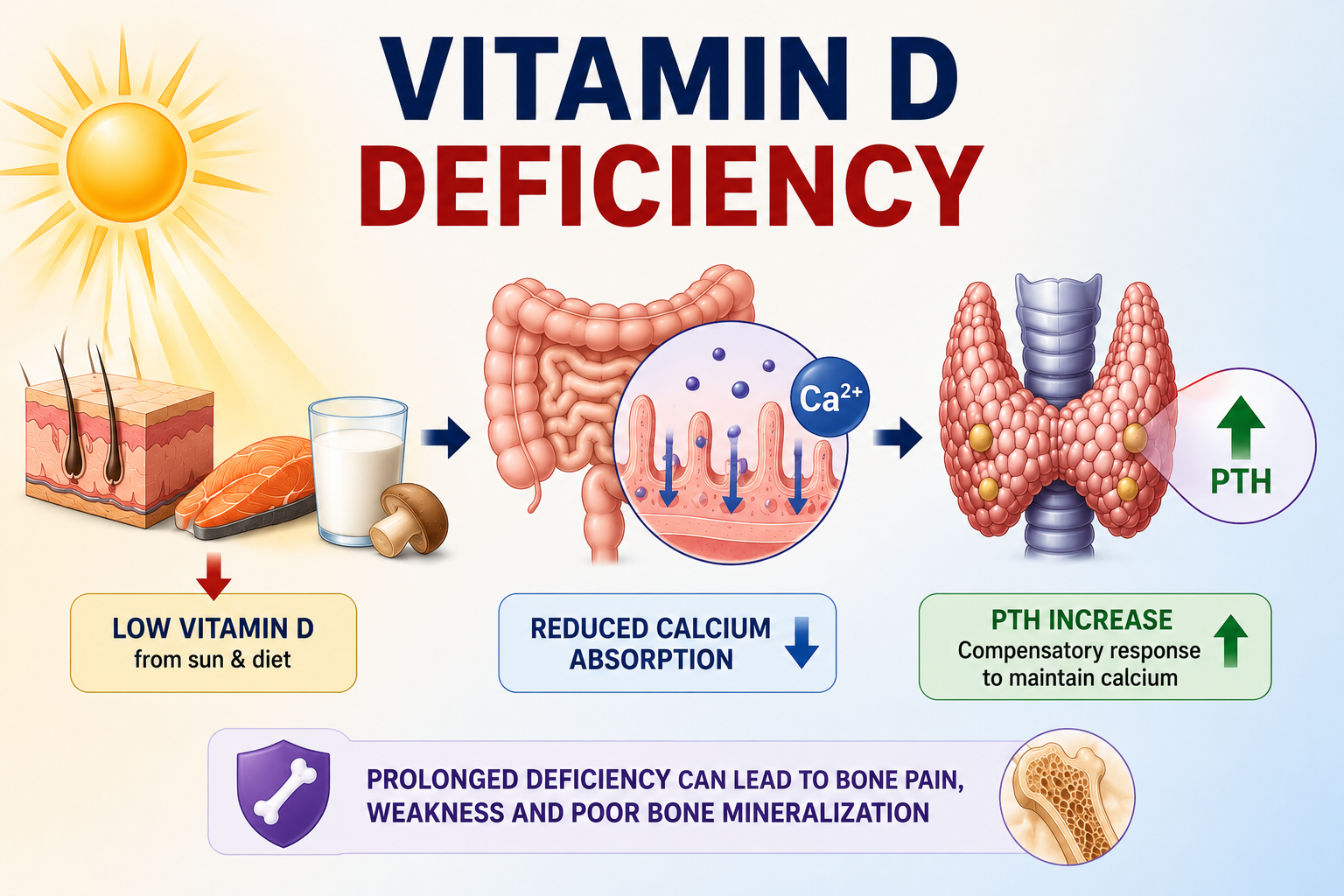
What Is Vitamin D Deficiency?
Vitamin D deficiency refers to inadequate vitamin D status sufficient to create concern about normal vitamin D-dependent physiology, particularly calcium-phosphate and skeletal mineral metabolism.
The main laboratory marker used to assess vitamin D status is serum 25-hydroxyvitamin D, or 25-OH D.
Calcitriol is the active hormone, but it is tightly regulated by PTH, FGF23, calcium-phosphate physiology and kidney function. A normal calcitriol concentration does not exclude nutritional vitamin D deficiency.
For the physiology behind this distinction, see Vitamin D Metabolism Explained.
Vitamin D Deficiency Is Not the Same as SHPT or Osteomalacia
| Term | Meaning | Key Point |
|---|---|---|
| Vitamin D deficiency | Inadequate vitamin D status. | A biochemical/nutritional state. |
| Secondary hyperparathyroidism | PTH rises as a physiological response to another stimulus. | May occur when calcium availability falls. |
| Osteomalacia | Defective mineralization of adult bone. | A skeletal consequence, not a synonym for low vitamin D. |
Not every person with low vitamin D has secondary hyperparathyroidism, and not every person with vitamin D deficiency has osteomalacia.
Which Vitamin D Test Should Be Used?
When vitamin D deficiency is clinically suspected, serum 25-OH vitamin D is the main test used to assess vitamin D status.
Do not routinely use calcitriol to diagnose nutritional vitamin D deficiency. In some patients with deficiency and preserved renal function, low 25-OH D can stimulate PTH, and PTH can stimulate renal calcitriol production.
Should Everyone Be Tested for Vitamin D Deficiency?
No. Current Endocrine Society guidance recommends against routine 25-OH vitamin D screening in generally healthy adults without an established indication for testing.
The same guidance emphasizes that outcome-specific universal 25-OH D thresholds for disease prevention have not been established in clinical trials.
This is different from clinically indicated testing. 25-OH D measurement remains relevant when vitamin D deficiency or a mineral disorder is suspected, such as an appropriate patient with hypocalcemia, suspected osteomalacia or rickets, relevant malabsorption, CKD-MBD assessment, or another clinical indication.
What Causes Vitamin D Deficiency?
Causes are best organized by mechanism rather than memorized as a long list.
| Mechanism | Examples | Clinical Question |
|---|---|---|
| Reduced skin production | Limited UVB exposure, reduced outdoor exposure, covering clothing, season, latitude, age and other factors. | Is cutaneous production low? |
| Inadequate intake | Low dietary vitamin D intake or restrictive diet patterns. | Is supply inadequate? |
| Malabsorption | Fat malabsorption or gastrointestinal disease affecting absorption. | Is vitamin D entering the body properly? |
| Altered metabolism | Liver, kidney or hormonal physiology affecting vitamin D metabolites. | Is activation or handling abnormal? |
| Medication-related | Medications that alter vitamin D metabolism or increase catabolism. | Could medication review explain persistence? |
Do not prescribe unsafe sun exposure. The clinical point is mechanism recognition, not a universal sunlight prescription.
Who Is at Risk?
Risk contexts include reduced sun exposure, dietary insufficiency, malabsorption, chronic illness, medication effects and disorders of mineral metabolism. These risk associations help clinical reasoning, but they do not automatically mean every generally healthy adult needs routine screening.
Risk association and routine screening are different. Test when there is an established clinical indication or a suspected disorder, not simply as a universal population rule.
What Happens When Vitamin D Falls?
Vitamin D deficiency reduces intestinal calcium absorption. Reduced calcium availability can stimulate PTH secretion.

Why PTH increases
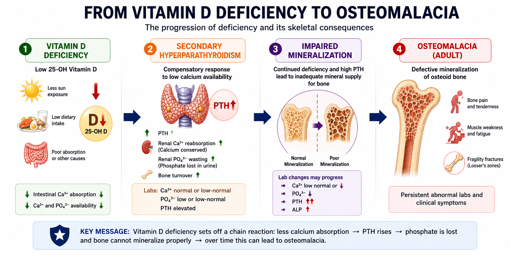
Vitamin D deficiency reduces intestinal calcium absorption. Reduced calcium availability stimulates the parathyroid glands to increase PTH secretion, producing secondary hyperparathyroidism in some patients.
For a focused explanation, see why vitamin D deficiency increases PTH.
Why calcium can remain normal
Compensatory PTH elevation can preserve serum calcium by increasing renal calcium conservation and mobilizing compensatory mineral mechanisms. Therefore normal serum calcium does not exclude vitamin D deficiency.
What happens to phosphate?
Phosphate may fall because intestinal phosphate absorption decreases and secondary hyperparathyroidism increases renal phosphate loss. However, normal phosphate does not exclude vitamin D deficiency.
What happens to ALP?
ALP may rise when defective mineralization increases osteoblastic activity, but ALP is not specific for vitamin D deficiency. Liver sources and other bone disorders should be considered. Normal ALP does not exclude early or uncomplicated deficiency.
Vitamin D Deficiency vs Osteomalacia
Vitamin D deficiency is a biochemical or nutritional state. Osteomalacia is defective mineralization of adult bone.
Severe or prolonged vitamin D deficiency can contribute to osteomalacia, but the two are not synonymous. Rickets is the corresponding mineralization problem in growing bone and is kept brief here because this page focuses on adult-style clinical reasoning.
Symptoms and clinical features
Mild biochemical deficiency may be asymptomatic. More significant or prolonged deficiency can be associated with bone pain, proximal muscle weakness, fragility fractures or features suggesting osteomalacia, but these symptoms are not specific.
How Vitamin D Deficiency Is Diagnosed
Diagnosis starts with the clinical reason for testing. If testing is indicated, 25-OH vitamin D assesses vitamin D status. The result should then be interpreted with context and, where appropriate, calcium, phosphate, PTH, ALP and kidney function.
| Pattern | Possible Meaning | Teaching Point |
|---|---|---|
| Low 25-OH D, normal Ca2+ | Deficiency may be compensated. | Normal calcium does not exclude deficiency. |
| Low 25-OH D, high PTH | Secondary hyperparathyroid response may be present. | Interpret with calcium, phosphate and kidney function. |
| Low 25-OH D, low PO4 | PTH-mediated phosphaturia may contribute. | Low phosphate is not required for diagnosis. |
| High ALP with bone symptoms | Consider mineralization disorder among differentials. | ALP is supportive context, not specific proof. |
| CKD with high PTH | CKD-MBD physiology and nutritional deficiency may coexist. | Do not reduce CKD vitamin D problems to one mechanism. |
Vitamin D Deficiency in CKD
CKD can reduce calcitriol production through altered renal activation and FGF23-related regulation. This is different from nutritional vitamin D deficiency measured by 25-OH D, although both abnormalities can coexist.
KDIGO guidance supports considering calcium, phosphate and PTH together in CKD-MBD, measuring 25-OH D depending on clinical context, correcting identified vitamin D deficiency or insufficiency using general-population strategies, and evaluating vitamin D deficiency among modifiable factors when PTH is persistently or progressively elevated.
In adults with CKD G3a-G5 not on dialysis, calcitriol and vitamin D analogues are not used routinely for every elevated PTH pattern; they are reserved for selected CKD-related situations. For the broader framework, see CKD-Mineral and Bone Disorder Explained.
Treatment Principles
This article does not provide vitamin D doses, loading regimens, pediatric schedules, pregnancy regimens, CKD protocols or target thresholds. Treatment should be mechanism-based.
- Replace nutritional vitamin D when deficiency is clinically identified and treatment is appropriate.
- Assess calcium adequacy when calcium availability or bone mineralization is relevant.
- Treat malabsorption, medication-related causes or reduced supply when they are driving the problem.
- Distinguish nutritional vitamin D replacement from calcitriol or vitamin D receptor-directed therapy.
- In CKD, interpret vitamin D treatment within CKD-MBD physiology rather than as a simple deficiency-only problem.
Current Endocrine Society prevention guidance suggests daily lower-dose vitamin D rather than non-daily higher-dose regimens in adults aged 50 years and older who have an indication for vitamin D treatment or supplementation, but that should not be converted into a universal rule for every clinical situation.
Why Vitamin D May Stay Low Despite Treatment
Persistent low 25-OH D should prompt a practical reassessment rather than automatic escalation.
- Was the diagnosis and indication correct?
- Was the preparation taken as intended?
- Is intake or absorption inadequate?
- Is malabsorption present?
- Are medications increasing catabolism?
- Is CKD or another disorder changing the mineral physiology?
Monitoring and Prevention Principles
Follow-up should match the clinical reason for testing and treatment. Do not write that everyone needs routine repeat 25-OH D testing. Current Endocrine Society guidance recommends against routine follow-up 25-OH D testing to guide supplementation in generally healthy populations without another established indication.
Prevention is also context-specific. Generally healthy adults should not automatically be framed as needing testing or intakes above established dietary reference intakes solely for disease prevention. This page is about clinically relevant deficiency, not a supplement policy debate.
Worked Clinical Cases
Case 1: Low 25-OH D with normal calcium
This can represent compensated vitamin D deficiency. PTH may be defending serum calcium, so normal calcium does not exclude deficiency.
Case 2: Low 25-OH D with high PTH and low phosphate
Vitamin D deficiency can reduce intestinal calcium absorption. Secondary PTH elevation can increase renal phosphate loss, producing low phosphate in some patients.
Case 3: CKD with high PTH
Do not assume this is simple nutritional deficiency. CKD can alter calcitriol, FGF23, phosphate and calcium physiology. Check 25-OH D when clinically relevant, but interpret it inside the CKD-MBD framework.
Common Mistakes
- Recommending routine vitamin D screening for all generally healthy adults.
- Inventing a universal optimal 25-OH D threshold for disease prevention.
- Using calcitriol as the routine nutritional deficiency test.
- Assuming normal calcium excludes vitamin D deficiency.
- Assuming every vitamin D deficiency has secondary hyperparathyroidism.
- Assuming every vitamin D deficiency has osteomalacia.
- Using ALP alone to diagnose or exclude deficiency.
- Confusing nutritional vitamin D replacement with calcitriol therapy.
Vitamin D Deficiency in One Minute
Vitamin D deficiency is inadequate vitamin D status that may disturb calcium, phosphate, PTH and skeletal mineral physiology. The principal laboratory marker is 25-OH vitamin D, not calcitriol.
Routine population screening is not recommended for generally healthy adults without another indication. When testing is clinically indicated, interpret 25-OH D with the clinical context and, when appropriate, calcium, phosphate, PTH, ALP and kidney function.
Vitamin D deficiency can reduce calcium absorption, raise PTH, preserve serum calcium through compensation and sometimes lower phosphate. Severe or prolonged deficiency can contribute to osteomalacia, but deficiency and osteomalacia are not synonyms.
Frequently Asked Questions
Key Take-Home Messages
- 25-OH vitamin D is the principal test for vitamin D status.
- Calcitriol is not the routine test for nutritional vitamin D deficiency.
- Routine screening is not recommended for generally healthy adults without another indication.
- Vitamin D deficiency, secondary hyperparathyroidism and osteomalacia are related but not synonymous.
- Normal serum calcium does not exclude vitamin D deficiency.
- Phosphate and ALP patterns provide context but are not required diagnostic features.
- Treatment should address both vitamin D deficiency and the cause.
Next in the Cluster
Next, review Osteomalacia Explained to connect prolonged vitamin D or phosphate problems with defective mineralization of newly formed adult bone.
This article is intended for medical education only. It explains vitamin D deficiency reasoning and management principles, not patient-specific medical advice, diagnostic cutoffs, treatment doses, loading schedules, pregnancy or pediatric regimens, CKD protocols, calcitriol doses or monitoring intervals.