Hypophosphatemia at a Glance
Hypophosphatemia means serum phosphate is below the laboratory reference interval. The practical question is whether phosphate moved into cells, too little entered the body, or phosphate was lost.
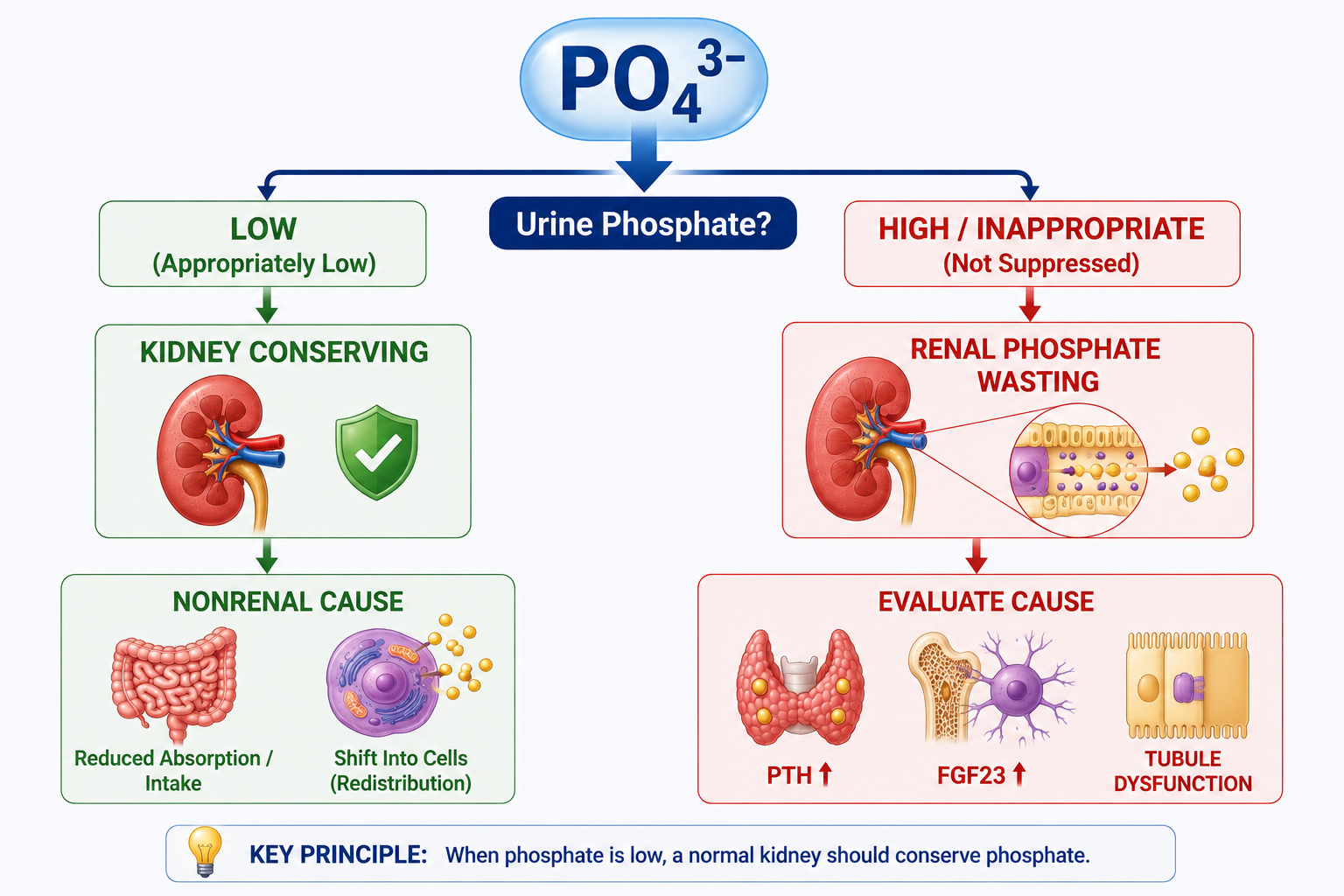
When serum phosphate is low, a normal kidney should conserve phosphate. If urinary phosphate loss remains inappropriate, think renal phosphate wasting.
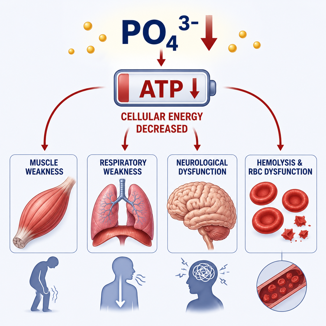
Severe depletion: PO4 low leads to ATP availability falling, which can impair muscle, respiratory, neurological, myocardial and red-cell function.

What Is Hypophosphatemia?
Hypophosphatemia is a serum phosphate concentration below the laboratory reference interval. It can result from intracellular phosphate redistribution, reduced intake or absorption, increased phosphate loss, or a combination of mechanisms.
The exact reference interval varies with laboratory method, age and clinical context, so interpretation should use the local laboratory reference interval rather than one universal value.
The clinical importance of a low phosphate result depends on severity, rate of development, duration, total-body phosphate depletion, symptoms, comorbid illness and the underlying mechanism.
For the physiology behind normal phosphate regulation, see Phosphate Homeostasis Explained.
Why Phosphate Matters
Most body phosphate is in bone and cells. Only a small fraction is extracellular, so serum phosphate is useful but does not directly measure total-body phosphate stores.
Phosphate is essential for ATP, phosphorylation reactions, intracellular signalling, DNA and RNA, phospholipid membranes, red-cell 2,3-BPG and skeletal mineralization.
A fall in serum phosphate may reflect true depletion, redistribution into cells, or both. This distinction is especially important in refeeding and respiratory alkalosis.
Symptoms of Hypophosphatemia
Mild hypophosphatemia may cause no symptoms or only nonspecific symptoms. The underlying illness may dominate the clinical picture.
Severe or prolonged phosphate depletion can affect multiple systems because ATP-dependent cellular function becomes impaired.
| System | Potential Manifestations | Teaching Point |
|---|---|---|
| Skeletal muscle | Generalized weakness, severe neuromuscular dysfunction | Muscle contraction and repair require cellular energy. |
| Respiratory muscles | Respiratory insufficiency, difficulty with ventilator weaning in relevant settings | Severe depletion may impair diaphragm and respiratory muscle function. |
| Muscle injury | Rhabdomyolysis in severe cases | Do not assume hypophosphatemia is the only cause of rhabdomyolysis. |
| Brain | Irritability, confusion, altered mental status, seizures or coma in extreme cases | Neurological features usually reflect substantial depletion or severe illness. |
| Heart | Reduced myocardial performance in severe depletion | There is no unique diagnostic hypophosphatemia ECG pattern. |
| Red blood cells | Reduced 2,3-BPG effect, hemolysis in severe cases | Severe depletion can impair red-cell energy metabolism. |
| White blood cells | Impaired leukocyte function in severe depletion | Avoid claiming hypophosphatemia alone causes infection. |
Why Severe Hypophosphatemia Is Dangerous
The central severity concept is ATP. Severe phosphate depletion reduces phosphate availability for ATP-related cellular energy processes.
Severe hypophosphatemia can therefore contribute to generalized weakness, respiratory muscle weakness, rhabdomyolysis, myocardial dysfunction, neurological abnormalities, seizures, altered consciousness, hemolysis and impaired leukocyte function.
Causes of Hypophosphatemia
Hypophosphatemia results from three major mechanisms: phosphate shifting into cells, reduced phosphate intake or intestinal absorption, and increased phosphate loss through the kidneys or other routes.
The article should not be learned as one long list of causes. Start by naming the mechanism, then identify the cause.
| Mechanism | Examples | Diagnostic Reflex |
|---|---|---|
| Redistribution into cells | Refeeding, insulin-mediated shifts, respiratory alkalosis | Serum phosphate falls because phosphate moves intracellularly. |
| Reduced intake or absorption | Severe malnutrition, malabsorption, vitamin D deficiency, phosphate-binding agents | Kidney should conserve phosphate if renal response is intact. |
| Increased loss | Nonrenal losses or renal phosphate wasting | If kidney is responsible, urinary phosphate remains inappropriate despite low serum phosphate. |
Intracellular Phosphate Redistribution
Serum phosphate can fall rapidly when phosphate moves from extracellular fluid into cells. This can happen without immediate external phosphate loss.
Important redistribution settings include refeeding, insulin-mediated cellular uptake and respiratory alkalosis.
Refeeding Syndrome
During refeeding, increased insulin drives phosphate into cells for ATP production and phosphorylation. In patients with depleted phosphate stores, serum phosphate can fall markedly.
Before feeding, a severely malnourished patient may already have depleted total-body phosphate, potassium and magnesium with reduced physiological reserve. Carbohydrate feeding then increases insulin and anabolic cellular metabolism.
This is not simply inadequate dietary phosphate. It is a shift plus increased intracellular metabolic demand in a patient who may already be depleted.
Respiratory Alkalosis
Respiratory alkalosis raises intracellular pH, stimulates glycolytic activity and increases intracellular phosphate utilization, causing phosphate to shift from extracellular fluid into cells.
This is a high-yield mechanism because hypophosphatemia here may reflect redistribution rather than pure external phosphate loss. For acid-base context, see Respiratory Alkalosis Explained.
Reduced Phosphate Intake and Absorption
Dietary phosphate is widely available, so isolated short-term low intake is not usually the only explanation for severe hypophosphatemia. Reduced intake becomes more important in severe malnutrition, prolonged poor intake and multiple simultaneous causes.
Malabsorption can reduce intestinal phosphate availability. If renal response is intact, urinary phosphate should fall as the kidney conserves phosphate.
Vitamin D deficiency can lower phosphate through reduced intestinal phosphate absorption and, in some settings, secondary hyperparathyroid physiology. Calcitriol normally promotes intestinal phosphate absorption; for calcium-phosphate integration, see Calcium Homeostasis Explained.
Phosphate-binding agents or relevant antacid exposure can also reduce phosphate absorption in susceptible patients.
Renal Phosphate Wasting
Renal phosphate wasting occurs when the kidneys continue excreting an inappropriate amount of phosphate despite hypophosphatemia, when they should normally be conserving phosphate.
When serum phosphate is low, a normal kidney should increase phosphate reabsorption and reduce urinary phosphate loss. Persistent urinary phosphate loss is therefore diagnostically important.
| Pattern | Meaning | Think About |
|---|---|---|
| Low PO4 with appropriate renal conservation | Urinary phosphate is appropriately reduced | Redistribution, reduced intake, reduced absorption or nonrenal loss depending on context |
| Low PO4 with inappropriate urinary phosphate loss | Kidney is not conserving phosphate appropriately | Renal phosphate wasting from PTH, FGF23, proximal tubular dysfunction or medication effects |
PTH and Hypophosphatemia
PTH is phosphaturic. It reduces proximal tubular phosphate reabsorption, increasing urinary phosphate excretion and tending to lower serum phosphate.
This is why hyperparathyroid physiology can produce or contribute to low phosphate, especially when urinary phosphate loss is inappropriate for the serum phosphate concentration.
FGF23 and Hypophosphatemia
FGF23 is also phosphaturic. It reduces renal phosphate reabsorption and increases urinary phosphate excretion.
FGF23 also suppresses calcitriol production. This differs from PTH and may reduce intestinal phosphate absorption.
Therefore FGF23-associated hypophosphatemia can involve both renal phosphate wasting and reduced intestinal phosphate absorption.
Fanconi Syndrome and Phosphate Wasting
Fanconi syndrome is generalized proximal tubular dysfunction, not isolated phosphate loss.
Because phosphate is normally reabsorbed in the proximal tubule, Fanconi syndrome can cause renal phosphate wasting and hypophosphatemia. Other proximal tubular losses may include glucose, amino acids, bicarbonate and uric acid.
The diagnostic clue is a broader proximal tubular pattern rather than phosphate alone.
How to Diagnose Hypophosphatemia
The diagnostic approach begins by confirming the low serum phosphate result in clinical context, then asking whether the patient is clinically affected and why phosphate is low.
- Assess symptoms and severe-depletion features: muscle weakness, respiratory function, neurological status, cardiac context and red-cell features when clinically relevant.
- Identify the mechanism: redistribution into cells, reduced intake or absorption, increased loss, or a combination.
- If phosphate loss is possible, assess urinary phosphate handling relative to the serum phosphate.
- If renal wasting is present, investigate PTH context, FGF23 context, vitamin D, proximal tubular dysfunction, medications and renal function.
How to Interpret Urinary Phosphate
Urinary phosphate is interpreted relative to serum phosphate. A random urinary phosphate concentration alone does not diagnose renal phosphate wasting.
When serum phosphate is low, a normal kidney should conserve phosphate.
If urinary phosphate is appropriately low, the kidney is responding correctly and nonrenal or redistribution mechanisms become more likely. If urinary phosphate loss remains inappropriate, renal phosphate wasting is present.
Fractional Excretion and TmP/GFR
Fractional excretion of phosphate estimates the fraction of filtered phosphate that is excreted in urine. It is useful conceptually because it helps assess whether the kidney is conserving phosphate.
TmP/GFR estimates the renal tubular maximum phosphate reabsorption relative to glomerular filtration. A reduced renal phosphate reabsorptive capacity supports renal phosphate wasting in the correct clinical context.
Do not memorize a single universal cutoff without the laboratory method, clinical setting and local reference framework. The key principle remains whether renal phosphate handling is appropriate for low serum phosphate.
Treatment of Hypophosphatemia
Hypophosphatemia treatment is not only phosphate replacement. Management depends on the mechanism, severity, symptoms and clinical context.
Examples include treating refeeding risk carefully, addressing respiratory alkalosis where appropriate, correcting malnutrition or malabsorption, reviewing medications, treating vitamin D-related problems and investigating renal phosphate wasting.
Oral Phosphate Replacement
Oral phosphate replacement may be appropriate when hypophosphatemia is mild to moderate, the patient can absorb enteral therapy and there is no urgent severe manifestation requiring another route.
Oral replacement still requires attention to renal function, calcium, magnesium, potassium, gastrointestinal tolerance and the underlying cause. For magnesium context during electrolyte correction, see Magnesium Homeostasis Explained.
IV Phosphate Replacement
IV phosphate is considered for selected severe, symptomatic or clinically urgent situations, especially when oral therapy is not suitable or rapid correction is required under supervision.
IV phosphate is not risk-free. Potential complications include hypocalcemia, calcium-phosphate precipitation risk, renal complications, potassium or sodium load depending on preparation, and overcorrection. Monitoring is part of treatment, not an optional extra.
This educational article explains principles only. It does not provide universal phosphate doses, infusion rates or monitoring intervals.
Worked Clinical Cases
Case 1: Refeeding
A malnourished patient restarts carbohydrate feeding and serum phosphate falls. Insulin rises, phosphate shifts into cells and phosphate is used for ATP and phosphorylated intermediates. Total-body stores may already be depleted.
Case 2: Respiratory Alkalosis
A hyperventilating patient has acute hypophosphatemia. The mechanism is intracellular redistribution driven by alkalosis-related glycolytic activity, not necessarily pure phosphate loss.
Case 3: Low Phosphate With Low Urinary Phosphate
The kidney is conserving phosphate appropriately. Think redistribution, reduced intake, reduced absorption or nonrenal loss depending on the clinical setting.
Case 4: Low Phosphate With Persistent Urinary Loss
The kidney is wasting phosphate. Investigate PTH effect, FGF23 effect, proximal tubular dysfunction and medication-related causes.
Case 5: Fanconi Pattern
Low phosphate occurs with other proximal tubular abnormalities. The diagnosis is not isolated phosphate loss; it is generalized proximal tubular dysfunction.
Common Mistakes
- Misconception: Every low phosphate means total-body phosphate depletion. Reality: redistribution can lower serum phosphate without equivalent immediate external loss.
- Misconception: All hypophosphatemia is symptomatic. Reality: mild hypophosphatemia may be asymptomatic.
- Misconception: Any phosphate in urine means renal wasting. Reality: urinary phosphate must be interpreted relative to serum phosphate and clinical context.
- Misconception: Low phosphate should increase urinary phosphate. Reality: a normal kidney should conserve phosphate.
- Misconception: PTH and FGF23 retain phosphate. Reality: both are phosphaturic.
- Misconception: Refeeding hypophosphatemia is simply inadequate dietary phosphate. Reality: insulin-driven intracellular shift and anabolic use are central.
- Misconception: Hypophosphatemia has a diagnostic ECG pattern. Reality: severe depletion may affect the myocardium, but there is no unique diagnostic ECG pattern.
- Misconception: Every patient requires IV phosphate. Reality: route depends on severity, symptoms, urgency and clinical context.
One-Minute Revision
- Hypophosphatemia is serum phosphate below the laboratory reference interval.
- Serum phosphate is not the same as total-body phosphate.
- Three mechanisms cause low phosphate: redistribution into cells, reduced intake or absorption, and increased loss.
- When serum phosphate is low, a normal kidney should conserve phosphate.
- Low phosphate plus inappropriate urinary phosphate loss means renal phosphate wasting.
- PTH and FGF23 are both phosphaturic.
- FGF23 suppresses calcitriol; PTH increases calcitriol.
- Fanconi syndrome is generalized proximal tubular dysfunction, not isolated phosphate loss.
- Severe phosphate depletion can lower ATP availability and impair muscle, respiratory, neurological, myocardial and red-cell function.
- Treatment means correcting the mechanism, replacing phosphate when appropriate and monitoring for complications.
Frequently Asked Questions
Key Take-Home Messages
Low phosphate is not the diagnosis. The mechanism is the diagnosis.
Ask whether phosphate moved into cells, whether too little phosphate entered the body, or whether phosphate was lost. If phosphate loss is possible, ask whether the kidney is conserving phosphate appropriately.
Low serum phosphate with appropriate renal conservation points toward redistribution, reduced intake, reduced absorption or nonrenal loss. Low serum phosphate with inappropriate urinary phosphate loss points toward renal phosphate wasting.
Severe hypophosphatemia matters because phosphate is needed for ATP. Treatment corrects the cause, replaces phosphate when clinically appropriate and monitors carefully for replacement complications.
Next in the Cluster
Next, use Renal Phosphate Wasting Explained to decide whether low phosphate is due to appropriate renal conservation or inappropriate renal phosphate loss. You can also compare the opposite pattern in Hyperphosphatemia Explained.
This article is intended for medical education only. It explains hypophosphatemia physiology, diagnosis and treatment principles, not patient-specific medical advice or a dosing protocol.