Phosphate Homeostasis at a Glance
Gut absorption, bone/cell storage and renal excretion determine serum phosphate. PTH and FGF23 are both phosphaturic, while calcitriol increases intestinal phosphate absorption.
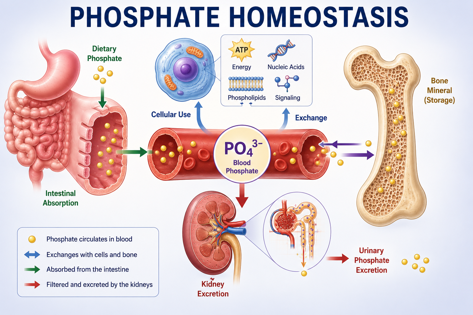
What Is Phosphate Homeostasis?
Phosphate homeostasis is the regulation of phosphate absorption, distribution, storage and excretion through coordinated actions of the intestine, bone, cells, kidneys and hormones including PTH, FGF23 and calcitriol.
Phosphorus is the chemical element. In biological fluids and tissues, phosphorus is largely present in phosphate-containing compounds. Clinically, laboratory measurements are usually discussed as serum phosphate.
The central question is: how does the body maintain phosphate balance, and how do the gut, bone and kidney respond to PTH, FGF23 and vitamin D?
Why Phosphate Is Important
Phosphate is sometimes taught mainly as a component of bone. That is incomplete.
Phosphate is essential for cellular energy transfer, ATP, phosphorylation reactions, nucleic acids, phospholipid cell membranes, intracellular signalling, red-cell 2,3-BPG and skeletal mineralization.
| Function | Why It Matters |
|---|---|
| ATP and energy | Phosphate-containing bonds participate in usable cellular energy transfer. |
| Phosphorylation | Phosphate groups regulate enzymes, receptors, signalling pathways and metabolic control. |
| DNA and RNA | Phosphate forms part of the structural backbone of nucleic acids. |
| Cell membranes | Phospholipids are major structural components of cell membranes. |
| 2,3-BPG | Red-cell 2,3-BPG influences hemoglobin oxygen affinity and tissue oxygen delivery. |
| Bone | Phosphate combines with calcium in skeletal mineral. |
Where Phosphate Is Stored
Most body phosphate is located in bone as mineral associated with calcium. A substantial portion is intracellular. Only a relatively small fraction is present in extracellular fluid.
Serum phosphate represents only a small part of total-body phosphate. A serum phosphate result is useful, but it does not directly measure the entire body's phosphate content.
Changes in serum phosphate may result from altered intestinal absorption, altered renal excretion, movement between extracellular and intracellular compartments, or movement between bone and extracellular fluid.
Intestinal Phosphate Absorption
Phosphate enters the body through dietary intake. Intestinal phosphate absorption occurs through passive/paracellular mechanisms and regulated transcellular transport.
The active vitamin D hormone, calcitriol, increases intestinal absorption of phosphate. Calcitriol also increases intestinal calcium absorption, creating an important link between calcium and phosphate homeostasis.
For the calcium side of this relationship, see Calcium Homeostasis Explained.
Bone and Phosphate
The majority of total-body phosphate is stored in the skeleton. Phosphate and calcium contribute to the mineral phase of bone, so bone acts as a large phosphate reservoir.
Bone is not simply a passive storage container. There is continuous exchange between bone and extracellular fluid through bone formation and resorption.
Bone is also an endocrine participant in phosphate regulation because FGF23 is produced predominantly by bone cells, particularly osteocytes and osteoblast-lineage cells.
Renal Phosphate Handling
For day-to-day phosphate balance, the kidney is a major regulatory organ. Phosphate is filtered at the glomerulus and then partly reabsorbed by the nephron.
The phosphate that is not reabsorbed is excreted in urine. Therefore the kidney can adjust phosphate balance by changing how much filtered phosphate is reclaimed.
The Proximal Tubule and NaPi Transporters
The proximal tubule is the major site of regulated renal phosphate reabsorption. This is a high-yield fact.
Phosphate reabsorption in the proximal tubule depends importantly on sodium-phosphate cotransporters on the apical membrane of proximal tubular cells. Important transporters include NaPi-IIa and NaPi-IIc.
These use the sodium gradient to facilitate phosphate uptake from tubular fluid into proximal tubular cells.

What Is Phosphaturia?
Phosphaturia means increased urinary phosphate excretion. A phosphaturic signal reduces renal phosphate reabsorption.
Two major phosphaturic hormones are PTH and FGF23.
PTH and Phosphate
PTH has a major effect on renal phosphate handling. PTH reduces proximal tubular phosphate reabsorption, increasing urinary phosphate excretion.
PTH reduces the activity or availability of proximal tubular sodium-phosphate cotransporters. Phosphate remains in tubular fluid and urinary phosphate rises.
PTH also stimulates renal production of calcitriol through increased 1-alpha-hydroxylase activity. This increases intestinal calcium absorption and intestinal phosphate absorption.
PTH is phosphaturic: PTH rises, renal phosphate reabsorption falls, urinary phosphate rises.
FGF23 and Phosphate
FGF23 means fibroblast growth factor 23. It is a major hormone regulating phosphate and vitamin D metabolism.
FGF23 is produced predominantly by bone cells, particularly osteocytes and osteoblast-lineage cells. This establishes bone as not only a mineral reservoir but also an endocrine organ involved in phosphate regulation.
FGF23 reduces proximal tubular phosphate reabsorption. FGF23 also suppresses renal calcitriol production and increases pathways of vitamin D inactivation, lowering calcitriol and reducing the drive for intestinal phosphate absorption.
FGF23 and Klotho
Many important renal actions of FGF23 depend on signalling involving Klotho as a co-receptor.
FGF23 plus Klotho-dependent signalling reduces renal phosphate reabsorption and alters vitamin D metabolism.
This article does not expand into detailed receptor molecular biology.
PTH vs FGF23
Both PTH and FGF23 are phosphaturic. Both reduce renal phosphate reabsorption. But their effects on calcitriol differ.
| Effect | PTH | FGF23 |
|---|---|---|
| Proximal tubular phosphate reabsorption | Falls | Falls |
| Urinary phosphate | Rises | Rises |
| Phosphaturia | Rises | Rises |
| NaPi transporter activity/availability | Falls | Falls |
| Calcitriol production | Rises | Falls |
PTH and FGF23 are both phosphaturic, but they have opposite effects on calcitriol production.

Vitamin D and Phosphate
Calcitriol is the active hormonal form of vitamin D. Its important action in phosphate homeostasis is increased intestinal phosphate absorption.
Calcitriol also increases intestinal calcium absorption. Therefore calcitriol supports availability of both minerals required for skeletal mineralization.
Calcium and Phosphate
Calcium and phosphate are both important components of skeletal mineral. Their homeostasis is interconnected through PTH, vitamin D, kidney and bone.
However, they are not regulated identically. A hormone may increase calcium while lowering phosphate. PTH is the classic example.
PTH tends to increase calcium while decreasing phosphate. This is a high-yield clinical pattern.
Excessively high calcium and phosphate concentrations together can favor unwanted calcium-phosphate precipitation in soft tissues, especially in advanced CKD with disturbed mineral metabolism. This article does not expand into treatment targets or calcium-phosphate product thresholds.
How the Kidney Responds to Low Phosphate
When phosphate availability is low, the kidney should conserve phosphate.
This is the phosphate equivalent of asking whether the kidney is responding appropriately to an electrolyte deficiency.
Renal Phosphate Wasting
If phosphate is low but the kidney continues losing an inappropriate amount of phosphate, renal phosphate wasting is present.
Potential hormonal mechanisms include excessive PTH effect and excessive FGF23 effect. Detailed causes belong in the future hypophosphatemia article.
A urinary phosphate value should be interpreted relative to serum phosphate and clinical context, not simply as high or low.
Phosphate Regulation in CKD
With increased phosphate availability and adequate renal function, the kidney can increase phosphate excretion. When renal function declines substantially, phosphate elimination becomes inadequate and phosphate retention can develop.
As functioning nephron mass falls, phosphate homeostasis is initially supported by compensatory hormonal responses. FGF23 rises and PTH may rise to increase phosphate excretion per remaining functioning nephron.
In later CKD, compensatory mechanisms may become insufficient, phosphate retention becomes more pronounced and serum phosphate may rise. This contributes to CKD-mineral and bone disorder, but this article does not become a CKD-MBD management guide.
How to Interpret an Abnormal Phosphate
When serum phosphate is abnormal, ask which part of phosphate balance is abnormal.
| Question | Mechanism |
|---|---|
| Is intestinal input altered? | Phosphate absorption may be reduced or increased. |
| Is renal handling appropriate? | The kidney may conserve phosphate, waste phosphate or fail to eliminate phosphate. |
| Are cellular shifts involved? | Phosphate may move into or out of cells. |
| Is bone/systemic metabolism involved? | Altered mineral metabolism may affect phosphate exchange. |
When serum phosphate is low, ask whether the kidney is conserving phosphate. Low urinary phosphate suggests appropriate conservation. Inappropriate urinary phosphate loss suggests renal phosphate wasting.
When phosphate is high, ask whether the kidney can eliminate phosphate adequately. Detailed differential diagnosis belongs in the future hyperphosphatemia article.
Worked Clinical Cases
Case 1: PTH Excess
A patient has increased PTH and low serum phosphate. Urinary phosphate remains inappropriate for the low serum phosphate. PTH reduces proximal tubular NaPi activity, phosphate reabsorption falls, urinary phosphate rises and serum phosphate falls. PTH is phosphaturic.
Case 2: FGF23 Excess
A patient has low serum phosphate, inappropriate renal phosphate loss and low or inappropriately low calcitriol for the physiological context. FGF23 lowers phosphate through renal phosphaturia plus suppression of calcitriol.
Case 3: Increased Calcitriol Activity
A patient has increased calcitriol activity. The expected intestinal effect is increased phosphate absorption and increased calcium absorption. Vitamin D physiology links calcium and phosphate absorption.
Case 4: Advanced CKD
A patient with advanced CKD develops increasing serum phosphate. Functional nephron mass falls, phosphate excretory capacity falls and phosphate retention rises. FGF23 and PTH may compensate, but eventually compensation becomes insufficient.
Case 5: Low Phosphate With Low Urinary Loss
A patient has low serum phosphate and appropriately reduced urinary phosphate excretion. The kidney is conserving phosphate appropriately, so look toward nonrenal mechanisms such as reduced availability, impaired absorption or intracellular redistribution depending on clinical context.
Case 6: Low Phosphate With Renal Loss
A patient has low serum phosphate but urinary phosphate remains inappropriately high. Renal phosphate wasting is present. Possible physiological drivers include PTH, FGF23 and renal tubular mechanisms.
Common Mistakes
- Misconception: Phosphate is only important for bone. Reality: phosphate is also important for ATP, signalling, nucleic acids, membranes and 2,3-BPG.
- Misconception: Serum phosphate represents total-body phosphate. Reality: most phosphate is in bone and cells.
- Misconception: The kidney only filters phosphate. Reality: the kidney dynamically regulates filtered phosphate reabsorption and excretion.
- Misconception: The distal tubule is the main site of phosphate regulation. Reality: the major regulated site is the proximal tubule.
- Misconception: PTH retains phosphate. Reality: PTH is phosphaturic.
- Misconception: FGF23 retains phosphate. Reality: FGF23 promotes phosphaturia.
- Misconception: PTH and FGF23 have identical effects. Reality: PTH raises calcitriol while FGF23 lowers calcitriol.
- Misconception: Vitamin D only affects calcium. Reality: calcitriol also increases intestinal phosphate absorption.
- Misconception: Low serum phosphate should cause increased urinary phosphate. Reality: the normal kidney should conserve phosphate.
- Misconception: Bone is only a passive phosphate store. Reality: bone is both a reservoir and an endocrine participant through FGF23.
One-Minute Revision
- Most body phosphate is not in serum.
- Bone contains the largest phosphate reservoir, and a substantial phosphate pool is intracellular.
- Phosphate is essential for ATP, signalling, nucleic acids, membranes, 2,3-BPG and skeletal mineralization.
- Calcitriol increases intestinal phosphate absorption and intestinal calcium absorption.
- The kidney determines final phosphate excretion.
- The proximal tubule is the major regulated site of renal phosphate reabsorption.
- NaPi-IIa and NaPi-IIc participate in proximal tubular phosphate transport.
- PTH is phosphaturic and increases calcitriol.
- FGF23 is phosphaturic and suppresses calcitriol.
- When serum phosphate is low, a normal kidney should conserve phosphate.
Key Clinical Pearls
- PTH and FGF23 are both phosphaturic.
- PTH increases calcitriol production.
- FGF23 suppresses calcitriol production.
- FGF23 is produced mainly by bone cells.
- FGF23 signalling involves Klotho.
- Low phosphate should trigger renal phosphate conservation.
- Persistent urinary phosphate loss during hypophosphatemia suggests renal phosphate wasting.
- Renal failure reduces phosphate elimination.
- FGF23 and PTH may compensate during earlier CKD.
- Advanced CKD can eventually produce phosphate retention and hyperphosphatemia.
Frequently Asked Questions
Key Take-Home Messages
Phosphate homeostasis becomes easier when reduced to the interaction between gut, bone, kidney and hormones.
Calcitriol increases intestinal phosphate and calcium absorption. Most body phosphate is stored in bone, while a substantial amount is intracellular. The kidney determines final phosphate excretion through regulated proximal tubular reabsorption.
The central hormonal framework is: PTH increases urinary PO4 and increases calcitriol; FGF23 increases urinary PO4 and decreases calcitriol; calcitriol increases gut PO4 absorption.
When phosphate becomes low, the kidney should conserve it. Low phosphate with inappropriate urinary phosphate loss suggests renal phosphate wasting. When renal function becomes severely impaired, phosphate elimination falls and phosphate retention can develop.
This article is intended for medical education only. It explains phosphate physiology and interpretation principles, not treatment protocols, phosphate replacement, CKD-MBD management or patient-specific care.