Vitamin D Metabolism at a Glance
Vitamin D metabolism converts vitamin D from skin or diet first to 25-OH vitamin D in the liver and then principally to the active hormone calcitriol in the kidney. Calcitriol regulates calcium, phosphate and PTH physiology.
Calcitriol increases intestinal calcium absorption, increases intestinal phosphate absorption and contributes to suppression of PTH secretion.

The Most Important Laboratory Distinction
25-OH vitamin D is not the same as 1,25-(OH)2 vitamin D.
| Measurement | Meaning | Main Use |
|---|---|---|
| 25-OH vitamin D | Main circulating vitamin D status marker. | Assessing nutritional vitamin D status. |
| 1,25-(OH)2 vitamin D | Biologically active hormone, also called calcitriol. | Selected clinical contexts; not the routine nutritional deficiency test. |
Serum 1,25-(OH)2 vitamin D is not the routine test for nutritional vitamin D deficiency. A patient can have low 25-OH D while calcitriol is normal or even increased if secondary hyperparathyroidism stimulates renal calcitriol production and renal function is preserved.
What Is Vitamin D?
Vitamin D is a precursor to a biologically active steroid hormone system that plays a central role in calcium and phosphate homeostasis.
Its major physiological importance comes from its active hormonal form, 1,25-dihydroxyvitamin D, also called calcitriol.
Calcitriol acts through the vitamin D receptor, or VDR, and influences multiple tissues involved in mineral metabolism.
- It increases intestinal calcium absorption.
- It increases intestinal phosphate absorption.
- It supports normal bone mineralization.
- It participates in negative feedback regulation of PTH.
Vitamin D Is Not Active Immediately
Vitamin D obtained from skin or diet must undergo metabolic activation before it becomes the main active hormone.
The liver step mainly creates the circulating status marker. The kidney step creates the active hormone under tight hormonal control.

Where Vitamin D Comes From
Vitamin D comes from two broad sources: skin synthesis and dietary intake.
Skin synthesis
In skin, UVB exposure converts 7-dehydrocholesterol to previtamin D3, which undergoes thermal conversion to vitamin D3, also called cholecalciferol.
Cutaneous vitamin D synthesis varies with UVB exposure, latitude, season, skin pigmentation, age, clothing, sunscreen use and time spent outdoors.
Dietary vitamin D
Diet can provide vitamin D as vitamin D2, or ergocalciferol, and vitamin D3, or cholecalciferol. Both can enter the vitamin D metabolic pathway.
This article focuses on pathway physiology rather than comparing supplement preparations.
Vitamin D-Binding Protein
Vitamin D metabolites circulate largely bound to vitamin D-binding protein, with albumin also contributing to binding.
This binding helps transport vitamin D metabolites through the circulation. The clinically important idea for this article is that vitamin D physiology is a circulating endocrine system, not only a local nutrient effect.
The Liver Step: 25-Hydroxylation
The liver converts vitamin D to 25-hydroxyvitamin D, commonly written as 25-OH vitamin D.
25-OH vitamin D is the main circulating marker used to assess vitamin D supply and nutritional vitamin D status.
Vitamin D supply/status is mainly assessed with 25-OH vitamin D.
The Kidney Step: 1 Alpha Hydroxylation
The kidney converts 25-OH vitamin D to 1,25-(OH)2 vitamin D, also called calcitriol.
This reaction is mediated by renal 1 alpha hydroxylase. It is not merely a passive supply step; it is regulated by calcium, phosphate, PTH, FGF23 and kidney function.
| Regulator | Effect on Calcitriol Physiology |
|---|---|
| PTH | Stimulates renal 1 alpha hydroxylase and increases calcitriol production when renal function permits. |
| FGF23 | Suppresses calcitriol production and promotes vitamin D catabolism. |
| Calcium/phosphate state | Feeds into PTH and FGF23 signals and therefore influences calcitriol physiology. |
| Kidney function | Determines how well renal activation can respond to regulatory signals. |
PTH and FGF23: The Two Major Hormonal Regulators
PTH
PTH rises when calcium availability is inadequate. One of its important actions is to stimulate renal calcitriol production.
FGF23
FGF23 is a phosphate-regulating hormone produced mainly by osteocytes and osteoblasts. It promotes renal phosphate excretion and suppresses calcitriol production.
How Calcitriol Affects Calcium and Phosphate
Calcitriol increases intestinal absorption of both calcium and phosphate. This is central to understanding why vitamin D is linked to mineral homeostasis and bone mineralization.
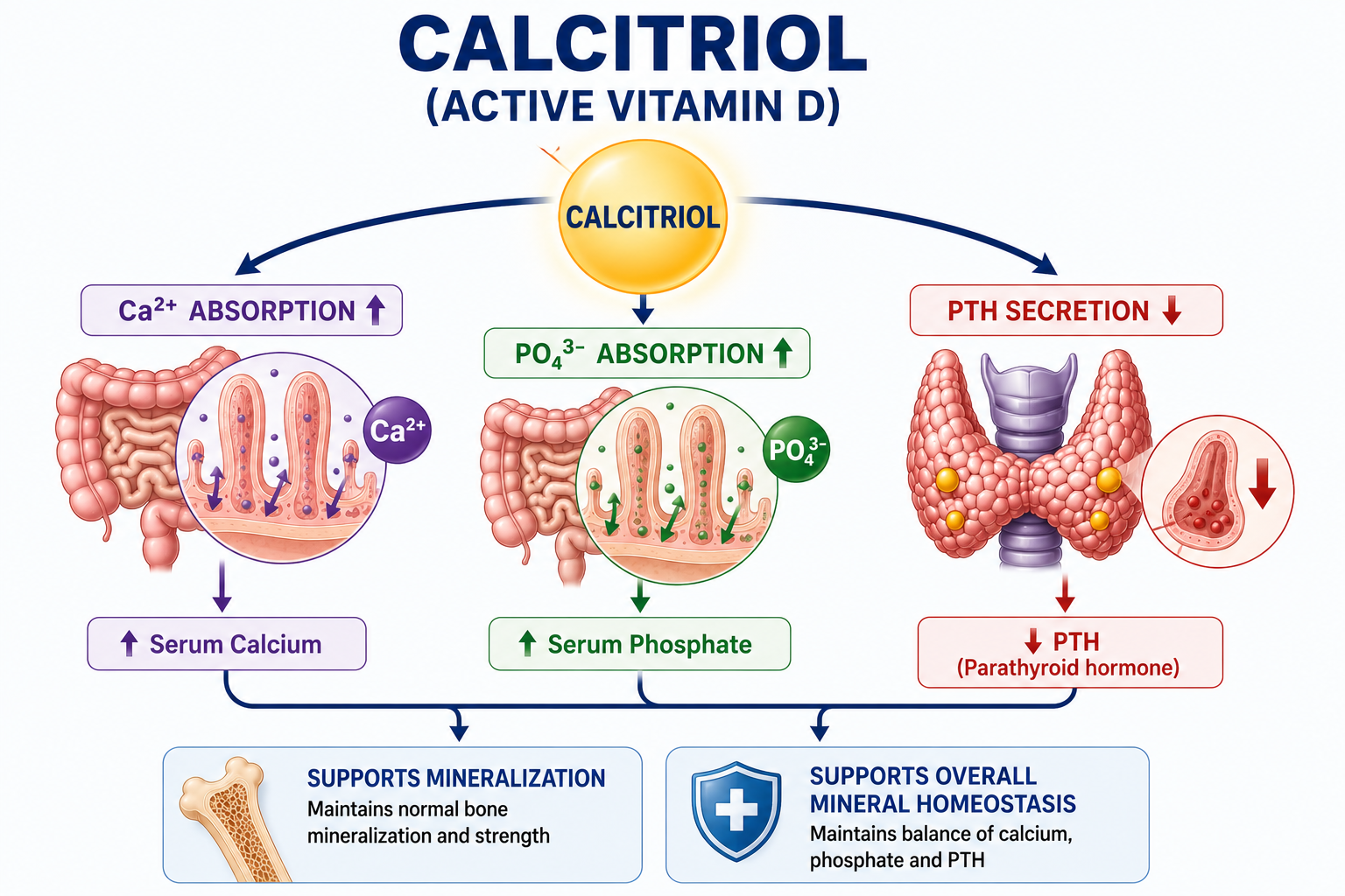
| Target | Main Effect | Clinical Meaning |
|---|---|---|
| Intestine | Increased calcium absorption. | Improves calcium availability. |
| Intestine | Increased phosphate absorption. | Supports mineral availability but interacts with phosphate burden in CKD. |
| Parathyroid gland | Contributes to suppression of PTH. | Forms part of vitamin D-PTH negative feedback. |
| Bone | Supports mineralization through calcium and phosphate availability. | Deficiency can impair mineralization physiology. |
Vitamin D and Bone Mineralization
Bone mineralization requires adequate calcium and phosphate availability. Vitamin D supports this indirectly and directly by maintaining intestinal mineral absorption and participating in endocrine feedback.
When vitamin D supply is inadequate, reduced calcium absorption can stimulate PTH. Persistent PTH excess can shift bone and renal handling in ways that defend serum calcium but may be costly for skeletal mineral physiology.
What Happens in Vitamin D Deficiency?
In nutritional vitamin D deficiency, the key laboratory abnormality is low 25-OH vitamin D. Calcitriol does not have to be low.
Why calcium can remain normal
Serum calcium can remain normal because PTH increases renal calcium conservation and mobilizes compensatory mechanisms to defend extracellular calcium.
Why phosphate can fall
PTH promotes renal phosphate excretion. Therefore, vitamin D deficiency with secondary hyperparathyroidism can produce a low or low-normal phosphate pattern, especially when renal function is preserved.
Why calcitriol can remain normal
Calcitriol can remain normal or increased because PTH stimulates renal 1 alpha hydroxylase when kidney function permits.
This is why low calcitriol is not required for nutritional vitamin D deficiency.
For the PTH response, see why vitamin D deficiency causes secondary hyperparathyroidism.
Vitamin D Metabolism in CKD
CKD changes vitamin D physiology in a different way from simple nutritional deficiency. Declining kidney function reduces the kidney's ability to generate calcitriol appropriately.
FGF23 often rises early in CKD and suppresses calcitriol production. Later, reduced functioning nephron mass and phosphate retention contribute further to disturbed calcium, phosphate and PTH physiology.
For the wider mineral pathway, see CKD-MBD explained.
25-OH Vitamin D vs Calcitriol
The most common testing mistake is ordering or interpreting calcitriol as though it were the same as nutritional vitamin D status.
| Clinical Question | More Relevant Test | Reason |
|---|---|---|
| What is the patient's vitamin D supply/status? | 25-OH vitamin D | It is the main circulating status marker. |
| Is active hormone physiology altered in a selected context? | 1,25-(OH)2 vitamin D | Calcitriol is tightly regulated by PTH, FGF23, calcium/phosphate physiology and kidney function. |
| Can nutritional deficiency exist with normal calcitriol? | Yes | PTH can stimulate calcitriol production if kidney response is preserved. |
Nutritional vitamin D status points to 25-OH D. Vitamin D activation points to calcitriol. Vitamin D regulation points to PTH, FGF23, calcium, phosphate and kidney function.
Basic Laboratory Patterns
Laboratory interpretation should combine 25-OH vitamin D, calcium, phosphate, PTH, kidney function and the clinical setting.
| Pattern | Possible Interpretation | Teaching Point |
|---|---|---|
| Low 25-OH D, high PTH, normal calcium | Vitamin D deficiency with compensated secondary hyperparathyroidism. | Normal calcium does not exclude vitamin D-driven PTH stimulation. |
| Low 25-OH D, high PTH, low phosphate | PTH-mediated phosphaturia may be contributing. | Phosphate can fall when PTH increases renal phosphate excretion. |
| CKD, high PTH, high phosphate, low/low-normal calcitriol physiology | CKD-MBD physiology. | Reduced renal activation and FGF23 effects matter. |
| Low 25-OH D with normal calcitriol | Nutritional deficiency can still be present. | Calcitriol is regulated, not a simple store marker. |
Treatment Principles
This article is not a dosing guide. Treatment depends on the clinical problem being treated: nutritional vitamin D deficiency, CKD-related altered calcitriol physiology, malabsorption, phosphate burden or another cause of abnormal mineral metabolism.
- For nutritional status, 25-OH vitamin D is the key marker.
- For high PTH, interpret the driver before treating a number.
- For CKD-MBD physiology, assess calcium, phosphate, PTH, vitamin D status, kidney function and the broader CKD context.
- Avoid confusing nutritional vitamin D replacement principles with active vitamin D therapy principles.
Worked Clinical Cases
Case 1: Low 25-OH D, high PTH, normal calcium
This pattern can occur when vitamin D deficiency reduces intestinal calcium absorption, but PTH rises enough to defend serum calcium. The normal calcium should not make the low 25-OH D irrelevant.
Case 2: Low 25-OH D with normal calcitriol
This does not exclude nutritional vitamin D deficiency. PTH-driven renal activation may preserve or increase calcitriol when renal function is intact.
Case 3: CKD with high phosphate and high PTH
This points toward CKD-MBD physiology. Phosphate retention, FGF23 elevation, impaired calcitriol production and reduced calcium absorption may all contribute.
Common Mistakes
- Using 25-OH vitamin D and active vitamin D as interchangeable terms.
- Thinking calcitriol must be low to diagnose nutritional vitamin D deficiency.
- Assuming the kidney is the only determinant of calcitriol concentration.
- Ignoring PTH when calcium is normal.
- Forgetting that FGF23 suppresses calcitriol production.
- Treating this physiology article as a supplement dosing guide.
Vitamin D Metabolism in One Minute
Vitamin D comes from skin synthesis and diet. It is converted in the liver to 25-OH vitamin D, the main marker of vitamin D status. It is then converted principally in the kidney to calcitriol, the active hormone.
PTH stimulates renal calcitriol production when renal function permits. FGF23 suppresses calcitriol production and promotes vitamin D catabolism. Calcitriol increases intestinal calcium and phosphate absorption and contributes to PTH suppression.
25-OH D tells you about vitamin D status. Calcitriol is the regulated active hormone. Do not confuse the two.
Frequently Asked Questions
Key Take-Home Messages
- Vitamin D from skin or diet must be activated before becoming calcitriol.
- 25-OH vitamin D is the main nutritional vitamin D status marker.
- Calcitriol is 1,25-(OH)2 vitamin D, the regulated active hormone.
- PTH stimulates renal calcitriol production when renal function permits.
- FGF23 suppresses calcitriol production and promotes vitamin D catabolism.
- Vitamin D deficiency can raise PTH even when serum calcium is normal.
- CKD changes calcitriol physiology through FGF23, reduced renal activation and phosphate handling.
Next in the Cluster
Next, review Vitamin D Deficiency Explained to connect low 25-OH vitamin D with calcium, phosphate, PTH, osteomalacia risk and treatment principles.
This article is intended for medical education only. It explains vitamin D metabolism and mineral physiology, not patient-specific medical advice, universal vitamin D thresholds, supplement schedules, CKD-specific active vitamin D dosing, target PTH values or treatment regimens.