Bone can die before the femoral head collapses
Perfusion failure → ischemia → osteocyte and marrow-cell death → necrotic segment.
Necrosis ≠ collapse. Subchondral fracture marks structural failure.
What Is Osteonecrosis of the Femoral Head?
Terminology
Use:
OSTEONECROSIS OF THE FEMORAL HEAD — ONFHas the preferred term.
Explain once that commonly used alternative terms include:
- avascular necrosis;
- AVN;
- ischemic necrosis;
- aseptic necrosis.
ARCO adopted osteonecrosis as standardized terminology for these necrotic femoral-head lesions. (PubMed Central (PMC))
For SEO, naturally use both:
osteonecrosis
and:
avascular necrosis / AVN
Central Teaching Concept
BONE CAN DIE BEFORE THE FEMORAL HEAD COLLAPSESCentral pathway:
VASCULAR / PERFUSION FAILURE ISCHEMIA MARROW-CELL + OSTEOCYTE DEATH NECROTIC FEMORAL-HEAD SEGMENT REPARATIVE RESPONSE AT THE INTERFACE SUBCHONDRAL MECHANICAL WEAKNESS SUBCHONDRAL FRACTURE FEMORAL-HEAD COLLAPSE SECONDARY OSTEOARTHRITISThe most important distinction is:
NECROSIS ≠ COLLAPSEOsteonecrosis can exist while the femoral head is still structurally preserved.
This creates the therapeutic window for:
JOINT-PRESERVING TREATMENTOpening — Use This Content
A patient receiving long-term glucocorticoid treatment develops deep groin pain.
The hip radiograph looks normal.
Does that exclude major structural bone disease?
NO.The patient may already have:
OSTEONECROSIS OF THE FEMORAL HEADIn early ONFH, bone and marrow cells have died within part of the femoral head, but the overall shape of the femoral head may remain intact.
Plain radiography may therefore still appear normal.
MRI can detect the disease at this stage.
If mechanical failure later develops, the sequence becomes:
SUBCHONDRAL FRACTURE COLLAPSE SECONDARY OSTEOARTHRITISThe key clinical goal is therefore not merely to ask:
“Is osteonecrosis present?”
It is also to ask:
“Is the femoral head still structurally preservable?”
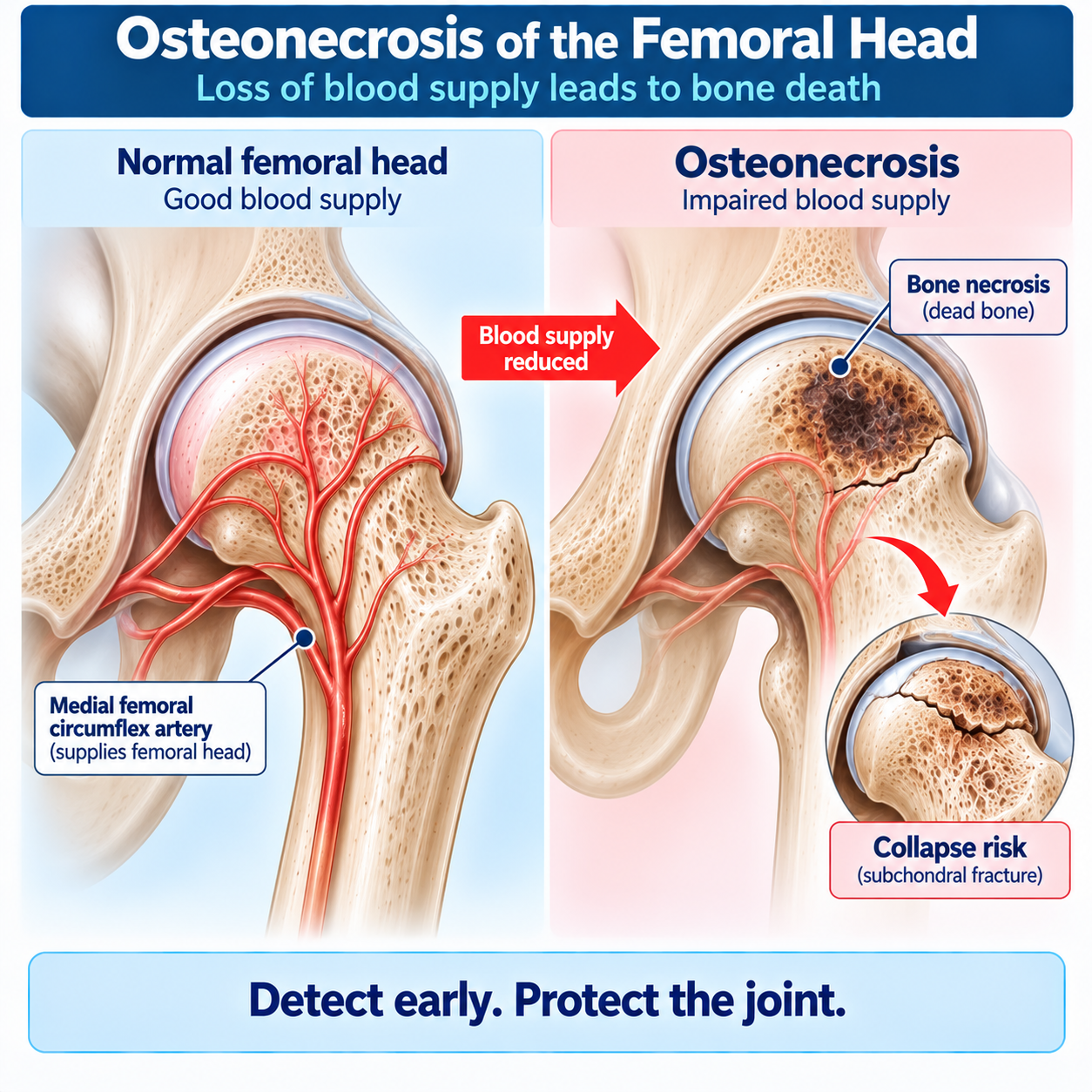
Normal Femoral-Head Blood Supply
The femoral head depends on a relatively vulnerable vascular network.
In adults, an important blood supply is provided by retinacular vessels arising predominantly from branches of the:
MEDIAL FEMORAL CIRCUMFLEX ARTERYAdditional vascular contributions exist, but the educational point is:
THE FEMORAL HEAD IS VULNERABLE TO INTERRUPTION OF ITS BLOOD SUPPLYThis is especially obvious after:
- femoral-neck fracture;
- hip dislocation.
What Is Osteonecrosis?
Osteonecrosis is:
DEATH OF BONE AND MARROW CELLS FOLLOWING FAILURE OF THE LOCAL BIOLOGICAL ENVIRONMENT TO MAINTAIN VIABILITYIn ONFH, this occurs within a segment of the femoral head.
Histological features include:
- marrow necrosis;
- osteocyte death;
- reparative fibrovascular tissue surrounding the necrotic region. (PubMed Central (PMC))
The necrotic bone does not instantly disappear.
That is crucial.
Dead Bone May Initially Retain Its Shape
Immediately after cellular death, the mineralized structural framework can remain present.
Therefore:
DEAD BONE CAN STILL LOOK STRUCTURALLY INTACTfor a period.
The clinical problem develops because normal living bone must withstand repeated mechanical loading.
As repair progresses around necrotic bone, mechanical weakness can develop.
Eventually:
SUBCHONDRAL FRACTUREmay occur.
That is the major transition toward collapse.
Why The Femoral Head Collapses
The femoral head is repeatedly loaded during:
- standing;
- walking;
- running;
- stair climbing.
The subchondral region beneath the articular cartilage must withstand substantial mechanical stress.
In ONFH:
NECROTIC BONE REPARATIVE REMODELING REPEATED MECHANICAL LOADINGcan eventually exceed structural capacity.
Then:
SUBCHONDRAL FRACTUREdevelops.
Further loading can lead to:
DEPRESSION / FLATTENING OF THE FEMORAL HEADand ultimately:
SECONDARY OSTEOARTHRITIS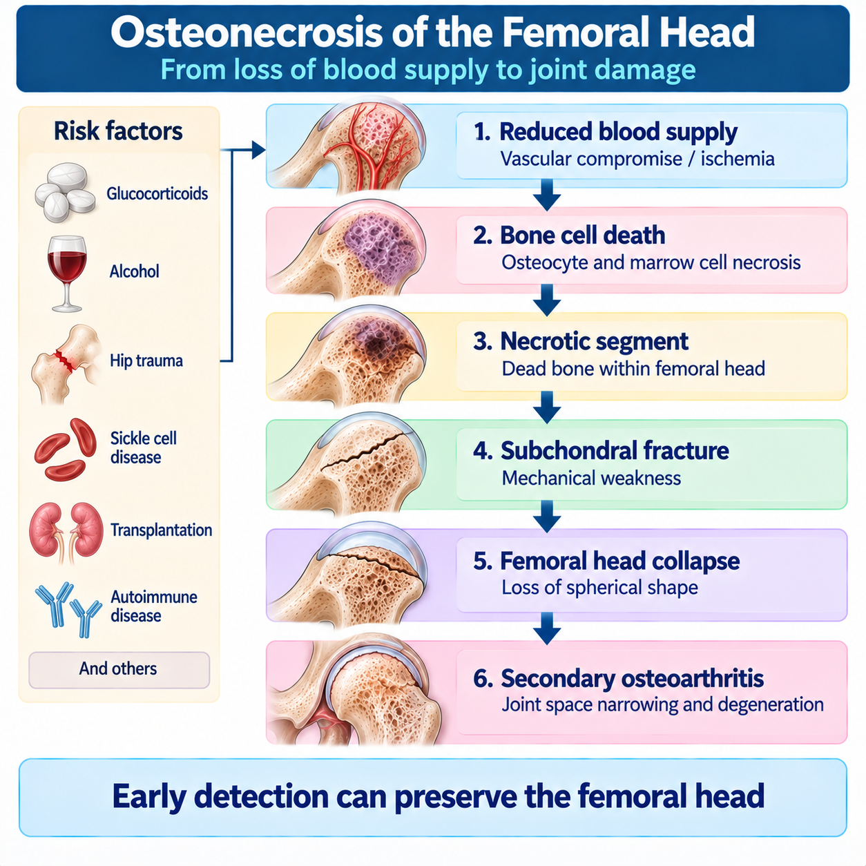
Causes and Risk Factors
Two Broad Categories
Teach ONFH using:
Traumatic osteonecrosis
A clear mechanical vascular insult occurs.
Examples:
- femoral-neck fracture;
- hip dislocation.
Non-traumatic osteonecrosis
No single major traumatic vascular event explains the disease.
Important associations include:
- glucocorticoid exposure;
- heavy alcohol exposure;
- sickle-cell disease and other hemoglobinopathies;
- autoimmune disease;
- organ transplantation;
- selected coagulation disorders;
- decompression sickness;
- other systemic/metabolic disorders;
- idiopathic disease.
The 2026 ARCO evidence review lists corticosteroids, excessive alcohol exposure, hemoglobinopathies, autoimmune disease, solid-organ transplantation and idiopathic disease among major non-traumatic contexts. (PubMed Central (PMC))
Traumatic ONFH
After a femoral-neck fracture or hip dislocation, vessels supplying the femoral head may be:
- torn;
- compressed;
- disrupted.
Therefore:
TRAUMA VASCULAR INTERRUPTION ISCHEMIA OSTEONECROSISThe risk depends on the injury pattern and vascular damage.
Non-Traumatic ONFH Is Multifactorial
Non-traumatic ONFH is not explained by thrombosis alone.
or:
“All steroid ONFH is caused by fat emboli.”
Current understanding is more complex.
Potential interacting mechanisms include:
- vascular dysfunction;
- thrombosis/hypercoagulability;
- hypofibrinolysis;
- impaired angiogenesis;
- lipid/fat-cell abnormalities;
- increased intraosseous pressure;
- cellular injury;
- impaired repair;
- genetic susceptibility.
Therefore:
MULTIPLE PATHWAYS → IMPAIRED PERFUSION / REPAIR → OSTEONECROSISARCO reviews emphasize multifactorial pathogenesis and individual susceptibility. (PubMed Central (PMC))
Glucocorticoids
Glucocorticoid exposure is one of the most important non-traumatic associations.
Potential mechanisms include:
- lipid metabolism changes;
- marrow adipocyte enlargement;
- vascular dysfunction;
- altered coagulation;
- cellular injury;
- impaired osteogenic repair.
Important:
STEROID EXPOSURE ≠ INEVITABLE OSTEONECROSISRisk depends on multiple factors, including exposure and individual susceptibility.
Alcohol
Heavy alcohol exposure is another established association.
Potential mechanisms overlap with steroid-associated disease, including:
- altered lipid metabolism;
- marrow changes;
- vascular dysfunction;
- impaired osteogenic differentiation.
Again:
ALCOHOL ASSOCIATION ≠ ONE UNIVERSAL DIAGNOSTIC THRESHOLDSickle-Cell Disease
Sickle-cell disease can cause repeated microvascular obstruction.
Therefore:
SICKLING MICROVASCULAR OCCLUSION BONE ISCHEMIA OSTEONECROSISThe femoral head is an important site.
This creates a strong connection between hematology and orthopedic pathology.
Autoimmune Disease
ONFH may occur in patients with systemic autoimmune disease.
Interpret carefully because risk may involve:
- the underlying systemic disease;
- glucocorticoid exposure;
- vascular/coagulation factors;
- combinations of these.
ONFH in a patient with SLE should not automatically be attributed solely to steroid exposure.
Organ Transplantation
Transplant recipients can develop ONFH.
Relevant contributors may include:
- glucocorticoid exposure;
- underlying disease;
- metabolic changes;
- other systemic factors.
Again, avoid reducing a multifactorial problem to one mechanism.
Idiopathic ONFH
Sometimes no definite cause is identified.
This is commonly termed:
IDIOPATHIC OSTEONECROSIS“Idiopathic” means:
NO CAUSE IDENTIFIED AFTER APPROPRIATE EVALUATIONnot:
NO BIOLOGICAL CAUSE EXISTSBilateral Disease
Non-traumatic ONFH may involve both hips.
Therefore:
UNILATERAL PAIN ≠ NECESSARILY UNILATERAL DISEASEA patient can have:
- symptomatic ONFH on one side;
- asymptomatic ONFH on the other.
Contralateral evaluation depends on clinical context.
Symptoms and Examination
Symptoms
Early ONFH may be:
ASYMPTOMATICWhen symptoms develop, the typical complaint is:
HIP / GROIN PAINPain may also be perceived in:
- buttock;
- thigh;
- occasionally around the knee.
Symptoms often worsen with weight bearing.
Later disease may produce:
- reduced range of motion;
- limp;
- progressive functional limitation.
Physical Examination
Early disease may have relatively limited examination findings.
As disease progresses, findings can include:
- painful hip movement;
- restricted internal rotation;
- reduced range of motion;
- antalgic gait.
Physical examination does not reliably exclude early ONFH.
Therefore:
CLINICAL SUSPICION + IMAGING MATTERImaging and MRI Diagnosis
Initial Imaging
For clinically suspected osteonecrosis:
PLAIN RADIOGRAPHY IS THE USUAL INITIAL IMAGING STUDYThe ACR Appropriateness Criteria rate radiography of the area of interest as usually appropriate for initial evaluation. (ACSearch)
For the hip, obtain the standard clinically appropriate hip radiographic views according to local practice.
The Major Limitation Of X-Ray
The most important imaging lesson is:
A NORMAL RADIOGRAPH DOES NOT EXCLUDE EARLY ONFHThis is literally incorporated into ARCO stage I:
X-RAY NORMALbut:
MRI ABNORMALTherefore a normal X-ray cannot end the investigation when clinical suspicion remains substantial.
When MRI Is Needed
When:
- osteonecrosis is clinically suspected;
- and radiographs are normal or suspicious but nondiagnostic;
the next study should generally be:
MRI WITHOUT IV CONTRASTThe ACR rates MRI without contrast as usually appropriate in this situation. (ACSearch)
Therefore:
SUSPECT ONFH + NORMAL/NONDIAGNOSTIC X-RAY → MRIThis should be one of the article's strongest memory statements.
MRI
MRI is the key imaging modality for:
EARLY ONFHIt can identify the necrotic segment before plain radiographic abnormalities develop.
MRI also helps assess:
- lesion extent;
- lesion location;
- surrounding marrow reaction;
- bilateral disease where imaged.
The 2026 systematic review supporting ARCO guideline development concluded that MRI had the strongest diagnostic performance among evaluated imaging modalities. (PubMed Central (PMC))
MRI is highly useful but not infallible.
T1 Low-Signal Band
A characteristic MRI finding is:
A LOW-SIGNAL BAND ON T1-WEIGHTED MRIaround the necrotic region.
This feature forms part of the revised ARCO stage-I definition. (PubMed Central (PMC))
The band helps demarcate the viable-necrotic interface.
Double-Line Sign
On T2-weighted imaging, the classic:
DOUBLE-LINE SIGNmay be seen.
Conceptually, it represents changes around the reactive interface between necrotic and viable tissue.
Important:
THE DOUBLE-LINE SIGN IS A CLASSIC CLUE — NOT A REQUIREMENT IN EVERY CASEThis sign alone cannot diagnose or exclude ONFH.
Bone-Marrow Edema
Bone-marrow edema can occur, particularly in symptomatic or more mechanically stressed disease.
But:
BONE-MARROW EDEMA IS NOT SPECIFIC FOR ONFHIt also occurs in:
- insufficiency fracture;
- transient osteoporosis/bone-marrow-edema syndrome;
- arthritis;
- trauma;
- infection;
- other disorders.
Interpret it with lesion morphology and clinical context.
CT
CT is particularly useful for evaluating:
SUBCHONDRAL FRACTUREand:
STRUCTURAL COLLAPSEIt can complement radiography/MRI when detailed bony architecture is important.
ARCO stage III specifically uses subchondral fracture demonstrated on:
X-RAY OR CTas a defining feature. (PubMed Central (PMC))
The Crescent Sign
The classic:
CRESCENT SIGNrepresents:
SUBCHONDRAL FRACTUREIt reflects structural failure beneath the articular surface.
“Crescent sign = simply another early MRI sign.”
Instead teach:
CRESCENT SIGN = MECHANICAL FAILURE HAS BEGUNThis has major prognostic and therapeutic implications.
ARCO Staging
ARCO Staging
Use the 2019 revised ARCO staging system.
Create this table exactly in concept:
| ARCO stage | Key imaging finding | Structural interpretation |
|---|---|---|
| I | X-ray normal; MRI abnormal | Osteonecrosis present, no radiographic structural change |
| II | X-ray abnormal; no subchondral fracture/collapse | Structural changes, still pre-collapse |
| III | Subchondral fracture ± collapse | Structural failure |
| IIIA | Femoral-head depression ≤2 mm | Early collapse |
| IIIB | Femoral-head depression >2 mm | More advanced collapse |
| IV | Secondary hip osteoarthritis | End-stage joint degeneration |
The revised system removed the old stage 0 and divided stage III at 2 mm of head depression. (PubMed Central (PMC))
ARCO Stage I
X-RAY NORMALbut:
MRI ABNORMALwith a characteristic low-signal band.
This is the clearest demonstration that:
NORMAL RADIOGRAPH ≠ NO OSTEONECROSISAt this stage:
THE FEMORAL HEAD HAS NOT YET RADIOGRAPHICALLY COLLAPSEDARCO Stage Ii
Stage II has:
ABNORMAL RADIOGRAPHPotential findings include:
- sclerosis;
- cystic change;
- focal osteoporotic change.
But critically:
NO SUBCHONDRAL FRACTUREand:
NO FEMORAL-HEAD FLATTENING/COLLAPSETherefore stage II remains:
PRE-COLLAPSE DISEASEARCO Stage III
Stage III is defined by:
SUBCHONDRAL FRACTUREon X-ray or CT, with or without depression/flattening.
This is the major structural turning point.
STAGE III = THE FEMORAL HEAD HAS BEGUN TO FAIL MECHANICALLYARCO IIIa Versus IIIb
Use:
IIIA
FEMORAL-HEAD DEPRESSION ≤2 mmIIIB
FEMORAL-HEAD DEPRESSION >2 mmThis distinction comes from the revised 2019 ARCO staging system. (PubMed Central (PMC))
This classification threshold is not an automatic treatment threshold.
ARCO Stage IV
Stage IV means:
SECONDARY OSTEOARTHRITISPotential findings include:
- joint-space narrowing;
- acetabular changes;
- progressive femoral-head deformity;
- degenerative destruction.
At this point the disease is no longer only a necrotic bone problem.
It has become:
A WHOLE-JOINT DEGENERATIVE PROBLEM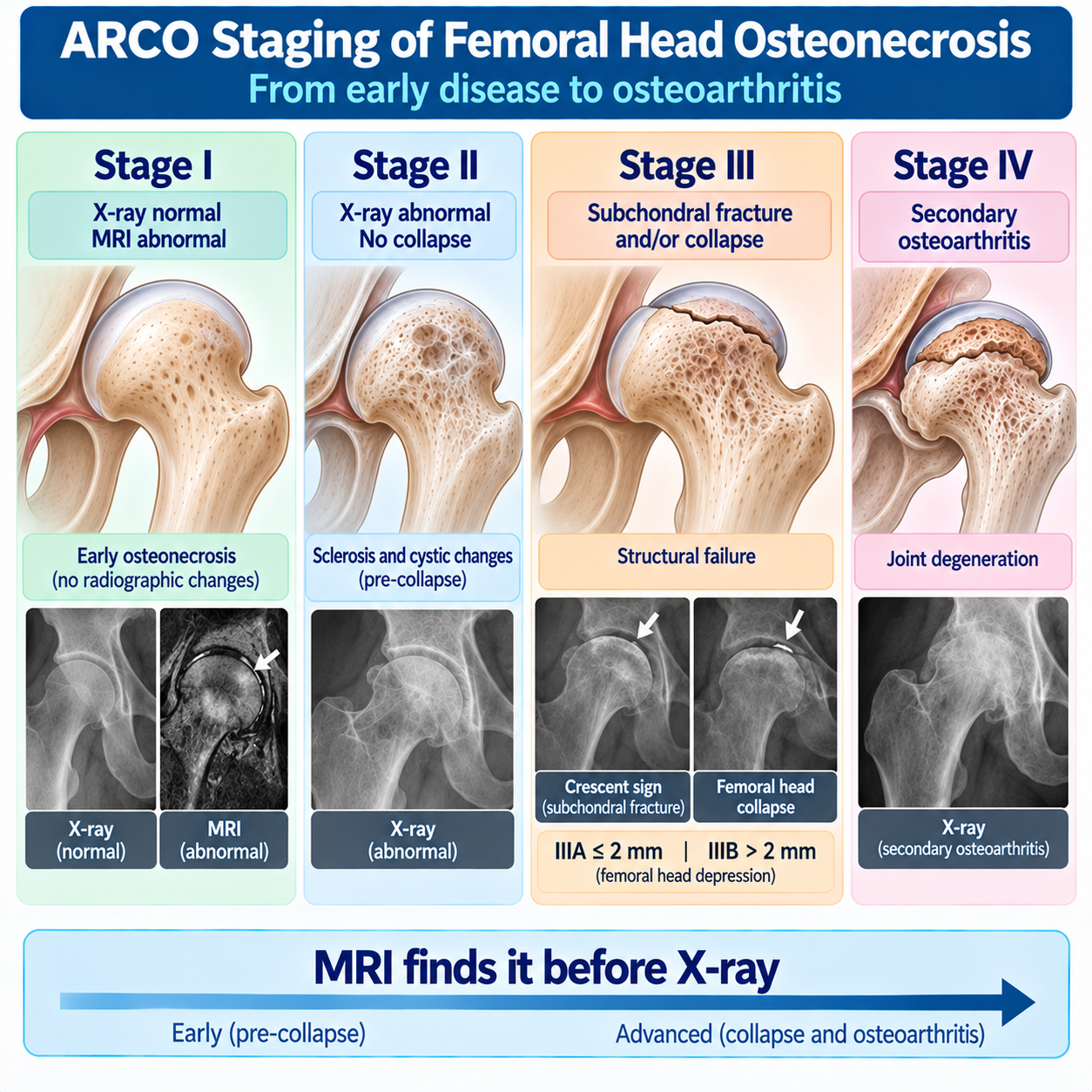
Lesion Size, Location and Prognosis
Stage Is Not Enough
This is an essential section.
ARCO II lesions do not all behave the same.
Prognosis also depends strongly on:
SIZE OF THE NECROTIC LESIONand:
LOCATION OF THE NECROTIC LESIONARCO reviews identify size/location as major predictors of collapse. (PubMed Central (PMC))
Therefore:
STAGE + SIZE + LOCATION = MUCH BETTER CLINICAL INFORMATIONWhy Lesion Location Matters
The lateral portion of the femoral head contributes heavily to the:
WEIGHT-BEARING DOMEA lesion extending into this region experiences substantial mechanical loading.
Therefore:
LARGE LATERAL WEIGHT-BEARING LESIONgenerally has greater collapse risk than:
SMALL MEDIAL LESIONeven when both are nominally the same ARCO stage.
ARCO 2021 Size/Location Classification
For advanced students, briefly include the 2021 ARCO early-stage classification described in the updated ARCO literature.
Type 1
Small lesion whose lateral necrotic margin remains medial to the femoral-head apex.
Type 2
Medium lesion with the lateral margin between the apex and lateral acetabular edge.
Type 3
Large lesion extending laterally beyond the lateral acetabular edge.
This classification reinforces:
LATERAL EXTENSION MATTERSThe ARCO review recommends this approach as a useful unified early-stage size/location classification. (PubMed Central (PMC))
This classification is distinct from the 2019 stage I–IV system.
Modified Kerboul Angle
Mention as an established alternative method for quantifying lesion extent on MRI.
Concept:
Measure the necrotic arc on:
- mid-coronal MRI;
- mid-sagittal MRI;
then combine the angles.
Larger combined necrotic angles correlate with greater collapse risk. (PubMed Central (PMC))
No single classification system is universally mandatory.
The educational message is:
QUANTIFYING LESION EXTENT IMPROVES PROGNOSTICATIONNatural History
Once the osteonecrotic lesion has formed, evidence suggests that its initial:
SIZE / LOCATIONlargely determines its subsequent structural risk.
Small lesions may remain stable.
Large, laterally located lesions are much more likely to progress toward collapse. (PubMed Central (PMC))
Important distinction:
PROGRESSION DOES NOT NECESSARILY MEAN THE NECROTIC AREA KEEPS SPREADINGThe same necrotic segment can progress from:
INTACTto:
FRACTUREDto:
COLLAPSEDSmall Lesions
Some small lesions have low collapse risk and may not require operative treatment.
Therefore:
MRI-POSITIVE ≠ AUTOMATIC SURGERYManagement depends on:
- symptoms;
- lesion size;
- lesion location;
- stage;
- patient factors.
The ARCO review specifically cautions against unnecessary treatment of small lesions with favorable natural history. (PubMed Central (PMC))
Large Lesions
Large lesions, especially those extending laterally into the weight-bearing region, have substantially greater risk of structural progression.
Therefore:
EARLY STAGE DOES NOT AUTOMATICALLY MEAN LOW RISKA large ARCO-I/II lesion can be clinically more concerning than a very small lesion at the same stage.
Treatment Principles
Treatment Philosophy
Treatment should answer two questions:
1. HAS STRUCTURAL COLLAPSE OCCURRED?and:
2. HOW LIKELY IS THIS LESION TO COLLAPSE?This creates two broad treatment pathways.
Pre-Collapse Disease
For appropriately selected:
ARCO I–IIdisease, the therapeutic objective is:
PRESERVE THE NATIVE FEMORAL HEADPotential strategies include:
- observation in selected low-risk lesions;
- addressing modifiable risk factors;
- symptom management;
- selected joint-preserving surgery;
- core decompression;
- selected grafting/biologic augmentation procedures;
- selected osteotomy approaches.
Choice depends on lesion characteristics and patient factors.
The 2026 evidence review describes core decompression and other joint-preserving interventions as approaches used in pre-collapse disease, while emphasizing that no single intervention has demonstrated superiority across all subgroups. (PubMed Central (PMC))
Observation
Observation may be appropriate for selected:
SMALL / LOW-RISK LESIONSparticularly when asymptomatic.
Not every MRI-positive hip requires intervention.
Conversely:
OBSERVATION SHOULD NOT BE EQUATED WITH IGNORING A HIGH-RISK LARGE LATERAL LESIONManagement must be risk-based.
Risk-Factor Modification
Where clinically appropriate:
- minimize avoidable glucocorticoid exposure in coordination with the treating specialist;
- address harmful alcohol exposure;
- optimize management of associated systemic disease;
- address relevant modifiable vascular/metabolic factors.
Important:
DO NOT ABRUPTLY STOP MEDICALLY NECESSARY GLUCOCORTICOIDS BECAUSE ONFH IS SUSPECTEDThe underlying disease may require them.
Medication decisions belong with the relevant treating clinician.
Weight Bearing
Mechanical loading matters, but:
WEIGHT-BEARING RESTRICTION ALONE IS NOT A PROVEN UNIVERSAL CUREActivity/weight-bearing recommendations should be individualized according to:
- pain;
- stage;
- lesion;
- fracture/collapse risk;
- orthopedic plan.
Core Decompression
Core decompression is a joint-preserving procedure most often considered in:
PRE-COLLAPSE ONFHThe conceptual aim is to:
- reduce intraosseous pressure;
- create channels through necrotic bone;
- encourage vascular/biological repair.
The most important principle is:
CORE DECOMPRESSION IS NOT EQUALLY EFFECTIVE IN EVERY LESIONOutcome is influenced by:
- lesion size;
- lesion location;
- disease stage;
- patient selection.
Core Decompression Is Not A Universal Answer
ARCO stage I or II does not automatically require core decompression.
Some small lesions may have favorable natural history without intervention.
Conversely, very large high-risk lesions may have poorer results despite decompression.
The ARCO review concluded that conventional core decompression has inconsistent outcomes and that effectiveness decreases in larger lesions. (PubMed Central (PMC))
Therefore:
PROCEDURE + WRONG LESION = POOR STRATEGYBiologic Augmentation
Core decompression has been combined with approaches such as:
- bone-marrow aspirate concentrate;
- cellular therapy;
- grafting;
- other biologic strategies.
Evidence remains heterogeneous.
BIOLOGIC AUGMENTATION IS AN EVOLVING FIELDnot:
STEM CELLS ARE PROVEN TO CURE ONFHThe 2026 evidence review found no single intervention clearly superior across patient groups/stages. (PubMed Central (PMC))
Bone Grafting
Various vascularized and nonvascularized bone-grafting techniques have been used in selected joint-preservation strategies.
The theoretical goals include:
- removing/supporting necrotic bone;
- improving structural support;
- providing biological repair potential.
These are specialist procedures.
Osteotomy
Selected osteotomies attempt to:
MOVE THE NECROTIC SEGMENT AWAY FROM THE MAJOR WEIGHT-BEARING REGIONand place more viable femoral-head surface under load.
These procedures require careful selection and substantial expertise.
They are not universal treatment for ONFH.
The ARCO literature describes them primarily as selected joint-preserving options. (PubMed Central (PMC))
Pharmacological Treatment
Many medications have been investigated, including:
- bisphosphonates;
- statins;
- anticoagulants;
- vasodilator approaches;
- antiplatelet/vascular strategies;
- other agents.
But:
NO PHARMACOLOGICAL THERAPY HAS BEEN ESTABLISHED AS A UNIVERSAL TREATMENT THAT RELIABLY PREVENTS COLLAPSEThe ARCO review concluded that pharmacological prevention/treatment had not been proven sufficiently effective for general recommendation. (PubMed Central (PMC))
The newer evidence synthesis similarly shows continuing uncertainty rather than one universally superior treatment. (PubMed Central (PMC))
Bisphosphonates
Bisphosphonates have been studied because suppression of bone resorption might theoretically reduce mechanical weakening during repair.
However evidence is inconsistent.
Therefore:
BISPHOSPHONATES ARE NOT A UNIVERSAL STANDARD CURE FOR ONFHAvailable evidence does not support promising prevention of collapse.
Anticoagulation
Anticoagulation has been studied in selected patients, particularly where thrombophilic mechanisms may be relevant.
But:
ONFH ≠ AUTOMATIC INDICATION FOR ANTICOAGULATIONThis belongs to individualized specialist assessment.
Statins
Statins have been investigated, particularly around steroid-associated disease and lipid pathways.
Current evidence does not justify teaching:
STATINS PREVENT ONFH IN EVERY STEROID-TREATED PATIENTUniversal prophylactic statin treatment is not recommended.
After Subchondral Fracture
Once:
SUBCHONDRAL FRACTUREhas developed, the mechanical situation changes fundamentally.
The femoral head has begun to fail.
Joint-preserving procedures generally become less predictable.
Therefore:
PRE-COLLAPSE AND POST-FRACTURE DISEASE SHOULD NOT BE TREATED AS THE SAME PROBLEMArthroplasty
For advanced symptomatic disease with:
- substantial collapse;
- deformity;
- secondary osteoarthritis;
is the definitive reconstructive treatment for many patients.
Modern THA outcomes in ONFH have improved substantially with contemporary implants and bearing surfaces. (PubMed Central (PMC))
Do Not Oversimplify The Treatment Threshold
Avoid:
“Stage III always means THA.”
and:
“Stage II always means core decompression.”
Treatment depends on:
STAGE LESION SIZE LESION LOCATION SYMPTOMS AGE / FUNCTIONAL REQUIREMENTS PATIENT-SPECIFIC FACTORSThe 2026 ARCO evidence synthesis emphasizes individualized care because no single intervention has proved best across all subgroups. (PubMed Central (PMC))
Differential Diagnosis
Differential Diagnosis
Important differential diagnoses include:
- subchondral insufficiency fracture;
- transient osteoporosis/bone-marrow-edema syndrome;
- hip osteoarthritis;
- occult fracture;
- femoroacetabular impingement;
- inflammatory arthritis;
- septic arthritis;
- stress injury;
- referred pain.
because imaging can overlap.
ONFH Versus Subchondral Insufficiency Fracture
| Feature | ONFH | Subchondral insufficiency fracture |
|---|---|---|
| Primary problem | Bone necrosis | Mechanical insufficiency fracture |
| Typical context | Risk factors such as steroids/alcohol/trauma/hemoglobinopathy | Often weakened bone/mechanical overload context |
| MRI | Necrotic segment with characteristic boundary | Fracture-related subchondral signal abnormality |
| Mechanism | Ischemia/cell death → structural failure | Mechanical fracture first |
| Collapse | Can occur | Can occur |
| Treatment logic | Stage + necrotic size/location | Fracture biology + mechanical context |
Bottom:
BOTH CAN COLLAPSE — BUT THEY DO NOT START WITH THE SAME PATHOLOGYONFH Versus Transient Osteoporosis / Bmes
| Feature | ONFH | Transient osteoporosis/BMES |
|---|---|---|
| Main lesion | Focal necrotic segment | Predominant marrow-edema process |
| MRI boundary | Characteristic necrotic interface may be present | Diffuse edema dominates |
| Collapse risk | Important depending on lesion | Different natural history |
| Treatment | ONFH-specific risk/stage strategy | Different supportive/etiologic approach |
Marrow edema itself does not equal osteonecrosis.
ONFH Versus Osteoarthritis
| Feature | Early ONFH | Primary hip OA |
|---|---|---|
| Initial pathology | Femoral-head necrosis | Articular degeneration |
| Early X-ray | May be normal | May show degenerative changes |
| MRI | Can diagnose early ONFH | Different pattern |
| Joint space early | Often preserved | Progressive narrowing |
| Final relationship | ONFH may cause secondary OA | OA is primary joint degeneration |
ARCO IV represents the point where ONFH has progressed to secondary osteoarthritis.
ONFH Versus Osteonecrosis Elsewhere
Osteonecrosis can affect other skeletal sites.
But this article owns:
OSTEONECROSIS OF THE FEMORAL HEAD- knee osteonecrosis;
- humeral-head osteonecrosis;
- jaw osteonecrosis;
- medication-related osteonecrosis of the jaw.
Those are distinct topics and would dilute search intent.
Master Pathophysiology Algorithm
TRAUMA / SYSTEMIC RISK FACTORS / SUSCEPTIBILITY FEMORAL-HEAD PERFUSION IMPAIRED ISCHEMIA MARROW + OSTEOCYTE DEATH NECROTIC SEGMENT REPARATIVE INTERFACETwo possibilities:
Favorable structural course
FEMORAL HEAD REMAINS INTACTespecially with favorable lesion characteristics.
Progressive mechanical failure
SUBCHONDRAL FRACTURE COLLAPSE SECONDARY OSTEOARTHRITISBottom:
NECROSIS OCCURS BEFORE COLLAPSEMaster Diagnostic Algorithm
HIP / GROIN PAIN ± ONFH RISK FACTORS PLAIN RADIOGRAPHDiagnostic structural ONFH changes?
STAGE DISEASENormal or nondiagnostic but suspicion persists?
MRI WITHOUT CONTRAST ONFH CONFIRMED? ARCO STAGE LESION SIZE LESION LOCATIONPre-collapse?
Assess collapse risk and joint-preserving strategy.
Subchondral fracture/collapse?
Assess structural failure and reconstructive options.
Bottom:
NORMAL X-RAY DOES NOT EXCLUDE EARLY ONFHACR supports MRI without contrast as the next study when radiographs are normal or suspicious in clinically suspected osteonecrosis. (ACSearch)
Master Treatment Algorithm
CONFIRMED ONFH HAS SUBCHONDRAL FRACTURE / COLLAPSE OCCURRED?NO — ARCO I–II
Assess:
Lesion size
Lesion location
Symptoms
Patient factors
Small / favorable low-risk lesion
OBSERVATION / INDIVIDUALIZED MANAGEMENTClinically important higher-risk pre-collapse lesion
CONSIDER JOINT-PRESERVING OPTIONSincluding selected core decompression ± specialist augmentation strategies.
YES — ARCO III–IV
Assess:
Amount of collapse
Symptoms
Age
Joint degeneration
Patient factors
Selected joint-preservation procedure in appropriate circumstances
or:
TOTAL HIP ARTHROPLASTY FOR ADVANCED SYMPTOMATIC DISEASEBottom:
TREAT THE LESION — NOT JUST THE STAGE NUMBERMost Important Prognostic Equation
Create a callout:
COLLAPSE RISK ≈ SIZE + LOCATION + STRUCTURAL STAGENot a literal mathematical equation.
It is a memory tool.
Students should understand:
Small + medial + intact
Generally more favorable.
Large + lateral + intact
Higher risk despite pre-collapse stage.
Subchondral fracture
Structural failure has already begun.
Worked Clinical Cases
Case 1 — Normal X-ray after steroids
A patient treated with systemic glucocorticoids develops persistent groin pain.
X-ray is normal.
Wrong conclusion
“No AVN.”
Correct next thought
MRIEarly ARCO-I disease can have a normal radiograph.
Case 2 — ARCO I
MRI shows a characteristic low-signal boundary around a necrotic segment.
Radiograph remains normal.
Stage
ARCO IKey:
NECROSIS PRESENT — COLLAPSE ABSENTCase 3 — Asymptomatic other hip
Patient has symptomatic non-traumatic ONFH on the right.
MRI also demonstrates a small lesion on the asymptomatic left side.
Lesson
SYMPTOMS DO NOT DEFINE WHETHER NECROSIS EXISTSManagement of the second hip depends on its lesion characteristics and risk.
Case 4 — ARCO II
Radiograph shows sclerosis and cystic change.
Femoral-head contour is preserved and no subchondral fracture is identified.
Stage
ARCO IIThis is still:
PRE-COLLAPSECase 5 — Crescent sign
Radiograph demonstrates a crescent-shaped subchondral lucency.
Meaning
SUBCHONDRAL FRACTUREThis is not merely another nonspecific sign of AVN.
It means:
MECHANICAL FAILURE HAS BEGUNCase 6 — IIIA versus IIIB
Two patients have subchondral fracture.
Patient A has 1.5-mm head depression.
Patient B has 3-mm depression.
Classification
Patient A:
ARCO IIIAPatient B:
ARCO IIIBbecause the revised ARCO dividing point is 2 mm. (PubMed Central (PMC))
Case 7 — ARCO IV
Patient has femoral-head collapse, joint-space narrowing and acetabular degenerative changes.
Stage
ARCO IVThe process has progressed to:
SECONDARY OSTEOARTHRITISCase 8 — Same stage, different prognosis
Two patients have ARCO-II ONFH.
Patient A has a small medial lesion.
Patient B has a large lesion extending into the lateral weight-bearing region.
Wrong conclusion
“They have the same prognosis because both are stage II.”
Correct principle
SIZE AND LOCATION MATTERPatient B has substantially greater structural concern.
Case 9 — Trauma
Patient sustains a major femoral-neck injury and later develops ONFH.
Mechanism
TRAUMATIC VASCULAR DISRUPTIONThis differs etiologically from steroid-associated disease even though the final femoral-head pathology is osteonecrosis.
Case 10 — Sickle-cell disease
Young adult with sickle-cell disease develops persistent hip pain.
Mechanism to remember
MICROVASCULAR OCCLUSION → ISCHEMIA → OSTEONECROSISPain should not be dismissed as a transient pain crisis when clinical features suggest structural hip disease.
Case 11 — Insufficiency fracture
Older patient without typical ONFH risk factors develops acute hip pain and a subchondral MRI abnormality.
Wrong action
Automatically label every subchondral low-signal band as ONFH.
Correct thought
CONSIDER SUBCHONDRAL INSUFFICIENCY FRACTUREInterpret morphology and clinical context.
Case 12 — “Can medicine restore the dead bone?”
Patient asks for medication guaranteed to reverse ONFH.
Answer
No pharmacological therapy has been established to universally reverse ONFH or reliably prevent collapse in all patients.
Treatment is based on:
STAGE + LESION SIZE/LOCATION + SYMPTOMS + PATIENT FACTORSCommon Mistakes
Mistake 1
Osteonecrosis and osteoporosis are the same disease. Wrong.
Mistake 2
AVN means infection of bone. Wrong.
Mistake 3
Normal X-ray excludes early ONFH. Wrong.
Mistake 4
MRI is unnecessary when X-ray is normal. Wrong when meaningful suspicion persists.
Mistake 5
Every steroid-treated patient develops ONFH. Wrong.
Mistake 6
All non-traumatic ONFH is caused by steroids. Wrong.
Mistake 7
Alcohol is the only other major cause. Wrong.
Mistake 8
ONFH must be symptomatic. Wrong.
Mistake 9
Unilateral pain means disease is necessarily unilateral. Wrong.
Mistake 10
Bone-marrow edema alone diagnoses ONFH. Wrong.
Mistake 11
The double-line sign must be present in every patient. Wrong.
Mistake 12
The crescent sign is merely an early nonspecific sign. Wrong — it indicates subchondral fracture.
Mistake 13
All ARCO-II lesions have the same prognosis. Wrong.
Mistake 14
ARCO stage alone determines treatment. Wrong.
Mistake 15
Every small MRI lesion requires surgery. Wrong.
Mistake 16
Every pre-collapse lesion requires core decompression. Wrong.
Mistake 17
Core decompression works equally well after major collapse. Wrong.
Mistake 18
Bisphosphonates reliably prevent collapse in every patient. Wrong.
Mistake 19
Weight-bearing restriction alone cures ONFH. Wrong.
Mistake 20
Once necrosis occurs, arthroplasty is immediately inevitable. Wrong.
ONFH in One Minute
ONFH IN ONE MINUTE PERFUSION FAILURE ISCHEMIA OSTEOCYTE + MARROW DEATH NECROTIC FEMORAL-HEAD SEGMENT MECHANICAL WEAKNESS SUBCHONDRAL FRACTURE COLLAPSE SECONDARY OAImportant risk contexts:
Trauma
Steroids
Alcohol
Sickle cell
Systemic disease
Imaging:
X-RAY FIRSTbut:
NORMAL X-RAY ≠ NO ONFHIf suspicion remains:
MRIARCO:
I = X-ray normal, MRI abnormal
II = X-ray abnormal, no fracture/collapse
III = subchondral fracture ± collapse
IIIA ≤2 mm
IIIB >2 mm
IV = secondary OA
Then remember:
STAGE IS NOT ENOUGHAlso assess:
SIZE + LOCATIONTreatment:
Pre-collapse
PRESERVE THE FEMORAL HEAD WHEN APPROPRIATEAdvanced collapse/OA
ARTHROPLASTY OFTEN BECOMES DEFINITIVEFinal:
NECROSIS ≠ COLLAPSEFrequently Asked Questions
What is osteonecrosis of the femoral head?
It is death of bone and marrow cells within part of the femoral head, usually related to impaired perfusion and failure of biological repair.
Is osteonecrosis the same as avascular necrosis?
Avascular necrosis, AVN, ischemic necrosis and aseptic necrosis have all been used for the condition. ARCO standardized osteonecrosis as the preferred terminology.
What causes ONFH?
Causes/associations include trauma, glucocorticoids, heavy alcohol exposure, hemoglobinopathies such as sickle-cell disease, autoimmune disease, organ transplantation and other systemic factors. Some cases remain idiopathic.
What are the symptoms?
The common symptom is deep hip or groin pain, often worsened by weight bearing. Early disease may be asymptomatic.
Can an X-ray be normal?
Yes. ARCO stage I specifically consists of normal radiographs with abnormal MRI findings.
What test is best when ONFH is suspected but the X-ray is normal?
MRI without IV contrast is generally the preferred next imaging study. The ACR rates it as usually appropriate after normal or suspicious radiographs when osteonecrosis remains clinically suspected.
What is the double-line sign?
It is a classic T2-weighted MRI appearance at the interface surrounding a necrotic segment. It is a useful clue but is not required in every case.
What is the crescent sign?
It represents a subchondral fracture and therefore indicates that structural failure has begun.
What is ARCO stage I?
Radiographs are normal, while MRI demonstrates osteonecrosis.
What is ARCO stage II?
Radiographs are abnormal, but there is no subchondral fracture or femoral-head collapse.
What is ARCO stage III?
A subchondral fracture is present, with or without femoral-head depression. IIIA has depression ≤2 mm; IIIB has depression >2 mm.
What is ARCO stage IV?
The hip has developed secondary osteoarthritis.
Does every early lesion need core decompression?
No. Treatment depends on symptoms, lesion size/location, stage and patient factors. Some small low-risk lesions may have a favorable natural history without surgery.
Can medicines cure ONFH?
No medication has been established as a universal treatment that reliably reverses ONFH or prevents collapse in all patients.
What determines whether the femoral head will collapse?
The most important considerations include the lesion's size, location and structural stage. Large lesions extending into the lateral weight-bearing region generally have greater collapse risk.
Key Take-Home Messages
Osteonecrosis of the femoral head begins with:
LOSS OF CELLULAR VIABILITY WITHIN A FEMORAL-HEAD SEGMENTThe key sequence is:
PERFUSION FAILURE ISCHEMIA OSTEOCYTE + MARROW-CELL DEATH NECROTIC BONEBut:
DEAD BONE DOES NOT IMMEDIATELY MEAN COLLAPSED BONEThis creates an important:
PRE-COLLAPSE WINDOWEarly disease may have:
NORMAL X-RAYSTherefore:
NORMAL X-RAY DOES NOT EXCLUDE ONFHWhen suspicion remains:
MRI IS THE KEY NEXT TESTThe revised ARCO staging system teaches the structural progression:
I → MRI-ONLY EARLY DISEASE II → RADIOGRAPHIC CHANGE WITHOUT FRACTURE III → SUBCHONDRAL FRACTURE / COLLAPSE IV → SECONDARY OSTEOARTHRITISBut:
STAGE ALONE IS NOT ENOUGHAlways consider:
LESION SIZE + LESION LOCATIONbecause these strongly influence collapse risk. (PubMed Central (PMC))
The therapeutic question is:
CAN THE NATIVE FEMORAL HEAD STILL BE PRESERVED?In selected pre-collapse disease:
JOINT-PRESERVING MANAGEMENT MAY BE POSSIBLEOnce major structural collapse and secondary OA develop:
ARTHROPLASTY OFTEN BECOMES THE DEFINITIVE RECONSTRUCTIVE OPTIONNo medication should be presented as a universal cure.
Final memory:
IN ONFH, FIND THE NECROSIS EARLY, MEASURE ITS STRUCTURAL RISK, AND ACT BEFORE A PRESERVABLE FEMORAL HEAD BECOMES A COLLAPSED ONE.