OI is a collagen-matrix disorder
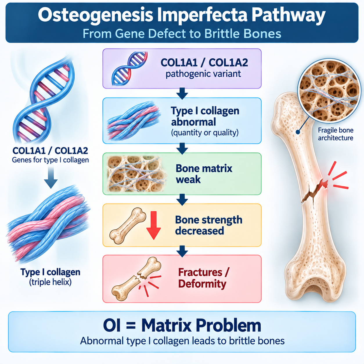
COL1A1 / COL1A2 defect → type I collagen abnormality → bone matrix weakness → recurrent fractures.
MATRIX PROBLEM ≠ MINERALIZATION PROBLEM
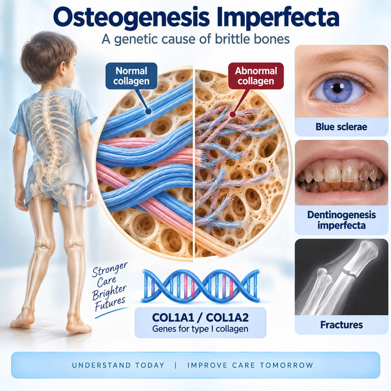
What Is Osteogenesis Imperfecta?
Central Teaching Distinction
This should be established near the beginning.
Hypophosphatasia primarily teaches:
DEFECTIVE MINERALIZATIONOsteogenesis imperfecta primarily teaches:
DEFECTIVE BONE MATRIXThe central mechanism is:
COL1A1 / COL1A2 DEFECT TYPE I COLLAGEN ABNORMALITY BONE MATRIX WEAKNESS SKELETAL FRAGILITY RECURRENT FRACTURES ± DEFORMITY OI = A COLLAGEN-MATRIX DISORDEROpening — Use This Content
A patient has repeated fractures after minimal trauma.
The immediate questions may include:
- osteoporosis?
- nutritional bone disease?
- non-accidental injury?
- another metabolic bone disorder?
But if the patient also has features such as:
- blue or gray sclerae;
- dentinogenesis imperfecta;
- short stature;
- skeletal deformity;
- ligamentous laxity;
- hearing loss;
- or a family history of similar fractures;
another diagnosis becomes particularly important:
OSTEOGENESIS IMPERFECTAOsteogenesis imperfecta, or OI, is a genetically heterogeneous group of disorders characterized principally by:
BONE FRAGILITYThe most familiar forms result from pathogenic variants involving:
COL1A1or:
COL1A2which encode components of:
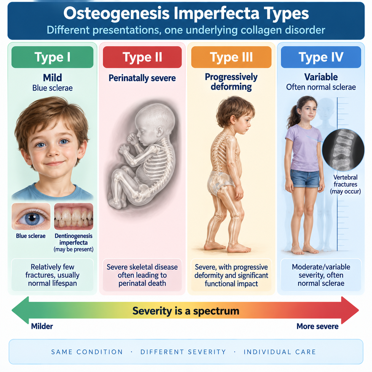
TYPE I COLLAGENCOL1A1/COL1A2-related OI ranges from mild disease with relatively few fractures and normal lifespan to severe progressively deforming disease and perinatally lethal forms. (NCBI)
The central clinical reasoning sequence is:
RECURRENT / LOW-TRAUMA FRACTURES LOOK FOR OI CLUES CLINICAL + RADIOGRAPHIC ASSESSMENT MOLECULAR GENETIC TESTING DEFINE PHENOTYPE AND SEVERITY MULTIDISCIPLINARY MANAGEMENTType I Collagen and Bone Matrix
What Is Type I Collagen?
Type I collagen is a major structural protein of:
- bone;
- dentin;
- sclera;
- tendons;
- ligaments;
- skin;
- other connective tissues.
This explains an important principle:
OI IS NOT ONLY A BONE DISEASEA collagen abnormality can produce manifestations in multiple tissues.
Type I Collagen Structure
Type I collagen is a triple-helical molecule composed primarily of:
TWO α1(I) CHAINSplus:
ONE α2(I) CHAINThe genes are:
COL1A1 → α1(I) COL1A2 → α2(I)These chains assemble into the type I collagen triple helix.
The required memory is:
2 × α1 + 1 × α2 = TYPE I COLLAGENBone Matrix Versus Mineral
This distinction is critical.
Normal bone contains:
Organic matrix
Dominated by:
TYPE I COLLAGENplus other proteins.
Mineral
Predominantly:
HYDROXYAPATITEThe collagen matrix provides a structural framework within which mineral is deposited.
Therefore bone strength depends on:
MATRIX + MINERALIn OI:
THE COLLAGEN MATRIX IS ABNORMALIn osteomalacia:
MINERALIZATION IS ABNORMALCOL1A1, COL1A2 and Bone Fragility
Molecular Mechanism
COL1A1 / COL1A2 PATHOGENIC VARIANT TYPE I COLLAGEN QUANTITY OR QUALITY ABNORMAL BONE MATRIX ABNORMAL BONE MATERIAL STRENGTH ↓ FRACTURE SUSCEPTIBILITY ↑ DEFORMITY / SHORT STATURE / OTHER CONNECTIVE-TISSUE FEATURESQuantitative Versus Qualitative Collagen Defects
This is an important advanced concept.
Some variants primarily result in:
REDUCED QUANTITY OF OTHERWISE RELATIVELY NORMAL COLLAGENOther variants produce:
STRUCTURALLY ABNORMAL COLLAGENThese mechanisms help explain some genotype–phenotype relationships.
A classic teaching simplification is:
Quantitative defect
Often associated with:
MILDER OIQualitative structural defect
Can produce:
MORE SEVERE OIBut immediately state:
GENOTYPE–PHENOTYPE RELATIONSHIPS ARE NOT ABSOLUTEThis is not a universal rule for every variant.
Genetics and Inheritance
Genetics
COL1A1/COL1A2-related OI is generally:
AUTOSOMAL DOMINANTThe diagnosis of COL1A1/COL1A2-related OI can be established in a patient with compatible clinical/radiographic manifestations by identifying an appropriate heterozygous pathogenic or likely pathogenic variant in COL1A1 or COL1A2. (NCBI)
However:
NOT ALL OI IS CAUSED BY COL1A1 OR COL1A2Other genes can produce OI or OI-like phenotypes through abnormalities involving:
- collagen processing;
- collagen modification;
- bone formation;
- osteoblast function;
- mineralization-related pathways.
De Novo Disease
An affected child may have:
UNAFFECTED PARENTSbecause a pathogenic variant may arise:
DE NOVOTherefore:
NO FAMILY HISTORY ≠ NO OIThis is particularly important when evaluating a child with unexplained fractures.
Parental Mosaicism
Recurrence can occasionally occur even when parents appear clinically unaffected and routine blood testing does not demonstrate the child's variant because of:
GERMLINE / SOMATIC MOSAICISMTherefore recurrence counseling should be individualized by genetics professionals.
Clinical Spectrum
OI IS A SPECTRUMSeverity ranges from:
Mild fracture predisposition
Moderate disease
Progressive skeletal deformity
Severe perinatal disease
Perinatal lethality
The 2025 GeneReviews update emphasizes that the traditional clinical types remain useful but overlap substantially. (NCBI)
Types of Osteogenesis Imperfecta
For COL1A1/COL1A2-related OI, teach the four familiar clinical phenotypes.
| Traditional type | Current descriptive name | General severity |
|---|---|---|
| Type I | Classic non-deforming OI with blue sclerae | Usually mild |
| Type II | Perinatally lethal OI | Extremely severe |
| Type III | Progressively deforming OI | Severe |
| Type IV | Common variable OI with normal sclerae | Moderate/variable |
Not every patient fits neatly into one category. (NCBI)
Type I OI
Type I — Classic Non-Deforming OI With Blue Sclerae
This is generally the mildest classic COL1A1/COL1A2 phenotype.
Features may include:
- fractures with minimal trauma;
- blue/gray sclerae;
- relatively mild or absent long-bone deformity;
- near-normal or mildly reduced stature;
- variable dentinogenesis imperfecta;
- ligamentous laxity;
- hearing loss later in life.
Fracture frequency may be greatest in childhood and may decrease after puberty, although fractures can continue throughout life.
Type I disease does not exclude severe complications in every patient.
Why Are The Sclerae Blue?
The sclera normally contains abundant type I collagen.
When scleral collagen is altered or the sclera is relatively thin/translucent, underlying pigmented structures become more visible.
Clinically this creates:
BLUE OR GRAY SCLERAE BLUE SCLERAE ARE A CLUE — NOT A DIAGNOSISThey may occur in other conditions.
Type II OI
Perinatally lethal OI represents an extremely severe phenotype.
Potential findings include:
- severe skeletal fragility;
- multiple fractures before birth;
- markedly abnormal bone mineralization/appearance;
- severe long-bone deformity;
- short limbs;
- small thorax;
- respiratory compromise.
The major life-threatening problem is often:
RESPIRATORY INSUFFICIENCYrelated to severe thoracic skeletal disease and pulmonary consequences.
Not every infant historically classified as Type II necessarily dies immediately; contemporary supportive care and phenotypic overlap make counseling more nuanced. GeneReviews emphasizes individualized neonatal counseling and uncertainty in outcome. (NCBI)
Type III OI
Type III is characterized by severe skeletal disease in individuals surviving the neonatal period.
Features may include:
- recurrent fractures from early life;
- progressive long-bone deformity;
- very short stature;
- scoliosis;
- vertebral compression;
- impaired mobility;
- chest-wall deformity;
- respiratory complications;
- variable scleral coloration;
- dentinogenesis imperfecta.
Memory:
TYPE III = PROGRESSIVELY DEFORMINGType IV OI
Type IV has:
VARIABLE MODERATE SEVERITYFeatures may include:
- recurrent fractures;
- short stature;
- variable long-bone deformity;
- vertebral compression;
- dentinogenesis imperfecta in some patients;
- sclerae that are usually normal or become less blue with age.
Memory:
TYPE IV = VARIABLE OI, OFTEN NORMAL SCLERAEFractures and Bone Deformity
Fractures
The hallmark of OI is:
INCREASED FRACTURE SUSCEPTIBILITYFractures may occur:
- after minimal trauma;
- after ordinary childhood activity;
- recurrently;
- in multiple bones.
Extremity fractures are particularly common in COL1A1/COL1A2-related OI. (NCBI)
But:
FRACTURE NUMBER ALONE DOES NOT DIAGNOSE OIThe whole phenotype matters.
Fracture Healing
Fractures generally can heal in OI.
The problem is not simply an inability to form callus.
However, repeated fractures plus abnormal mechanical loading can contribute to:
- deformity;
- refracture;
- loss of function;
- immobilization-related weakness.
Management therefore aims to restore function while minimizing unnecessary prolonged immobilization.
Bone Deformity
Repeated fractures and intrinsically weak bone can produce:
- bowing of long bones;
- angular deformity;
- limb-length abnormalities;
- scoliosis;
- kyphosis;
- chest-wall abnormalities.
Severe deformity is particularly important in progressively deforming OI.
Short Stature
Short stature may result from:
- abnormal skeletal growth;
- vertebral compression;
- long-bone deformity;
- repeated fractures;
- severe disease burden.
Severity varies markedly.
Mild Type I disease may have relatively normal stature.
Dentinogenesis Imperfecta
Dentinogenesis Imperfecta
OI may affect dentin because:
DENTIN CONTAINS TYPE I COLLAGENDentinogenesis imperfecta may produce teeth that are:
- gray;
- brown;
- translucent;
- structurally fragile;
- prone to wear;
- prone to breakage.
GeneReviews identifies DI as an important but variable feature of COL1A1/COL1A2-related OI. (NCBI)
Oi Dental Disease Versus HPP Dental Disease
This distinction is mandatory.
Osteogenesis imperfecta
Primary dental problem:
ABNORMAL DENTIN→ dentinogenesis imperfecta.
Hypophosphatasia
Characteristic dental mechanism:
DEFECTIVE CEMENTUM / TOOTH ATTACHMENT→ premature root-intact tooth loss.
Therefore:
OI → THINK DENTIN HPP → THINK CEMENTUM / PREMATURE TOOTH LOSSHypophosphatasia Explained
Hearing Loss and Joint Laxity
Hearing Loss
Hearing impairment may develop, particularly later in life.
It may be:
- conductive;
- sensorineural;
- mixed.
Therefore:
OI IS A BONE + CONNECTIVE-TISSUE DISORDER WITH OTIC CONSEQUENCESHearing symptoms should not be dismissed as unrelated.
GeneReviews identifies hearing loss, commonly developing in adulthood, as a recognized manifestation. (NCBI)
Ligamentous Laxity And Joints
Abnormal connective tissue may produce:
- ligamentous laxity;
- joint hypermobility;
- instability;
- pain;
- functional impairment.
This is another reason OI should not be conceptualized only as “bones that fracture.”
Muscle Weakness And Mobility
Patients may have:
- muscle weakness;
- reduced endurance;
- altered gait;
- impaired mobility;
- reduced physical activity after repeated fractures.
This can create a cycle:
FRACTURE IMMOBILIZATION MUSCLE WEAKNESS FUNCTION ↓ SKELETAL LOADING ↓Therefore rehabilitation is a core component of management. (NCBI)
Spine and Respiratory Disease
Spine
Spinal manifestations may include:
- vertebral compression fractures;
- scoliosis;
- kyphosis;
- reduced trunk height.
Progressive scoliosis is particularly important in severe disease and can contribute to:
RESPIRATORY COMPROMISEBasilar Impression
Some patients can develop craniovertebral abnormalities including:
BASILAR IMPRESSION / BASILAR INVAGINATIONPotential consequences can include neurological symptoms.
Symptomatic craniovertebral disease requires specialist evaluation in an experienced center.
GeneReviews recommends specialist surgical management when symptomatic. (NCBI)
Respiratory Disease
Respiratory problems can result from several interacting mechanisms:
- abnormal chest-wall geometry;
- scoliosis;
- reduced thoracic volume;
- respiratory muscle effects;
- intrinsic pulmonary/connective-tissue abnormalities in severe disease.
Therefore:
RESPIRATORY DISEASE IN OI IS NOT ALWAYS JUST A CONSEQUENCE OF FRACTURESSevere respiratory involvement is a major contributor to morbidity.
Cardiovascular/Connective-Tissue Context
Because type I collagen is widespread, cardiovascular manifestations can occur in OI.
When to Suspect OI
THINK OI WHEN FRACTURES ARE ACCOMPANIED BY:- minimal trauma;
- recurrent fractures;
- blue/gray sclerae;
- dentinogenesis imperfecta;
- short stature;
- long-bone deformity;
- scoliosis;
- ligamentous laxity;
- hearing loss;
- family history.
But:
NO SINGLE FEATURE IS REQUIRED IN EVERY PATIENTLaboratory Tests
Laboratory Tests
Routine biochemical testing is important mainly to:
ASSESS ALTERNATIVE / COEXISTING BONE DISEASEIn COL1A1/COL1A2-related OI, typical biochemical studies such as:
- calcium;
- phosphate;
- vitamin-D-related testing;
- alkaline phosphatase;
may be normal outside situations such as fracture healing or other coincident abnormalities.
Therefore:
NORMAL BONE BIOCHEMISTRY DOES NOT EXCLUDE OIThis contrasts strongly with HPP, where persistently low ALP is a central biochemical clue. (NCBI)
ALP — Important Comparison
Osteogenesis imperfecta
ALP IS NOT THE DEFINING DIAGNOSTIC BIOMARKERHypophosphatasia
PERSISTENTLY LOW ALP IS THE KEY BIOCHEMICAL CLUETherefore:
RECURRENT FRACTURES + LOW ALP → RECONSIDER HPPRadiographic Findings
Radiographs
Radiographic findings depend on severity and age.
Potential findings include:
- fractures at different stages of healing;
- osteopenic appearance;
- long-bone bowing;
- deformity;
- vertebral compression;
- scoliosis;
- thin cortices;
- skull abnormalities;
- wormian bones.
No single radiographic sign is diagnostic.
Wormian Bones
Wormian bones are accessory ossicles within cranial sutures.
They may be seen in OI, particularly when numerous.
However:
WORMIAN BONES ARE NOT SPECIFIC FOR OIThey must be interpreted with the overall phenotype.
DXA
DXA may demonstrate reduced BMD.
It can be useful for:
- baseline skeletal assessment;
- longitudinal follow-up in selected patients;
- monitoring response to bone-directed treatment.
But:
DXA DOES NOT DIAGNOSE OIand:
BMD DOES NOT FULLY CAPTURE BONE MATERIAL QUALITYA patient can have clinically important skeletal fragility not explained by a single DXA value.
NIAMS includes bone-density testing as part of clinical assessment, while molecular testing can identify the underlying genetic disorder. (NIAMS)
Diagnosis and Genetic Testing
Diagnosis
The diagnostic process should integrate:
HISTORY PHYSICAL EXAMINATION RADIOGRAPHIC FINDINGS FAMILY HISTORY MOLECULAR GENETIC TESTINGFor COL1A1/COL1A2-related disease, identification of an appropriate pathogenic/likely pathogenic heterozygous variant in a compatible patient establishes the molecular diagnosis. (NCBI)
Genetic Testing
When OI is suspected, molecular testing may involve:
- COL1A1;
- COL1A2;
- broader multigene skeletal-fragility/OI panels when appropriate;
- more extensive genomic testing in unresolved cases.
The exact testing strategy depends on phenotype and local genetics practice.
Negative Genetic Testing
NEGATIVE INITIAL GENETIC TEST ≠ AUTOMATICALLY NO OIReasons can include:
- testing limitations;
- genes not included;
- variant-detection limitations;
- alternative genetic skeletal-fragility disorders.
NIAMS similarly notes that a negative genetic test does not necessarily exclude an OI diagnosis. (NIAMS)
OI vs Hypophosphatasia
Implement this major table.
| Feature | Osteogenesis imperfecta | Hypophosphatasia |
|---|---|---|
| Main defect | Collagen matrix | Mineralization |
| Major genes | COL1A1/COL1A2 | ALPL |
| Type I collagen | Abnormal quantity/quality | Not primary defect |
| TNSALP | Not primary problem | Deficient |
| ALP | Usually not persistently low as defining feature | Persistently low |
| Blue sclerae | Classic clue | Not typical |
| Dentinogenesis imperfecta | Important | Not defining |
| Premature root-intact primary tooth loss | Not defining | Important clue |
| Pseudofractures | Not central classic feature | Important adult clue |
| DXA | May be low | Variable |
| Bisphosphonates | Commonly used in selected OI | Generally avoided in established HPP |
Bottom memory:
OI = MATRIX PROBLEM HPP = MINERALIZATION PROBLEMOI vs Osteomalacia
| Feature | OI | Osteomalacia |
|---|---|---|
| Primary defect | Bone matrix quality | Mineralization of osteoid |
| Classic cause | Genetic collagen disorder | Multiple mineral/metabolic causes |
| COL1A1/COL1A2 | Common classic genes | Not defining |
| Blue sclerae | May occur | Not typical |
| DI | May occur | Not typical |
| Pseudofractures | Not defining | Classic clue |
| ALP | Often not diagnostically abnormal | Frequently elevated depending on cause |
| Treatment | OI-specific multidisciplinary care | Treat mineralization cause |
Osteomalacia Explained
OI vs Osteoporosis
| Feature | OI | Conventional osteoporosis |
|---|---|---|
| Typical basis | Genetic matrix disorder | Acquired/multifactorial skeletal fragility |
| Onset | Often childhood/lifelong | Commonly later life |
| Blue sclerae | Can occur | Not typical |
| DI | Can occur | Not typical |
| Deformity | May be prominent | Usually not classic |
| Genetic testing | Often diagnostically important | Not routine for typical disease |
| DXA | May be low | Central fracture-risk tool |
| Management | Multidisciplinary genetic bone disease care | Standard osteoporosis framework |
Adults with OI can also develop age-related osteoporosis.
OI and Non-Accidental Injury
Multiple fractures in an infant or child may raise concern for:
NON-ACCIDENTAL TRAUMAOI may also cause multiple fractures.
Therefore clinicians must perform:
A COMPLETE AND OBJECTIVE EVALUATIONFeatures that may support OI include:
- compatible family history;
- blue sclerae;
- DI;
- generalized skeletal phenotype;
- characteristic deformity;
- molecular confirmation.
But:
OI SHOULD NOT BE USED AUTOMATICALLY TO EXPLAIN OTHERWISE CONCERNING INJURIESand:
ABSENCE OF CLASSIC OI FEATURES DOES NOT BY ITSELF PROVE ABUSEThese are separate diagnostic questions requiring careful multidisciplinary evaluation.
Treatment Principles
Management Principles
There is currently no cure that universally corrects the underlying genetic defect in COL1A1/COL1A2-related OI.
Management aims to:
- reduce fracture burden;
- preserve mobility;
- prevent or limit deformity;
- maximize muscle strength;
- manage pain;
- maintain independence;
- optimize dental health;
- identify hearing problems;
- manage spinal disease;
- address respiratory complications;
- support quality of life.
Management is best:
MULTIDISCIPLINARYGeneReviews emphasizes individualized multidisciplinary management. (NCBI)
Safe Handling
Infants and children with significant OI require:
GENTLE BUT NORMALIZING CARECaregivers should be educated in safe handling.
Important principles include:
- broad hand support;
- gentle movements;
- avoiding unnecessary twisting or focal stress;
- supporting development rather than excessive restriction.
Avoid:
“NEVER TOUCH OR MOVE THE CHILD”Excessive restriction can worsen weakness and functional limitation.
Physical Therapy and Mobility
Physical Therapy
Physical therapy is one of the most important management components.
Goals include:
- muscle strength;
- mobility;
- joint stability;
- motor development;
- functional independence;
- participation;
- prevention of contractures;
- recovery after fractures.
GeneReviews describes physical rehabilitation as a major therapeutic modality in OI. (NCBI)
Memory:
PROTECT THE BONE — BUT BUILD THE MUSCLEImmobilization
Fractures require appropriate stabilization.
However, unnecessarily prolonged immobilization may contribute to:
- muscle weakness;
- bone loss;
- delayed functional recovery.
Therefore:
IMMOBILIZE THE FRACTURE — NOT THE PATIENT FOR LONGER THAN NECESSARYGeneReviews recommends keeping immobilization as short as practical and restarting physical therapy promptly after cast removal. (NCBI)
Fracture and Orthopedic Management
Orthopedic Surgery
Orthopedic surgery may be required for:
- recurrent fractures;
- severe deformity;
- long-bone bowing;
- functional limitation;
- selected spinal disease.
A major technique in moderate/severe OI is:
INTRAMEDULLARY RODDINGThe purpose is to:
- improve alignment;
- support long bones;
- facilitate function;
- reduce consequences of deformity.
GeneReviews describes intramedullary rodding as a mainstay of orthopedic management where indicated. (NCBI)
Telescoping Rods
In growing children, specialized rods may be designed to:
EXPAND WITH BONE GROWTHThis can reduce the need for repeated replacement compared with fixed-length constructs in selected circumstances.
Bisphosphonates
Bisphosphonates
Create a major section.
Bisphosphonates are:
ANTIRESORPTIVE AGENTSThey are the most extensively used pharmacological bone-directed treatment in OI, particularly in patients with:
- vertebral fractures;
- frequent long-bone fractures;
- more severe disease.
They generally:
INCREASE BONE MINERAL DENSITYBut:
BISPHOSPHONATES DO NOT CORRECT THE COLLAGEN DEFECTand:
THEY DO NOT CURE OIGeneReviews' 2025 update emphasizes that although BMD generally improves, evidence for fracture reduction—especially across all ages and phenotypes—is less definitive than the BMD effect. (NCBI)
Do Bisphosphonates Definitely Prevent Fractures?
Clinical studies support skeletal benefits, including increased BMD, and some observational/trial data suggest fracture benefits in selected populations.
However, available studies have limitations.
Therefore:
BMD BENEFIT IS CLEARER THAN THE CERTAINTY OF FRACTURE-REDUCTION BENEFITAvoid the inaccurate statement:
“Bisphosphonates completely prevent fractures in OI.”
Bisphosphonates: OI Versus HPP
OI
Bisphosphonates:
COMMONLY USED IN SELECTED PATIENTSHPP
Antiresorptive therapy:
GENERALLY AVOIDED IN ESTABLISHED DISEASETherefore:
THE SAME FRACTURE PHENOTYPE CAN REQUIRE VERY DIFFERENT TREATMENTThis reinforces why correct diagnosis matters.
Calcium, Vitamin D and Pain
Calcium And Vitamin D
Patients should have appropriate nutritional assessment and avoid clinically important deficiency.
But:
CALCIUM OR VITAMIN D DOES NOT CORRECT THE COLLAGEN DEFECTAvoid unnecessary excess.
Pain Management
Pain may arise from:
- acute fractures;
- deformity;
- chronic musculoskeletal stress;
- spinal disease;
- joint problems.
Management may combine:
- treatment of the underlying skeletal problem;
- rehabilitation;
- physical strategies;
- appropriate pharmacological analgesia;
- psychosocial support.
Dental and Hearing Care
Dental Management
Dental care should aim to preserve:
- primary dentition where possible;
- permanent dentition;
- function;
- occlusion;
- gingival health;
- appearance.
Dentinogenesis imperfecta may require specialist pediatric/restorative dental care.
GeneReviews recommends ongoing dental care appropriate to the OI phenotype. (NCBI)
Hearing Management
Patients should be evaluated when hearing concerns arise, with appropriate audiologic/ENT management.
Hearing loss may be:
- conductive;
- sensorineural;
- mixed.
Management depends on type and severity.
Respiratory Management
Respiratory assessment becomes particularly important in:
- severe OI;
- significant scoliosis;
- chest-wall deformity;
- symptoms of respiratory limitation.
Management should involve appropriate respiratory expertise.
Emerging Therapies
Emerging And Investigational Therapies
EMERGING / INVESTIGATIONALThese are not established universal therapies.
Areas of study include:
- anabolic approaches;
- sclerostin inhibition;
- other pathways affecting bone formation/remodeling;
- molecular/genetic approaches.
GeneReviews' 2025 update describes several therapies under investigation, including anabolic approaches. (NCBI)
Teriparatide
Teriparatide has been studied particularly in adults with OI.
It has:
OSTEOANABOLIC ACTIVITYSome studies suggest differential response according to OI phenotype.
However:
TERIPARATIDE IS NOT A UNIVERSAL STANDARD TREATMENT FOR ALL OISclerostin Inhibition
Sclerostin inhibits Wnt-mediated bone formation.
Therefore blocking sclerostin is biologically attractive as an:
ANABOLIC STRATEGYSeveral sclerostin-directed approaches have been investigated in OI.
Investigational agents are not established treatment.
Genetic Therapy
Gene editing, gene silencing and cell-based approaches are scientifically attractive because OI is fundamentally a genetic disorder.
However:
GENETIC CORRECTION IS NOT CURRENT ROUTINE CLINICAL THERAPYGenetic Counseling
Genetic Counseling
Genetic counseling should address:
- molecular diagnosis;
- inheritance;
- family testing;
- recurrence considerations;
- phenotypic variability;
- reproductive options where appropriate.
For typical heterozygous COL1A1/COL1A2 disease:
AUTOSOMAL DOMINANT INHERITANCEis central.
But parental mosaicism and de novo disease mean counseling should be individualized.
Pregnancy
Adults with OI can have successful pregnancies.
Pregnancy may nevertheless create additional issues involving:
- maternal skeletal health;
- mobility;
- respiratory status in severe disease;
- delivery planning;
- fetal inheritance risk.
Management should be individualized by appropriate obstetric and OI specialists.
The updated GeneReviews notes increased pregnancy complications but successful pregnancy is common. (NCBI)
Surveillance Principle
Surveillance should be:
PHENOTYPE- AND AGE-DEPENDENTPotential domains include:
- fracture history;
- growth;
- deformity;
- scoliosis;
- mobility;
- pain;
- DXA where clinically useful;
- dental health;
- hearing;
- respiratory function;
- neurological symptoms suggesting craniovertebral disease;
- functional independence.
The current GeneReviews provides age- and phenotype-dependent surveillance rather than a one-size-fits-all approach. (NCBI)
Diagnostic Algorithm
Implement prominently.
RECURRENT / LOW-TRAUMA FRACTURES HISTORY + EXAMINATIONLook for:
Blue/gray sclerae
Dentinogenesis imperfecta
Short stature
Bone deformity
Joint laxity
Hearing loss
Family history
RADIOGRAPHSFractures?
Deformity?
Vertebral compression?
Wormian bones?
BASIC BONE BIOCHEMISTRYCa / PO₄ / ALP / vitamin-D context
Persistently low ALP?
THINK HPP / OTHER LOW-ALP CAUSESBiochemistry not diagnostic?
OI STILL POSSIBLE MOLECULAR GENETIC TESTINGCOL1A1 / COL1A2 ± broader panel
COMPATIBLE PATHOGENIC VARIANT? DEFINE OI PHENOTYPE / SEVERITY MULTIDISCIPLINARY MANAGEMENTBottom memory:
FRACTURES + CONNECTIVE-TISSUE CLUES → THINK OITreatment Algorithm
Implement separately.
CONFIRMED / STRONGLY SUSPECTED OI ASSESS SEVERITYFractures
Deformity
Mobility
Spine
Pain
Dental
Hearing
Respiratory
REHABILITATION / SAFE MOBILITY FRACTURE MANAGEMENT ORTHOPEDIC CORRECTION WHEN NEEDED SIGNIFICANT SKELETAL DISEASE? CONSIDER BONE-DIRECTED PHARMACOLOGICAL THERAPYMost established:
BISPHOSPHONATES IN SELECTED PATIENTS MONITOR FUNCTION + FRACTURES + SKELETAL HEALTH MANAGE EXTRASKELETAL DISEASEFinal:
THE GOAL IS FUNCTION — NOT JUST BMDWorked Clinical Cases
Case 1 — Child with recurrent fractures
A child has several low-trauma long-bone fractures plus blue sclerae.
Key thought
OSTEOGENESIS IMPERFECTAAssess the complete phenotype and proceed to appropriate molecular testing.
Case 2 — No family history
A child has fractures, DI and blue sclerae, but both parents are unaffected.
Wrong conclusion
“No family history, therefore not genetic.”
Correct principle
DE NOVO OI OCCURSCase 3 — Mild adult disease
A 35-year-old reports numerous childhood fractures, few fractures after puberty, blue sclerae and adult-onset hearing difficulty.
Likely phenotype
CLASSIC NON-DEFORMING OI WITH BLUE SCLERAECase 4 — Severe progressive disease
Child has fractures from infancy, marked long-bone bowing, very short stature and progressive scoliosis.
Likely phenotype
PROGRESSIVELY DEFORMING OICase 5 — Normal sclerae
Patient has recurrent fractures, moderate short stature and DI but normal sclerae.
Lesson
NORMAL SCLERAE DO NOT EXCLUDE OICase 6 — Low ALP
Adult with fractures has repeatedly low ALP.
Wrong approach
“Every genetic fracture disorder is OI.”
Better approach
PERSISTENTLY LOW ALP SHOULD RAISE HPPCase 7 — Dentinogenesis imperfecta
Child has gray translucent teeth plus recurrent fractures.
Lesson
DENTIN CAN REVEAL THE COLLAGEN DISORDERCase 8 — Multiple fractures and safeguarding concern
Infant has multiple fractures.
Correct principle
OBJECTIVELY EVALUATE BOTH MEDICAL BONE DISEASE AND NON-ACCIDENTAL INJURYOne diagnosis should not be assumed solely to exclude the other.
Case 9 — Low BMD
Adult with genetically confirmed OI has low DXA BMD.
Wrong statement
“The DXA diagnosed OI.”
Correct statement
DXA MEASURES BONE MASS — IT DOES NOT DIAGNOSE THE COLLAGEN DEFECTCase 10 — Bisphosphonate expectation
Parent asks whether bisphosphonate treatment will cure the child's OI.
Answer
NOIt may improve skeletal measures and is widely used in selected significant OI, but it does not correct the COL1A1/COL1A2 defect.
Case 11 — Fracture immobilization
Child with OI has a treated long-bone fracture and remains inactive long after stabilization.
Lesson
AVOID UNNECESSARILY PROLONGED IMMOBILIZATIONRehabilitation is important for restoring function.
Case 12 — Adult hearing loss
Adult with mild OI develops progressive hearing impairment.
Lesson
OI IS SYSTEMICHearing loss is a recognized extraskeletal manifestation.
Common Mistakes
Mistake 1
OI is simply osteoporosis in children.
Wrong.
Mistake 2
OI is primarily a calcium-deficiency disorder.
Wrong.
Mistake 3
OI is primarily defective mineralization.
Wrong.
Mistake 4
Every patient with OI has blue sclerae.
Wrong.
Mistake 5
Normal sclerae exclude OI.
Wrong.
Mistake 6
Every OI patient has dentinogenesis imperfecta.
Wrong.
Mistake 7
No family history excludes OI.
Wrong.
Mistake 8
All OI is caused by COL1A1/COL1A2.
Wrong.
Mistake 9
All COL1A1/COL1A2 variants produce identical severity.
Wrong.
Mistake 10
One fracture proves OI.
Wrong.
Mistake 11
Multiple fractures automatically prove OI.
Wrong.
Mistake 12
Normal calcium and phosphate exclude OI.
Wrong.
Mistake 13
Persistently low ALP is typical of classic OI.
Wrong.
Mistake 14
Low BMD on DXA diagnoses OI.
Wrong.
Mistake 15
A negative initial genetic test always excludes OI.
Wrong.
Mistake 16
Bisphosphonates correct the collagen mutation.
Wrong.
Mistake 17
Bisphosphonates cure OI.
Wrong.
Mistake 18
Children with OI should avoid all physical activity.
Wrong.
Mistake 19
Long immobilization always protects OI bone.
Wrong.
Mistake 20
OI affects only bone.
Wrong.
Osteogenesis Imperfecta in One Minute
OSTEOGENESIS IMPERFECTA IN ONE MINUTE COL1A1 / COL1A2 TYPE I COLLAGEN ABNORMAL BONE MATRIX WEAK FRACTURESLook for:
Blue sclerae
DI
Short stature
Deformity
Joint laxity
Hearing loss
GENETIC TESTINGPhenotypes:
Type I → mild / blue sclerae
Type II → perinatally lethal
Type III → progressively deforming
Type IV → variable / often normal sclerae
Treatment:
REHABILITATION ORTHOPEDIC CARE BISPHOSPHONATES IN SELECTED PATIENTS MULTISYSTEM CAREFinal memory:
OI = MATRIX PROBLEM HPP / OSTEOMALACIA = MINERALIZATION PROBLEMSFrequently Asked Questions
What is osteogenesis imperfecta?
Osteogenesis imperfecta is a genetically heterogeneous skeletal-fragility disorder. The classic forms are commonly caused by COL1A1 or COL1A2 variants affecting type I collagen.
Why do bones fracture in OI?
Abnormal type I collagen quantity or structure weakens the organic bone matrix, reducing bone material strength and increasing fracture susceptibility.
What genes commonly cause classic OI?
COL1A1 and COL1A2 are the major genes responsible for the classic forms, although other genes can produce OI and related skeletal-fragility phenotypes.
Is OI always inherited from an affected parent?
No. Pathogenic variants may arise de novo, so an affected child may have clinically unaffected parents.
Does every patient with OI have blue sclerae?
No. Blue/gray sclerae are characteristic of some phenotypes, particularly classic non-deforming OI, but normal sclerae do not exclude OI.
What is dentinogenesis imperfecta?
It is abnormal dentin formation that can produce discolored, translucent, fragile teeth. It occurs in some people with OI.
Does normal calcium exclude OI?
No. Routine calcium, phosphate and other bone biochemical tests may be normal because OI is fundamentally a structural collagen disorder.
Is low ALP characteristic of OI?
Persistently low ALP is not the defining biochemical feature of classic OI. In a patient with fractures and persistently low ALP, hypophosphatasia should be considered.
Does DXA diagnose OI?
No. DXA measures bone mineral density and may help with skeletal assessment and monitoring, but it does not diagnose the collagen disorder.
How is OI confirmed?
Diagnosis integrates the clinical and radiographic phenotype with molecular genetic testing. Identification of an appropriate pathogenic or likely pathogenic COL1A1/COL1A2 variant can establish COL1A1/COL1A2-related OI in a compatible patient.
What are the four classic OI types?
They correspond broadly to classic non-deforming OI with blue sclerae, perinatally lethal OI, progressively deforming OI and common variable OI with normal sclerae. The phenotypes overlap.
Are bisphosphonates used in OI?
Yes. They are widely used, particularly in patients with vertebral fractures, frequent long-bone fractures or more severe OI. They improve BMD, although the certainty of fracture reduction is less consistent across studies.
Do bisphosphonates cure OI?
No. They alter bone remodeling but do not correct the underlying collagen defect.
Why is physical therapy important?
Appropriate rehabilitation supports muscle strength, mobility, joint stability, functional independence and recovery after fractures.
Does OI affect organs other than bone?
Yes. Depending on phenotype, OI can involve teeth, hearing, joints, spine, respiratory function and other connective tissues.
Key Take-Home Messages
Osteogenesis imperfecta is fundamentally a disorder of:
BONE MATRIXThe classic molecular pathway is:
COL1A1 / COL1A2 DEFECT TYPE I COLLAGEN ABNORMALITY BONE MATRIX WEAKNESS SKELETAL FRAGILITY RECURRENT FRACTURESBut OI is not simply:
“BRITTLE BONES”Type I collagen is distributed throughout connective tissues.
Therefore look for:
BLUE SCLERAE DENTINOGENESIS IMPERFECTA JOINT LAXITY HEARING LOSS SHORT STATURE SKELETAL DEFORMITYThe clinical spectrum ranges from:
MILD FRACTURE PREDISPOSITIONto:
PROGRESSIVELY DEFORMING DISEASEto:
EXTREMELY SEVERE PERINATAL DISEASEDiagnosis integrates:
PHENOTYPE + RADIOGRAPHS + GENETICSRoutine bone biochemistry may be normal.
Therefore:
NORMAL CALCIUM / PHOSPHATE DOES NOT EXCLUDE OIAnd:
DXA DOES NOT DIAGNOSE OIThe distinction from hypophosphatasia is especially important:
OI = COLLAGEN MATRIX DEFECTwhereas:
HPP = TNSALP DEFICIENCY + DEFECTIVE MINERALIZATIONManagement is multidisciplinary:
SAFE ACTIVITY + REHABILITATION FRACTURE / ORTHOPEDIC CARE BONE-DIRECTED TREATMENT IN SELECTED PATIENTS DENTAL + HEARING + SPINAL + RESPIRATORY CAREBisphosphonates remain the most widely used pharmacological skeletal therapy in significant OI, but:
THEY DO NOT CORRECT THE COLLAGEN DEFECTand:
THEY DO NOT CURE OIThe ultimate goal is:
FEWER COMPLICATIONS + BETTER FUNCTION + MAXIMUM INDEPENDENCEFinal memory:
RECURRENT FRACTURES + CONNECTIVE-TISSUE CLUES = THINK OSTEOGENESIS IMPERFECTAand:
OI = A MATRIX PROBLEM, NOT SIMPLY A LOW-BMD PROBLEM