Arterial blood gas interpretation is an essential clinical skill for medical students, nursing students and junior doctors. ABGs help assess oxygenation, ventilation and acid-base status. A systematic approach prevents common mistakes and helps identify simple and mixed acid-base disorders. This article explains ABG interpretation step by step using pH, PaCO2, bicarbonate, compensation, anion gap, Winter's Formula and Delta Ratio.
Dr. Seneth Gajasinghe, MBBS, MD Published: 27 June 2026 Updated: 27 June 2026 30 min readReviewed Content
ABG interpretation becomes much easier when every result is approached in the same order. The aim is not to memorise isolated patterns, but to build a repeatable clinical workflow.
Use Winter's Formula and Delta Ratio appropriately
Interpret common clinical ABG examples
What Is an ABG?
An arterial blood gas is a blood test taken from an artery. It provides rapid information about oxygenation, ventilation, acid-base balance and metabolic status.
ABG interpretation is especially useful in DKA, sepsis, shock, COPD exacerbation, asthma, respiratory failure, renal failure, poisoning, ICU care and emergency medicine.
Oxygenation↓Ventilation↓Acid-base balance↓Metabolic status
Figure 1. An ABG helps assess oxygenation, ventilation and acid-base status.
What Does an ABG Measure?
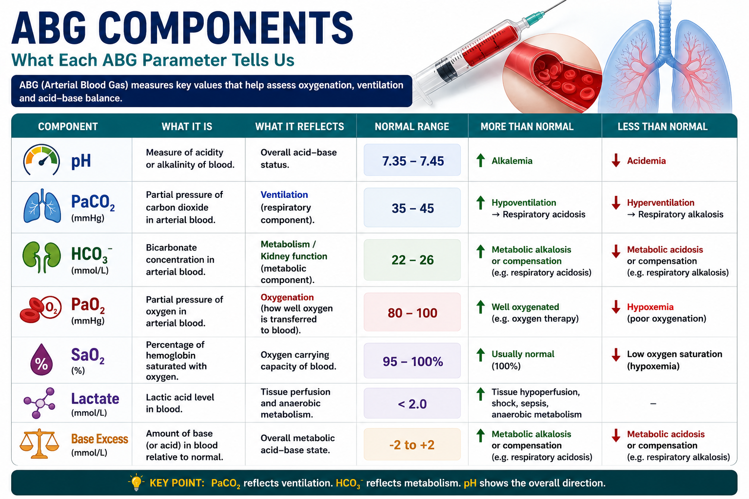
An ABG contains several values, but pH, PaCO2 and bicarbonate are the core acid-base variables. PaO2 and SaO2 help assess oxygenation. Lactate and base excess add important metabolic information.
Component
Meaning
pH
Acidaemia or alkalaemia
PaCO2
Respiratory component and ventilation
HCO3-
Metabolic component
PaO2
Oxygenation
SaO2
Oxygen saturation
Base excess
Metabolic acid-base burden
Lactate
Tissue hypoperfusion or anaerobic metabolism
Key Teaching Point
PaCO2 reflects ventilation. HCO3- reflects the metabolic or kidney component. pH tells the overall direction.
Figure 2. ABG components must be interpreted together rather than as isolated numbers.
Normal ABG Values
Parameter
Normal value
pH
7.35-7.45
PaCO2
35-45 mmHg
HCO3-
22-26 mmol/L
PaO2
80-100 mmHg
SaO2
95-100%
Lactate
Usually < 2 mmol/L
Important
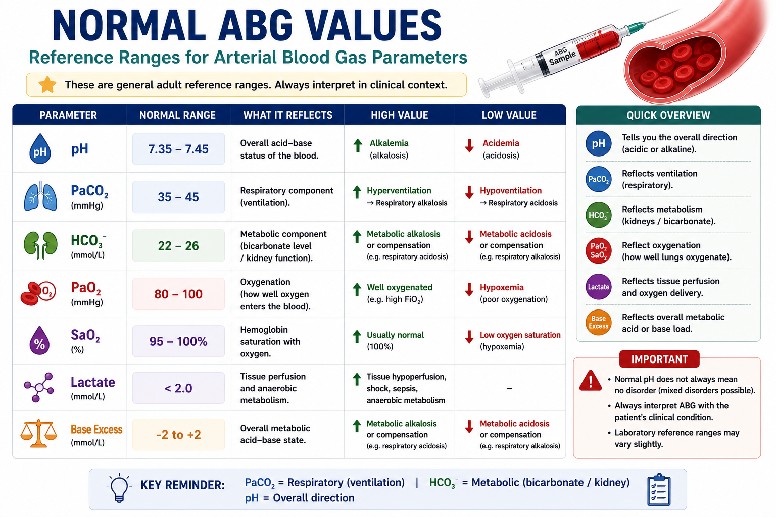
Normal ranges may vary slightly between laboratories. Always interpret ABG results with the clinical picture.
Figure 3. Normal ABG values provide the starting point for interpretation.
Step-by-Step ABG Interpretation Algorithm
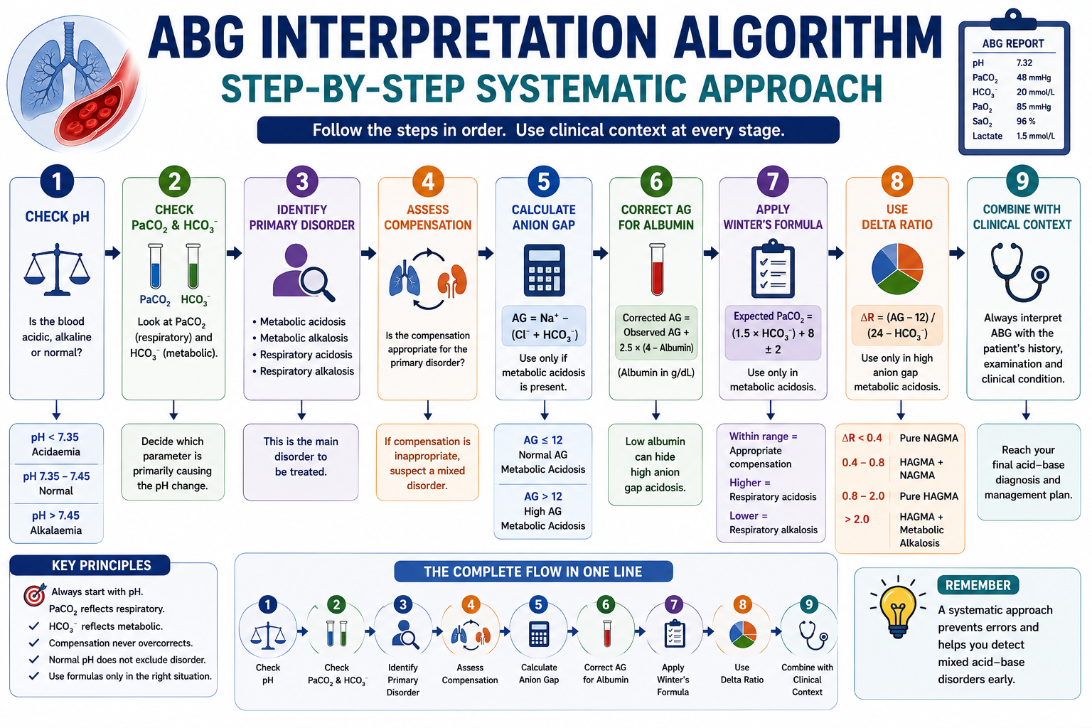
The safest way to read an ABG is to use the same order every time. This prevents common mistakes such as jumping to compensation before identifying the primary disorder.
Check pH.
Check PaCO2 and HCO3-.
Identify the primary disorder.
Assess compensation.
Calculate anion gap if metabolic acidosis is present.
Correct the anion gap for albumin.
Use Winter's Formula if metabolic acidosis is present.
Use Delta Ratio if high anion gap metabolic acidosis is present.
Figure 4. A structured algorithm prevents common ABG interpretation errors.
Step 1: Check pH
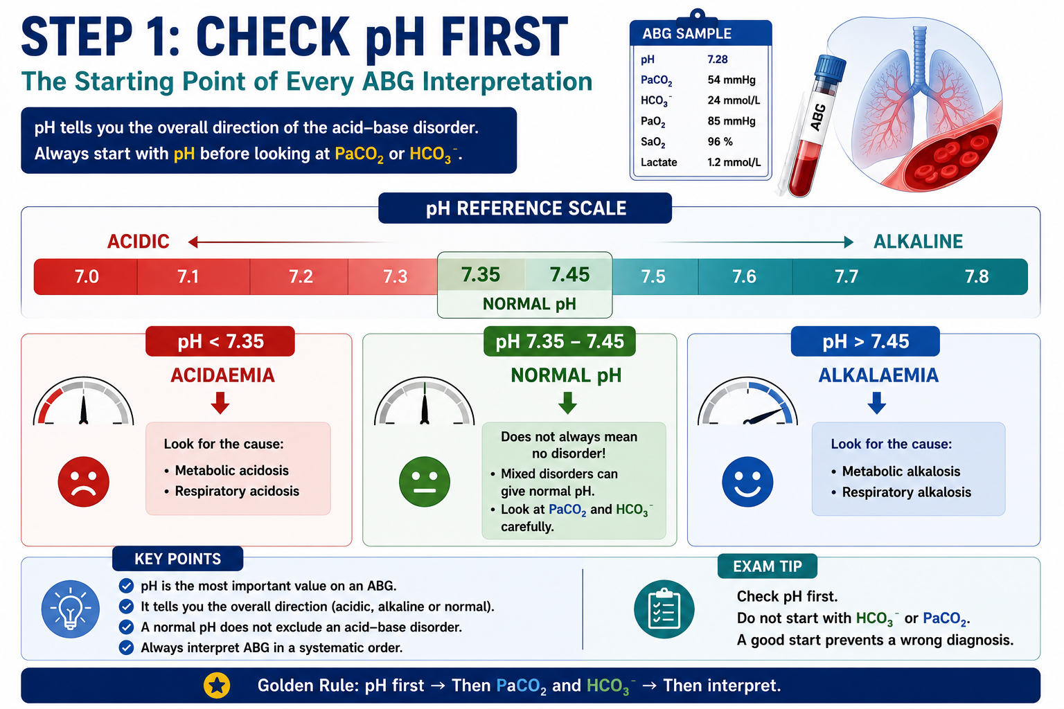
First decide whether the blood is acidic, alkaline or within the normal pH range.
pH
Meaning
< 7.35
Acidaemia
> 7.45
Alkalaemia
7.35-7.45
Normal pH range
A normal pH does not exclude an acid-base disorder. A mixed disorder may bring pH back toward normal.
Figure 5. The pH tells whether the overall blood gas direction is acidic, alkaline or near normal.
Step 2: Identify the Primary Disorder
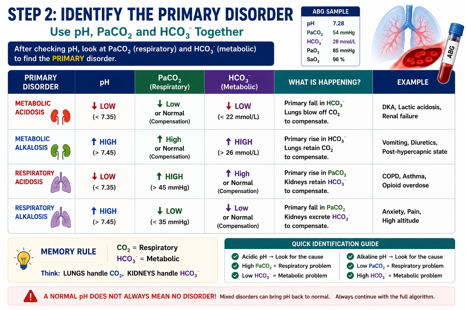
Use pH direction, PaCO2 and HCO3- together. CO2 is respiratory. HCO3- is metabolic.
Disorder
pH
PaCO2
HCO3-
Metabolic acidosis
Low
Low or normal
Low
Metabolic alkalosis
High
High or normal
High
Respiratory acidosis
Low
High
High or normal
Respiratory alkalosis
High
Low
Low or normal
Memory Rule
CO2 = respiratory. HCO3- = metabolic.
Figure 6. Primary disorder identification depends on pH direction, PaCO2 and bicarbonate.
Step 3: Assess Compensation
Compensation is the body's attempt to reduce the pH disturbance. Metabolic problems are compensated by the lungs. Respiratory problems are compensated by the kidneys.
Compensation does not fully correct the disorder and should not overshoot. If it appears to overshoot, suspect a mixed disorder.
In metabolic acidosis, use Winter's Formula Explained to assess whether respiratory compensation is appropriate.
Figure 7. Compensation moves pH toward normal but does not erase the primary disorder.
Step 4: Calculate the Anion Gap
Calculate the anion gap when metabolic acidosis is present. The anion gap helps separate high anion gap metabolic acidosis from normal anion gap metabolic acidosis.
Figure 8. The anion gap classifies metabolic acidosis into high gap and normal gap patterns.
Step 5: Correct the Anion Gap for Albumin
Low albumin lowers the anion gap and may hide high anion gap metabolic acidosis. This is especially important in sepsis, chronic illness, liver disease, malnutrition and critical care.
Corrected AG = Observed AG + 2.5 × (4 - Albumin)
Albumin is measured in g/dL.
Do Not Miss This
In critically ill patients, always consider albumin correction before deciding that the anion gap is normal.
Figure 9. Low albumin can hide a high anion gap metabolic acidosis.
Step 6: Use Winter's Formula
Winter's Formula is used only in metabolic acidosis. It predicts the expected PaCO2 for the degree of bicarbonate reduction.
Figure 11. Delta Ratio helps detect mixed metabolic disorders in high anion gap metabolic acidosis.
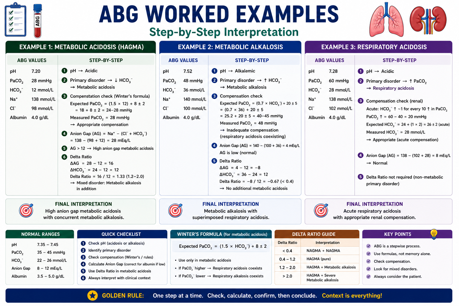
Worked ABG Examples
Examples help convert the algorithm into pattern recognition. Always start with pH, then identify the primary disorder, then assess compensation and context.
Example
ABG
Interpretation
Normal ABG
pH 7.40, PaCO2 40, HCO3- 24
Normal acid-base status
Simple metabolic acidosis
pH 7.25, PaCO2 30, HCO3- 14
Metabolic acidosis with respiratory compensation
DKA
pH 7.15, PaCO2 24, HCO3- 8, high AG
High anion gap metabolic acidosis, likely DKA
DKA + respiratory acidosis
pH 7.05, PaCO2 40, HCO3- 10
Expected PaCO2 is 23 ± 2, so measured PaCO2 is high: metabolic acidosis + respiratory acidosis
Sepsis / lactic acidosis
pH 7.28, PaCO2 20, HCO3- 10, high lactate, high AG
High anion gap metabolic acidosis with additional respiratory alkalosis
COPD exacerbation
pH 7.28, PaCO2 65, HCO3- 30
Respiratory acidosis with metabolic compensation
Vomiting
pH 7.50, PaCO2 48, HCO3- 36
Metabolic alkalosis with respiratory compensation
Panic attack
pH 7.52, PaCO2 25, HCO3- 22
Respiratory alkalosis
Salicylate toxicity
pH 7.44, PaCO2 20, HCO3- 13, high AG
Mixed respiratory alkalosis + high anion gap metabolic acidosis
DKA + vomiting
pH 7.30, PaCO2 28, HCO3- 14, AG 32, Delta Ratio > 2
High AG metabolic acidosis + metabolic alkalosis
Figure 12. Worked examples show how the ABG algorithm applies to common clinical scenarios.
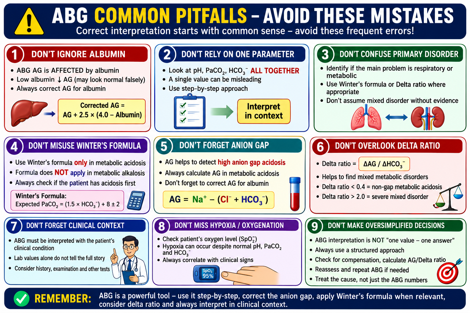
Common Pitfalls
Looking at HCO3- before pH.
Ignoring PaCO2.
Calling compensation a primary disorder.
Forgetting the anion gap.
Not correcting the anion gap for albumin.
Using Winter's Formula in the wrong disorder.
Using Delta Ratio when the anion gap is normal.
Ignoring oxygenation.
Ignoring clinical context.
Assuming normal pH means normal ABG.
Figure 13. Common ABG mistakes usually come from skipping a structured sequence.
One Minute Revision
Check pH.
Check PaCO2 and HCO3-.
Identify the primary disorder.
Assess compensation.
If metabolic acidosis is present, calculate the anion gap.
Correct the anion gap for albumin.
Use Winter's Formula when appropriate.
Use Delta Ratio when appropriate.
Interpret everything with clinical context.
Figure 14. ABG interpretation should follow the same structured order every time.
Frequently Asked Questions
What is ABG interpretation?
ABG interpretation is the systematic analysis of arterial blood gas values to assess oxygenation, ventilation and acid-base status.
What are the main ABG values?
The main ABG values are pH, PaCO2, HCO3, PaO2 and oxygen saturation. Lactate and base excess may also be reported.
How do you interpret ABG step by step?
First check pH, then PaCO2 and HCO3, identify the primary disorder, assess compensation, calculate the anion gap when appropriate and interpret everything with the clinical picture.
What does PaCO2 show?
PaCO2 reflects ventilation and the respiratory component of acid-base balance. High PaCO2 causes respiratory acidosis and low PaCO2 causes respiratory alkalosis.
What does HCO3 show?
HCO3 reflects the metabolic component of acid-base balance. Low HCO3 suggests metabolic acidosis and high HCO3 suggests metabolic alkalosis.
When should the anion gap be calculated?
The anion gap should be calculated when metabolic acidosis is present. It helps distinguish high anion gap from normal anion gap metabolic acidosis.
When should Winter's Formula be used?
Winter's Formula should be used in metabolic acidosis to assess whether respiratory compensation is appropriate.
When should Delta Ratio be used?
Delta Ratio should be used in high anion gap metabolic acidosis to look for additional metabolic disorders.
Can pH be normal in a mixed acid-base disorder?
Yes. A normal pH can occur when two opposing acid-base disorders coexist. Therefore pH should not be interpreted alone.
What is the most common mistake in ABG interpretation?
A common mistake is identifying one abnormal value without using a systematic approach. ABGs should always be interpreted step by step and with clinical context.
Medical Education Disclaimer
This article is intended for medical education only. ABG interpretation must be combined with clinical assessment, oxygen delivery settings, local laboratory ranges, medication history and senior or specialist input when patients are acutely unwell.