Acid-base compensation is the body's physiological attempt to reduce the change in blood pH caused by a primary acid-base disorder. It is always secondary, always predictable, and it never corrects the underlying disease. Comparing the expected compensatory response with the measured ABG is how mixed acid-base disorders are detected.
This article ties together Metabolic Acidosis Explained, Metabolic Alkalosis Explained, Respiratory Acidosis Explained and Respiratory Alkalosis Explained. If the four primary disorders feel unfamiliar, read those articles first — along with Acid-Base Disorders Explained and ABG Interpretation Explained — then return here to learn how compensation rules tie them together.
The lungs compensate for primary metabolic disorders, while the kidneys compensate for primary respiratory disorders. Compensation reduces the change in pH; it does not correct the underlying disease.
Learning Objectives
- Define acid-base compensation and distinguish it from correction
- Explain why compensation occurs and why it is always secondary
- Describe respiratory compensation and renal compensation
- Know when each type of compensation begins and reaches its maximum
- Apply the expected compensation rules during ABG interpretation
- Recognise when compensation is inappropriate
- Detect mixed acid-base disorders using worked examples
What Is Acid-Base Compensation?
Acid-base compensation is the body's physiological attempt to reduce changes in blood pH caused by a primary acid-base disorder. The key point is that compensation is always secondary — it never initiates the disturbance, but develops after the primary abnormality has occurred. In metabolic acidosis, the primary problem is a fall in bicarbonate, and the lungs respond by increasing ventilation to lower PaCO2. In respiratory acidosis, the primary problem is a rise in PaCO2, and the kidneys respond by retaining bicarbonate. The organ system that did not cause the disorder is the one that compensates.
Compensation vs Correction
Compensation is a physiological response that reduces the change in blood pH while the primary disorder remains present. Correction is elimination of the underlying disease, allowing pH to return completely to normal. A patient with diabetic ketoacidosis develops Kussmaul respiration — this lowers PaCO2 and partially raises pH, but the ketoacids remain elevated and the patient still has DKA. The hyperventilation is compensation, not correction; only fluids, insulin and electrolyte replacement correct the underlying disorder.
Compensation Never Overcorrects
This is one of the most important concepts in ABG interpretation. Physiological compensation does not overshoot and create the opposite acid-base disorder. Metabolic acidosis causes hyperventilation and lowers PaCO2, but this does not by itself produce respiratory alkalosis. If the pH becomes abnormal in the opposite direction, or the measured values exceed the expected compensatory response, a second primary acid-base disorder should be suspected.
Although compensation varies slightly between individuals, it follows well-established patterns. This predictability lets clinicians calculate what the compensatory response should be — and when the measured value differs substantially, it usually signals a mixed acid-base disorder. This principle is the foundation of modern ABG interpretation.

Why Compensation Occurs
The body's primary goal is to maintain blood pH within a narrow physiological range. Most cellular enzymes function optimally close to pH 7.40, and even small deviations can impair enzyme activity, alter protein function, disturb electrolyte balance and reduce cardiovascular performance. Chemoreceptors continuously monitor arterial pH and trigger corrective responses whenever a significant disturbance occurs.
Respiratory Disorders → Renal Compensation
In respiratory acidosis, the kidneys retain HCO3-. In respiratory alkalosis, the kidneys excrete HCO3-. Because renal adaptation requires changes in tubular transport and ammonium production, this compensation develops gradually over several days.
Metabolic Disorders → Respiratory Compensation
In metabolic acidosis, hyperventilation lowers PaCO2. In metabolic alkalosis, hypoventilation raises PaCO2. Respiratory compensation develops rapidly because ventilation can change within minutes.
Why Compensation Is Limited
Compensation itself is constrained by normal physiology. In metabolic acidosis, ventilation cannot increase indefinitely because respiratory muscles fatigue and extremely low PaCO2 would impair cerebral blood flow. In respiratory acidosis, the kidneys' capacity to generate and retain bicarbonate is finite and develops slowly. As a result, compensation reduces the severity of a pH disturbance but cannot fully normalise blood pH while the primary disorder persists.
When interpreting any ABG, always ask three questions: What is the primary disorder? Is the compensatory response appropriate? If not, is there a second primary disorder?
Respiratory Compensation
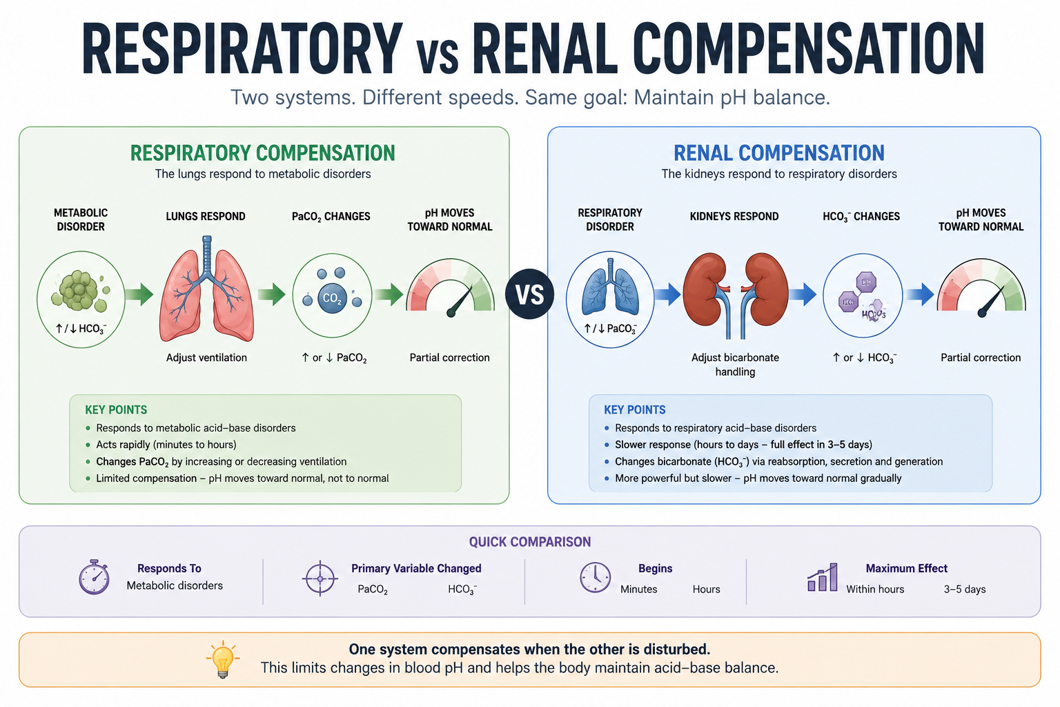
Respiratory compensation is the body's response to a primary metabolic disorder. The respiratory centre rapidly adjusts alveolar ventilation to modify PaCO2, which shifts hydrogen ion concentration and therefore blood pH. Unlike renal compensation, respiratory compensation develops within minutes and reaches its maximum effect within a few hours.
In Metabolic Acidosis
As bicarbonate falls, blood pH decreases. Chemoreceptors detect the rising hydrogen ion concentration and stimulate the respiratory centre, producing hyperventilation and a fall in PaCO2 that partially corrects pH. The deep, rapid breathing seen in severe metabolic acidosis — Kussmaul respiration — is commonly seen in diabetic ketoacidosis, severe lactic acidosis and advanced renal failure, and represents appropriate compensation rather than primary lung disease.
Example: a patient with DKA has pH 7.18, HCO3- 10 mmol/L, PaCO2 24 mmHg. The low bicarbonate indicates primary metabolic acidosis; the reduced PaCO2 demonstrates appropriate respiratory compensation.
In Metabolic Alkalosis
As bicarbonate rises, blood pH increases. The respiratory centre responds by reducing ventilation, raising PaCO2 and partially correcting pH. This compensation is much less effective than compensation for metabolic acidosis, because marked hypoventilation would cause hypoxaemia — so respiratory compensation stops before oxygenation becomes dangerously impaired. This is why patients with metabolic alkalosis often remain alkalemic despite appropriate compensation.
Example: a patient with prolonged vomiting has pH 7.52, HCO3- 36 mmol/L, PaCO2 48 mmHg — primary metabolic alkalosis with appropriate respiratory compensation.
Respiratory compensation responds to metabolic disorders, begins within minutes, reaches maximum effect within hours, changes PaCO2, and never completely normalises or overcorrects pH.
Renal Compensation
Renal compensation occurs in response to a primary respiratory disorder. Unlike the lungs, the kidneys require hours to days to modify bicarbonate handling through reabsorption of filtered bicarbonate, generation of new bicarbonate, secretion of hydrogen ions, and production and excretion of ammonium. Renal compensation is therefore slower but more powerful than respiratory compensation.
In Respiratory Acidosis
As PaCO2 rises, blood pH falls. The kidneys respond by increasing bicarbonate reabsorption, hydrogen ion secretion and ammonium production, retaining HCO3- and moving pH back toward normal.
Example: a patient with COPD has pH 7.36, PaCO2 62 mmHg, HCO3- 34 mmol/L — chronic respiratory acidosis with significant renal compensation.
In Respiratory Alkalosis
As PaCO2 falls, blood pH rises. The kidneys compensate by reducing bicarbonate reabsorption and hydrogen ion secretion while increasing bicarbonate excretion, moving pH back toward normal.
Example: a pregnant woman has pH 7.45, PaCO2 30 mmHg, HCO3- 20 mmol/L — chronic respiratory alkalosis with appropriate renal compensation.
Renal compensation responds to respiratory disorders, begins after several hours, usually requires 3–5 days for maximum effect, changes plasma bicarbonate, and never fully normalises or overcorrects pH.
Time Course of Compensation
The speed of compensation is determined by the organ responsible. The lungs can alter ventilation almost immediately, while the kidneys require cellular adaptation and changes in tubular transport, making renal compensation much slower.
| Feature | Respiratory Compensation | Renal Compensation |
|---|---|---|
| Responds to | Metabolic disorders | Respiratory disorders |
| Primary regulator | Lungs | Kidneys |
| Main variable changed | PaCO2 | HCO3- |
| Begins | Minutes | Hours |
| Maximum effect | Within hours | 3–5 days |
This timing allows clinicians to distinguish acute from chronic respiratory disorders. In acute respiratory acidosis, high PaCO2 has produced little renal compensation, so bicarbonate remains near-normal. In chronic respiratory acidosis, persistently high PaCO2 has allowed renal adaptation, so bicarbonate is markedly elevated. The same principle applies to respiratory alkalosis.
Expected Compensation Rules
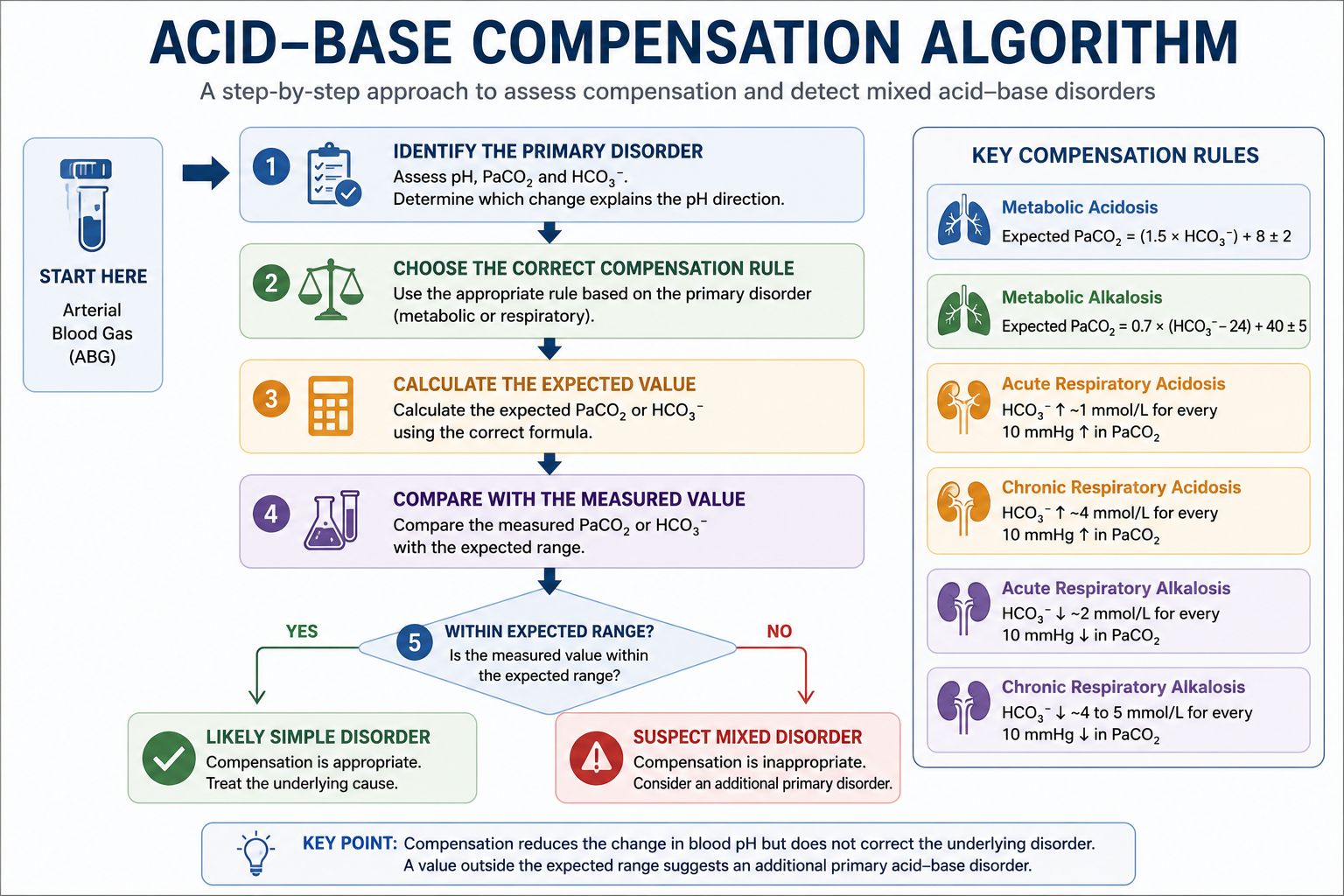
The compensatory response follows predictable physiological patterns. These rules help determine whether compensation is appropriate or whether an additional acid-base disorder is present. Remember: these are expected physiological responses, not treatment targets.
1. Metabolic Acidosis — Winter's Formula
Measured PaCO2 equal to expected → appropriate respiratory compensation. Higher than expected → additional respiratory acidosis. Lower than expected → additional respiratory alkalosis.
2. Metabolic Alkalosis
Higher than expected PaCO2 → additional respiratory acidosis. Lower than expected PaCO2 → additional respiratory alkalosis.
| Primary Disorder | Expected Compensation |
|---|---|
| Metabolic acidosis | Winter's Formula |
| Metabolic alkalosis | Expected PaCO2 formula |
| Acute respiratory acidosis | HCO3- up ~1 mmol/L per 10 mmHg PaCO2 rise |
| Chronic respiratory acidosis | HCO3- up ~4 mmol/L per 10 mmHg PaCO2 rise |
| Acute respiratory alkalosis | HCO3- down ~2 mmol/L per 10 mmHg PaCO2 fall |
| Chronic respiratory alkalosis | HCO3- down ~4–5 mmol/L per 10 mmHg PaCO2 fall |
Identify the primary disorder → choose the correct compensation rule → calculate the expected value → compare it with the measured value → if it's within range, it's a simple disorder; if not, suspect a mixed acid-base disorder.
Detecting Mixed Acid-Base Disorders
A mixed acid-base disorder occurs when two or more primary disturbances are present at the same time. This differs from compensation: compensation is a predictable physiological response, while a mixed disorder exists when the measured response falls outside the expected range.
A Structured Method
- Assess the pH. Acidaemia (below 7.35), alkalemia (above 7.45), or near-normal — which may reflect compensation or two opposing primary disorders.
- Identify the primary disorder by comparing pH, PaCO2 and bicarbonate.
- Choose the correct compensation rule — Winter's formula for metabolic acidosis, the expected PaCO2 formula for metabolic alkalosis, or the acute/chronic bicarbonate rules for respiratory disorders. Never apply a respiratory rule to a metabolic disorder or vice versa.
- Calculate the expected value using the accepted range rather than a single exact number.
- Compare expected and measured values. Within range → likely simple disorder. Outside range → mixed disorder likely.
| pH Direction | Primary Change | Likely Disorder |
|---|---|---|
| Low pH | Low HCO3- | Metabolic acidosis |
| Low pH | High PaCO2 | Respiratory acidosis |
| High pH | High HCO3- | Metabolic alkalosis |
| High pH | Low PaCO2 | Respiratory alkalosis |
Mixed Patterns to Recognise
- Metabolic acidosis + respiratory acidosis (PaCO2 above the Winter's formula range) — e.g. severe pneumonia with lactic acidosis, opioid toxicity, respiratory muscle fatigue in DKA, cardiorespiratory arrest.
- Metabolic acidosis + respiratory alkalosis (PaCO2 below the Winter's formula range) — the classic salicylate poisoning pattern; also sepsis with lactic acidosis, liver failure, pregnancy.
- Metabolic alkalosis + respiratory acidosis (PaCO2 above the expected range) — e.g. vomiting in a patient with COPD, diuretic-associated alkalosis with sedative toxicity.
- Metabolic alkalosis + respiratory alkalosis (PaCO2 below the expected range) — e.g. vomiting with pain or anxiety, metabolic alkalosis in pregnancy.
- Respiratory acidosis + metabolic disturbance — if bicarbonate is lower than the acute/chronic expected rise, suspect additional metabolic acidosis; if higher, suspect additional metabolic alkalosis.
- Respiratory alkalosis + metabolic disturbance — the same logic applies using the expected bicarbonate fall.
A near-normal pH may reflect a well-compensated chronic disorder, or it may hide two opposing primary disorders. For example, pH 7.40 with PaCO2 20 mmHg and HCO3- 12 mmol/L both markedly abnormal is not a normal acid-base state — always calculate expected compensation rather than trusting pH alone.
Six Worked ABG Cases
Simple Metabolic Acidosis — Diabetic Ketoacidosis
A 22-year-old patient: pH 7.21, PaCO2 25 mmHg, HCO3- 11 mmol/L.
Primary disorder: metabolic acidosis. Expected PaCO2 = (1.5 × 11) + 8 = 24.5 mmHg (range 22.5–26.5 mmHg). Measured PaCO2 (25 mmHg) is within range.
Metabolic Acidosis with Respiratory Acidosis — Septic Shock
A drowsy patient with respiratory fatigue: pH 7.08, PaCO2 40 mmHg, HCO3- 12 mmol/L.
Primary disorder: metabolic acidosis. Expected PaCO2 = (1.5 × 12) + 8 = 26 mmHg (range 24–28 mmHg). Measured PaCO2 (40 mmHg) is considerably higher than expected.
Metabolic Acidosis with Respiratory Alkalosis — Salicylate Poisoning
pH 7.46, PaCO2 18 mmHg, HCO3- 13 mmol/L.
Bicarbonate is markedly low (metabolic acidosis), but the pH is alkalemic, showing the low PaCO2 has an independent effect. Expected PaCO2 = (1.5 × 13) + 8 = 27.5 mmHg (range 25.5–29.5 mmHg). Measured PaCO2 (18 mmHg) is much lower than expected.
Simple Metabolic Alkalosis — Prolonged Vomiting
pH 7.50, PaCO2 48 mmHg, HCO3- 34 mmol/L.
Primary disorder: metabolic alkalosis. Expected PaCO2 = 0.7 × (34 − 24) + 40 = 47 mmHg (range 42–52 mmHg). Measured PaCO2 (48 mmHg) is within range.
Acute Respiratory Acidosis — Opioid Overdose
pH 7.24, PaCO2 60 mmHg, HCO3- 26 mmol/L.
Primary disorder: respiratory acidosis. PaCO2 has risen 20 mmHg above 40 mmHg. Acute rule: HCO3- rises ~1 mmol/L per 10 mmHg → expected HCO3- = 24 + 2 = 26 mmol/L. Measured HCO3- (26 mmol/L) matches.
Chronic Respiratory Acidosis with Metabolic Alkalosis — COPD on Diuretics
pH 7.43, PaCO2 60 mmHg, HCO3- 39 mmol/L.
PaCO2 has risen 20 mmHg above 40 mmHg. Chronic rule: HCO3- rises ~4 mmol/L per 10 mmHg → expected HCO3- = 24 + 8 = 32 mmol/L. Measured HCO3- (39 mmol/L) is substantially higher than expected.
Common Pitfalls
- Assuming every second abnormality is compensation — PaCO2 and bicarbonate often move in the expected direction, but the magnitude must still be calculated.
- Expecting compensation to normalise pH completely — a normal pH with markedly abnormal PaCO2 and bicarbonate suggests either a chronic compensated disorder or two opposing primary disorders.
- Believing compensation can overcorrect — if pH moves beyond normal into the opposite disturbance, suspect an additional primary disorder.
- Using the wrong formula — Winter's formula applies only to metabolic acidosis; each primary disorder has its own expected response.
- Ignoring whether a respiratory disorder is acute or chronic — the expected bicarbonate response differs significantly; clinical history is essential.
- Treating compensation as the disease — Kussmaul respiration and renal bicarbonate retention are adaptive responses, not separate disorders to suppress.
- Ignoring the clinical context — mathematical rules support interpretation but cannot replace history, examination and other laboratory findings.
- Treating expected values as exact — compensation formulas give estimated ranges; small deviations may reflect normal variation rather than proof of a mixed disorder.
One Minute Revision
- The organ that did not cause the primary disorder compensates: metabolic disorder → lungs; respiratory disorder → kidneys.
- Respiratory compensation begins within minutes; renal compensation takes hours to days (3–5 days for maximum effect).
- Metabolic acidosis: Expected PaCO2 = (1.5 × HCO3-) + 8 ± 2 (Winter's Formula).
- Metabolic alkalosis: Expected PaCO2 = 0.7 × (HCO3- − 24) + 40 ± 5.
- Respiratory acidosis: HCO3- rises ~1 mmol/L (acute) or ~4 mmol/L (chronic) per 10 mmHg rise in PaCO2.
- Respiratory alkalosis: HCO3- falls ~2 mmol/L (acute) or ~4–5 mmol/L (chronic) per 10 mmHg fall in PaCO2.
- If the measured value falls outside the expected range, suspect a mixed acid-base disorder.
Frequently Asked Questions
This article is intended for medical education only. Compensation formulas provide estimated ranges to support, not replace, clinical judgement. ABG interpretation should always be combined with history, examination and other investigations, with senior or specialist input where indicated.