Respiratory alkalosis develops when alveolar ventilation exceeds the body's metabolic requirement for carbon dioxide elimination, lowering PaCO2 and raising blood pH. It is not a disease in itself, but a physiological consequence of excessive ventilation that always deserves a search for its underlying cause.
This article builds on Acid-Base Disorders Explained and complements Respiratory Acidosis Explained, Metabolic Acidosis Explained and Metabolic Alkalosis Explained. If ABG interpretation feels unfamiliar, read ABG Interpretation Explained first, then return here for the respiratory alkalosis pathway.
Respiratory alkalosis occurs when PaCO2 falls because ventilation exceeds metabolic demand, causing blood pH to rise. The primary problem is excessive ventilation, not metabolism.
Learning Objectives
- Define respiratory alkalosis and identify the primary abnormality
- Explain why hyperventilation lowers PaCO2 and raises blood pH
- Distinguish acute from chronic respiratory alkalosis
- Recognise the characteristic ABG pattern and expected renal compensation
- Identify the major causes, including serious and benign causes
- Apply a structured diagnostic and management approach
What Is Respiratory Alkalosis?
Respiratory alkalosis is a primary acid-base disorder caused by a reduction in arterial carbon dioxide tension (PaCO2), resulting in an increase in blood pH. Unlike metabolic alkalosis, where the primary abnormality is an increase in bicarbonate concentration, respiratory alkalosis begins with excessive elimination of carbon dioxide through the lungs. When ventilation exceeds the body's metabolic production of carbon dioxide, arterial PaCO2 falls, fewer hydrogen ions are produced, and blood pH rises.
The reduced bicarbonate is a compensatory response rather than the primary abnormality — the kidneys respond by reducing bicarbonate reabsorption and increasing bicarbonate excretion, but this develops gradually over several days.
Every acid-base disorder should first be classified by the variable that changed first: a primary decrease in PaCO2 is respiratory alkalosis, while a primary increase in HCO3- is metabolic alkalosis. Identifying the primary disturbance is the first step in interpreting every ABG.
Why Carbon Dioxide Affects pH
Carbon dioxide participates in a reversible reaction with water that produces hydrogen ions and bicarbonate. When PaCO2 falls, the reaction shifts to the left, hydrogen ion concentration decreases, and blood pH rises — the opposite of what happens in respiratory acidosis.
Hypocapnia vs Respiratory Alkalosis
Hypocapnia simply means a reduced arterial carbon dioxide level. Respiratory alkalosis occurs when this reduction in PaCO2 raises blood pH. Patients with chronic respiratory alkalosis may have only a mildly elevated or near-normal pH because renal compensation has lowered bicarbonate.
| Feature | Respiratory Alkalosis | Metabolic Alkalosis |
|---|---|---|
| Primary abnormality | Decreased PaCO2 | Increased HCO3- |
| Problem | Hyperventilation | Excess bicarbonate |
| Main compensation | Kidneys | Lungs |
| Time to compensate | Days | Minutes to hours |
Respiratory alkalosis is frequently encountered in anxiety and panic attacks, pulmonary embolism, pneumonia, high altitude, sepsis, pregnancy, chronic liver disease, mechanical ventilation and early salicylate poisoning. Many of these conditions are serious illnesses, so respiratory alkalosis should never automatically be attributed to anxiety without careful clinical assessment.
Respiratory alkalosis is not a disease — it is a physiological consequence of excessive alveolar ventilation. Nearly every cause shares the same pathway: increased alveolar ventilation → decreased PaCO2 → decreased hydrogen ions → increased blood pH → respiratory alkalosis.

Carbon Dioxide Physiology
Carbon dioxide is continuously produced by metabolically active cells and eliminated through the lungs. Under normal circumstances, production and elimination are precisely balanced, maintaining an arterial PaCO2 of approximately 40 mmHg. When alveolar ventilation increases beyond metabolic demand, carbon dioxide is removed faster than it is produced, and PaCO2 falls.
Hyperventilation
Hyperventilation is defined as alveolar ventilation that exceeds the body's metabolic requirement for carbon dioxide elimination. This definition matters because hyperventilation is not determined by respiratory rate alone: a patient may breathe rapidly without hyperventilating if tidal volume is small, while a patient taking deep, effective breaths may significantly reduce PaCO2 with only a modest increase in respiratory rate. Hyperventilation should be understood as excessive alveolar ventilation, not simply "fast breathing."
Ventilation vs Oxygenation
Ventilation removes carbon dioxide; oxygenation transfers oxygen into the bloodstream. Respiratory alkalosis develops because ventilation is excessive, not because oxygen levels are necessarily abnormal. Many patients with respiratory alkalosis are hypoxaemic because hypoxaemia itself stimulates hyperventilation, while others, such as those with anxiety-induced hyperventilation, may have completely normal oxygen levels.
Acute vs Chronic Respiratory Alkalosis
Both conditions share a reduced PaCO2, but the body's response changes significantly over time. The key difference is renal compensation. In acute disease the kidneys have had little time to respond, so pH rises markedly while bicarbonate decreases only slightly. In chronic disease, sustained hypocapnia stimulates the kidneys to excrete bicarbonate and retain hydrogen ions, so blood pH is often only mildly elevated despite persistently low PaCO2.
Acute Respiratory Alkalosis
Develops over minutes to hours. Common causes include panic attack, anxiety, pain, early pulmonary embolism, early sepsis, mechanical overventilation and high altitude.
| Parameter | Typical Finding |
|---|---|
| pH | High |
| PaCO2 | Low |
| HCO3- | Slightly reduced |
Chronic Respiratory Alkalosis
Develops over several days or longer. Common causes include pregnancy, chronic liver disease, long-term high-altitude exposure and chronic hyperventilation syndromes. During this period the kidneys reduce bicarbonate reabsorption and increase bicarbonate excretion.
| Parameter | Typical Finding |
|---|---|
| pH | Mildly elevated or near normal |
| PaCO2 | Low |
| HCO3- | Markedly reduced |
Acute-on-Chronic Respiratory Alkalosis
Some patients with chronic hyperventilation develop an acute increase in ventilation during illness — for example, pregnancy with pneumonia, chronic liver disease with sepsis, or high-altitude residents developing pulmonary embolism. These patients may show a further fall in PaCO2 with only limited additional renal compensation.

Recognising acute versus chronic respiratory alkalosis helps interpret the ABG accurately, detect mixed acid-base disorders, estimate expected bicarbonate, and identify chronic physiological adaptation rather than misdiagnosing an acute problem.
ABG Findings
Respiratory alkalosis has a characteristic ABG pattern, but interpretation should always follow a structured sequence rather than relying on a single parameter.
| Parameter | Finding |
|---|---|
| pH | Up |
| PaCO2 | Down |
| HCO3- | Down (compensation) |
pH above 7.45 indicates alkalemia, though chronic respiratory alkalosis may have only a mildly elevated pH because of renal compensation. The defining abnormality is reduced PaCO2, confirming a respiratory primary disturbance. Bicarbonate then reflects renal compensation: slightly reduced suggests acute disease, markedly reduced suggests chronic disease, higher than expected suggests an associated metabolic alkalosis, and lower than expected suggests an associated metabolic acidosis.
Worked Examples
| Parameter | Example 1 | Example 2 |
|---|---|---|
| pH | 7.55 | 7.46 |
| PaCO2 | 26 mmHg | 28 mmHg |
| HCO3- | 22 mmol/L | 18 mmol/L |
| Interpretation | Acute respiratory alkalosis | Chronic compensated respiratory alkalosis |
In Example 1, bicarbonate has decreased only slightly. In Example 2, the kidneys have reduced bicarbonate concentration considerably, limiting the rise in pH.
Renal Compensation
The kidneys cannot immediately compensate for respiratory alkalosis. Instead, they gradually reduce bicarbonate reabsorption and increase bicarbonate excretion by reducing hydrogen ion secretion and ammonium production. Maximum compensation usually requires 3–5 days.
Compensation reduces the severity of alkalemia but does not eliminate the underlying cause. Only correcting the excessive ventilation will normalise PaCO2, so renal compensation never completely restores blood pH if hypocapnia persists.
Expected Compensation
These are approximate clinical values used to detect mixed acid-base disorders. For example, a patient with PaCO2 30 mmHg and HCO3- 22 mmol/L fits acute respiratory alkalosis, whereas a patient with the same PaCO2 but HCO3- 18 mmol/L fits chronic respiratory alkalosis.
Causes of Respiratory Alkalosis
Although respiratory alkalosis has many causes, nearly all share a common mechanism: excessive alveolar ventilation. Rather than memorising long lists, group causes by the physiological process driving hyperventilation.
- Increased respiratory drive — anxiety, panic attacks, pain and fever increase ventilation despite structurally normal lungs.
- Hypoxaemia — pulmonary embolism, pneumonia, pulmonary oedema, early severe asthma and high altitude stimulate peripheral chemoreceptors, increasing ventilation.
- Central nervous system stimulation — stroke, meningitis, encephalitis and head injury directly stimulate the respiratory centre.
- Drugs and hormones — early salicylate poisoning and progesterone (including pregnancy) stimulate ventilation; pregnancy commonly produces a mild chronic respiratory alkalosis as a normal adaptation.
- Mechanical ventilation — excessive minute ventilation settings. Always review ventilator settings when hypocapnia develops in an intubated patient.
| Mechanism | Examples |
|---|---|
| Increased respiratory drive | Anxiety, pain, fever |
| Hypoxaemia | Pulmonary embolism, pneumonia, high altitude |
| CNS stimulation | Stroke, meningitis, head injury |
| Drugs / hormones | Salicylates, progesterone, pregnancy |
| Mechanical ventilation | Excessive ventilator support |
Although the underlying diseases differ considerably, they all converge on one pathway: increased alveolar ventilation → decreased PaCO2 → decreased hydrogen ions → increased blood pH → respiratory alkalosis. Understanding this pathway is more valuable than memorising isolated causes.
Clinical Features
Clinical manifestations depend on how rapidly PaCO2 falls, the severity of hypocapnia, and the underlying disease. Patients with acute respiratory alkalosis usually have more prominent symptoms because there has been little time for physiological adaptation, while patients with chronic respiratory alkalosis often have few symptoms and may be diagnosed incidentally.
Many symptoms are not caused by the elevated pH itself but by the physiological effects of hypocapnia: cerebral vasoconstriction, increased calcium binding to albumin, reduced ionized calcium and increased neuromuscular excitability. Common symptoms include light-headedness, dizziness, headache, difficulty concentrating, anxiety, shortness of breath and chest tightness. Reduced cerebral blood flow may cause confusion, blurred vision or near-syncope, while reduced ionized calcium produces perioral numbness, tingling of the fingers and toes, muscle cramps, carpopedal spasm and, rarely, tetany. This albumin-binding effect is explained further in Calcium Homeostasis Explained, and its clinical low-calcium presentation is covered in Hypocalcemia Explained.
Examination findings depend largely on the underlying disease and may include tachypnoea, deep breathing, signs of anxiety, fever, hypoxaemia, or features of pulmonary embolism, pneumonia or liver disease. Always search for the underlying cause rather than attributing symptoms to anxiety alone.
A patient with respiratory alkalosis who is hypoxaemic, tachycardic and has pleuritic chest pain should immediately raise suspicion for pulmonary embolism rather than a simple panic attack.
Diagnostic Approach
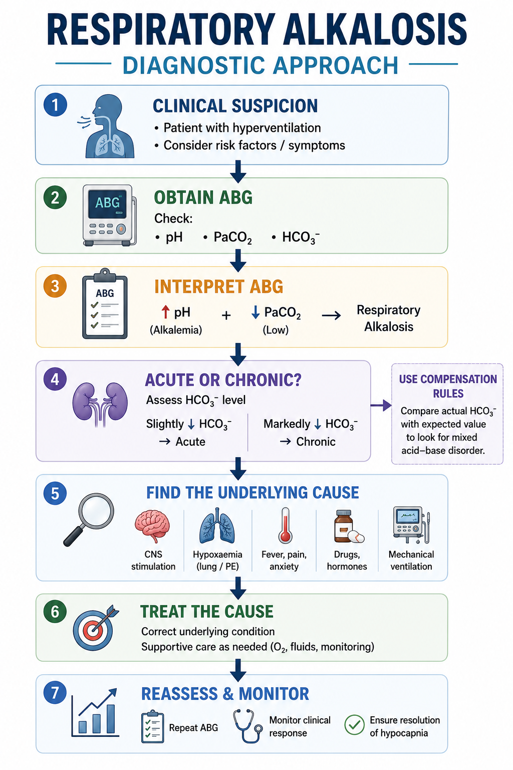
Diagnosis involves two goals: confirm the acid-base disorder, and determine why the patient is hyperventilating. The ABG confirms the diagnosis; the history and examination identify the cause.
- Confirm respiratory alkalosis. Review the ABG for elevated pH, reduced PaCO2 and appropriate bicarbonate reduction, and determine whether compensation is acute or chronic.
- Assess clinical severity. Look for hypoxaemia, chest pain, hypotension, altered consciousness, persistent tachycardia, high fever or severe respiratory distress — these patients require urgent assessment.
- Take a focused history. Ask about sudden onset, chest pain, anxiety, fever, pregnancy, liver disease, high-altitude exposure, salicylate ingestion or mechanical ventilation.
- Perform a focused examination of respiratory rate, oxygen saturation, lungs, cardiovascular system, neurological status, and signs of infection or deep vein thrombosis.
- Request appropriate investigations — ABG, full blood count, CRP, electrolytes, lactate, liver function tests, chest X-ray, CT pulmonary angiography when PE is suspected, ECG, D-dimer, pregnancy test, toxicology screening or salicylate level.
Management Principles
Respiratory alkalosis is treated by addressing the underlying cause. Attempting to normalise the blood gas without identifying the cause may delay life-saving treatment.
Airway: most patients do not require airway intervention. Breathing: assess respiratory rate, oxygen saturation, work of breathing and the ABG, and treat hypoxaemia appropriately. Circulation: assess blood pressure, heart rate and peripheral perfusion, and correct haemodynamic instability.
| Cause | Treatment |
|---|---|
| Anxiety | Reassurance, breathing techniques, psychological support |
| Pulmonary embolism | Anticoagulation ± reperfusion therapy |
| Pneumonia | Antibiotics |
| Sepsis | Early sepsis management |
| High altitude | Descent, oxygen, acetazolamide where appropriate |
| Mechanical ventilation | Adjust ventilator settings |
| Salicylate poisoning | Poison-specific management |
Patients were historically encouraged to breathe into a paper bag. This is no longer recommended, because paper-bag rebreathing may worsen hypoxaemia and delay recognition of life-threatening illnesses such as pulmonary embolism or myocardial infarction. Instead, identify and treat the underlying cause of hyperventilation.
Worked Clinical Cases
Case 1 — Panic Attack
A 24-year-old woman develops sudden shortness of breath during an examination. ABG: pH 7.54, PaCO2 28 mmHg, HCO3- 22 mmol/L.
Interpretation: acute respiratory alkalosis caused by hyperventilation. Management includes excluding serious pathology, reassurance, and controlled breathing techniques.
Case 2 — Pulmonary Embolism
A 62-year-old man develops sudden pleuritic chest pain and dyspnoea. ABG: pH 7.49, PaCO2 30 mmHg, HCO3- 23 mmol/L.
Interpretation: acute respiratory alkalosis. Management includes urgent investigation for pulmonary embolism and anticoagulation when appropriate.
Case 3 — Mechanical Ventilation
A ventilated ICU patient develops pH 7.56, PaCO2 24 mmHg, HCO3- 21 mmol/L.
Interpretation: respiratory alkalosis due to excessive minute ventilation. Management involves reviewing ventilator settings and reducing excessive ventilation where clinically appropriate.
Common Pitfalls
- Assuming every hyperventilating patient has anxiety — always exclude pulmonary embolism, sepsis, hypoxaemia, acute coronary syndrome and salicylate poisoning.
- Ignoring compensation — failure to recognise chronic renal compensation may lead to incorrect ABG interpretation.
- Confusing respiratory alkalosis with metabolic alkalosis — always determine whether the primary abnormality is low PaCO2 or high bicarbonate.
- Treating the blood gas instead of the patient — the ABG is a clue; the underlying disease requires treatment.
- Using paper-bag rebreathing — modern practice discourages this because it may worsen hypoxaemia in serious illness.
One Minute Revision
- Respiratory alkalosis is caused by a primary decrease in PaCO2 resulting from excessive alveolar ventilation.
- Common causes: anxiety, panic attack, pulmonary embolism, pneumonia, high altitude, pregnancy, chronic liver disease, mechanical ventilation, early salicylate poisoning.
- Typical ABG: pH up, PaCO2 down, HCO3- down (compensation).
- Acute disease shows minimal renal compensation and a higher pH; chronic disease shows significant compensation and near-normal pH.
- Management: confirm the diagnosis, search for and treat the underlying cause, correct hypoxaemia, and avoid routine paper-bag rebreathing.
Frequently Asked Questions
This article is intended for medical education only. Respiratory alkalosis may reflect a benign physiological response or the earliest clue to serious illness such as pulmonary embolism, sepsis or severe pneumonia. Clinical management requires proper assessment, local protocols and senior or specialist input.