Respiratory acidosis develops when the lungs fail to eliminate enough carbon dioxide, raising PaCO2 and lowering blood pH. Renal compensation then develops gradually, which is why acute and chronic respiratory acidosis look very different on an ABG despite sharing the same primary abnormality.
This article builds on Acid-Base Disorders Explained and complements Metabolic Acidosis Explained and Metabolic Alkalosis Explained. If ABG interpretation feels unfamiliar, read ABG Interpretation Explained first, then return here for the respiratory acidosis pathway.
Respiratory acidosis occurs when PaCO2 rises because the lungs cannot eliminate enough carbon dioxide, causing blood pH to fall. The primary problem is ventilation, not metabolism.
Learning Objectives
- Define respiratory acidosis and identify the primary abnormality
- Explain how carbon dioxide influences blood pH
- Describe normal carbon dioxide production, transport and elimination
- Distinguish acute from chronic respiratory acidosis
- Recognise the characteristic ABG pattern and expected renal compensation
- Identify the major causes of respiratory acidosis
- Apply a structured diagnostic and management approach
What Is Respiratory Acidosis?
Respiratory acidosis is a primary acid-base disorder characterised by an increase in arterial carbon dioxide tension (PaCO2), resulting in a reduction in blood pH. Unlike metabolic acid-base disorders, where the primary abnormality involves bicarbonate concentration, respiratory acidosis begins with a problem in ventilation. When the lungs fail to eliminate sufficient carbon dioxide, the gas accumulates in the bloodstream, reacts with water to form carbonic acid, and dissociates into hydrogen ions and bicarbonate. The rise in hydrogen ion concentration lowers blood pH, producing acidemia.
The kidneys respond by increasing hydrogen ion excretion and bicarbonate reabsorption, but this renal compensation requires hours to days to become fully effective. Acute respiratory acidosis therefore often presents with marked acidemia, whereas chronic respiratory acidosis may have a near-normal pH despite persistently elevated PaCO2.
The simplest way to classify acid-base disorders is to identify which variable changed first. A primary increase in PaCO2 is respiratory acidosis; a primary decrease in HCO3- is metabolic acidosis. The elevated bicarbonate seen in respiratory acidosis is a compensatory response, not the initiating abnormality.
Why Carbon Dioxide Behaves as an Acid
Carbon dioxide is not itself a strong acid, but when dissolved in water it participates in a reversible reaction that produces hydrogen ions and bicarbonate.
As PaCO2 rises, this reaction shifts to the right, producing more hydrogen ions and lowering blood pH. This relationship underpins every respiratory acid-base disorder.
Hypercapnia and Respiratory Acidosis
Hypercapnia simply means an elevated arterial carbon dioxide level. Respiratory acidosis occurs when hypercapnia lowers blood pH. The two terms are related but not synonymous: patients with chronic hypercapnia, such as long-standing COPD, may have a nearly normal pH because the kidneys have retained enough bicarbonate to partially compensate.
| Feature | Respiratory Acidosis | Metabolic Acidosis |
|---|---|---|
| Primary abnormality | Increased PaCO2 | Decreased HCO3- |
| Initial problem | Ventilation | Metabolism |
| Main compensation | Kidneys | Lungs |
| Time to compensate | Hours to days | Minutes to hours |
Respiratory acidosis is commonly encountered in COPD exacerbations, severe asthma, drug-induced respiratory depression, neuromuscular disorders, obesity hypoventilation syndrome, chest wall disorders, mechanical ventilation problems and cardiorespiratory arrest. Early recognition is essential because severe hypercapnia may lead to respiratory failure, altered mental status, arrhythmias and haemodynamic instability.

Carbon Dioxide Physiology
Carbon dioxide is continuously produced by every metabolically active cell as a by-product of aerobic metabolism. Under normal circumstances, production is precisely matched by elimination through the lungs, maintaining an arterial PaCO2 of approximately 40 mmHg.
Respiratory acidosis almost never develops because of excessive carbon dioxide production. Instead, it develops because the lungs fail to remove the normal amount being produced.
Transport of Carbon Dioxide
Carbon dioxide travels from tissues to the lungs in three forms: approximately 70% as bicarbonate ions, 20–25% bound to proteins such as haemoglobin (carbamino compounds), and 5–10% dissolved directly in plasma. Although only a small fraction is dissolved, this fraction determines the measured PaCO2 on an arterial blood gas.
Elimination and Ventilation vs Oxygenation
In the lungs, carbon dioxide diffuses from pulmonary capillary blood into the alveoli and is exhaled. This process depends primarily on alveolar ventilation, not oxygen concentration — any reduction in effective alveolar ventilation results in carbon dioxide retention. Ventilation and oxygenation are closely related but serve different functions: ventilation eliminates carbon dioxide, while oxygenation transfers oxygen into the blood. A patient may be severely hypoxaemic without hypercapnia if ventilation is maintained, and conversely a hypoventilating patient may develop significant hypercapnia before severe hypoxaemia becomes evident.
Nearly every cause of respiratory acidosis produces the same sequence: reduced alveolar ventilation → reduced CO2 elimination → increased PaCO2 → increased hydrogen ion concentration → reduced blood pH.
Acute vs Chronic Respiratory Acidosis
Both conditions share an elevated PaCO2, but the body's response differs dramatically depending on how long the hypercapnia has been present. The key difference is renal compensation. In acute disease the kidneys have had little time to respond, so pH falls markedly while bicarbonate rises only slightly. In chronic disease, sustained hypercapnia stimulates the kidneys to retain bicarbonate and excrete hydrogen ions, so pH is often close to normal despite a persistently elevated PaCO2.
Acute Respiratory Acidosis
Develops over minutes to hours. Common causes include opioid overdose, sedative toxicity, acute airway obstruction, severe asthma, acute respiratory muscle fatigue, mechanical ventilation failure and cardiorespiratory arrest.
| Parameter | Typical Finding |
|---|---|
| pH | Low |
| PaCO2 | High |
| HCO3- | Slightly increased |
Chronic Respiratory Acidosis
Develops over several days or longer. Typical causes include COPD, obesity hypoventilation syndrome, neuromuscular disease, chest wall disorders and chronic sleep-disordered breathing. During this period the kidneys increase bicarbonate reabsorption, so pH improves, bicarbonate rises considerably, and PaCO2 remains elevated.
| Parameter | Typical Finding |
|---|---|
| pH | Near normal or mildly low |
| PaCO2 | High |
| HCO3- | Significantly elevated |
Acute-on-Chronic Respiratory Acidosis
Many hospital patients do not fit neatly into either category. For example, a patient with stable COPD who already has chronic hypercapnia with renal compensation may develop pneumonia. The superimposed acute illness causes further CO2 retention that the kidneys cannot compensate for immediately, producing acute-on-chronic respiratory acidosis. These patients often present with increasing drowsiness, confusion, headache, worsening dyspnoea, markedly elevated PaCO2 and a pH lower than their usual baseline. Recognising this deterioration is clinically important because these patients frequently require urgent ventilatory support.
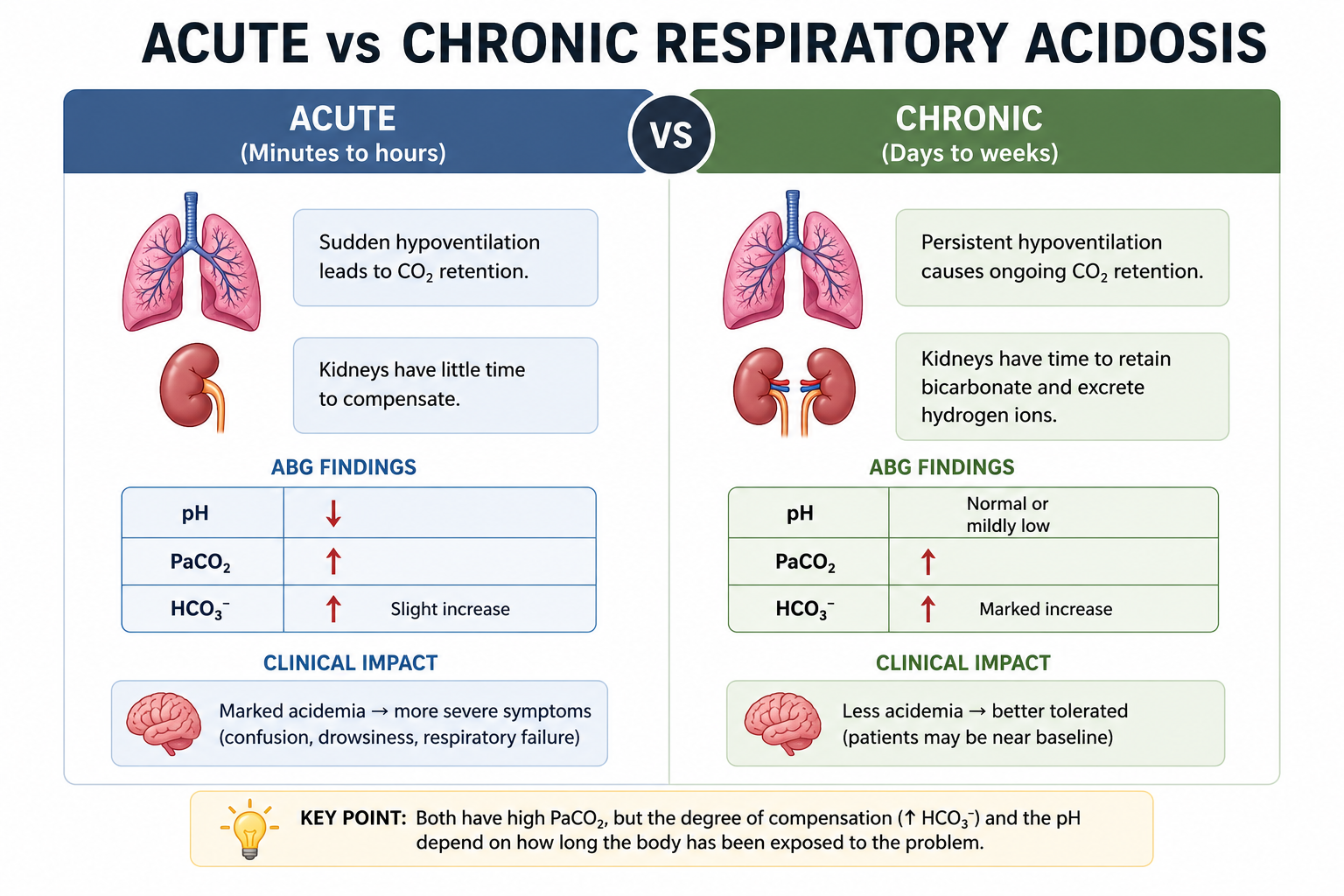
Correctly identifying acute versus chronic disease helps interpret the ABG accurately, estimate the expected bicarbonate, detect mixed acid-base disorders, and decide on the urgency of treatment.
ABG Findings
Respiratory acidosis has a characteristic arterial blood gas pattern, but the diagnosis should never rely on a single parameter — interpret pH, PaCO2 and HCO3- systematically.
| Parameter | Finding |
|---|---|
| pH | Down |
| PaCO2 | Up |
| HCO3- | Up (compensation) |
pH below 7.35 indicates acidemia, though pH may be nearly normal in chronic disease because of renal compensation. The defining abnormality is elevated PaCO2, confirming a respiratory primary disturbance. Bicarbonate then indicates the degree of renal compensation: slightly elevated suggests acute disease, markedly elevated suggests chronic disease, lower than expected suggests an associated metabolic acidosis, and higher than expected suggests an associated metabolic alkalosis.
Worked Examples
| Parameter | Example 1 | Example 2 |
|---|---|---|
| pH | 7.22 | 7.37 |
| PaCO2 | 68 mmHg | 62 mmHg |
| HCO3- | 27 mmol/L | 35 mmol/L |
| Interpretation | Acute respiratory acidosis | Chronic compensated respiratory acidosis |
In Example 1, bicarbonate has increased only slightly. In Example 2, despite the high PaCO2, the pH is almost normal because renal compensation has increased bicarbonate substantially.
Renal Compensation
The kidneys cannot immediately correct respiratory acidosis. Instead, they gradually increase bicarbonate reabsorption while excreting additional hydrogen ions. This process begins within several hours but usually requires 3–5 days to approach maximal compensation, achieved by increasing bicarbonate reabsorption and generation, hydrogen ion secretion, ammonium production and titratable acid excretion.
Compensation reduces the severity of acidemia but does not remove the underlying cause. Only improved ventilation can reduce PaCO2, so renal compensation can never completely normalise blood pH if hypercapnia persists.
Expected Compensation
These approximations help detect mixed acid-base disorders. For example, a patient with PaCO2 60 mmHg and HCO3- 25 mmol/L fits acute respiratory acidosis, whereas a patient with the same PaCO2 but HCO3- 33 mmol/L fits chronic respiratory acidosis.
Causes of Respiratory Acidosis
Although many diseases cause respiratory acidosis, almost all do so by reducing effective alveolar ventilation. Instead of memorising long lists, understand the underlying mechanism.
- Central respiratory depression — opioids, benzodiazepines, general anaesthesia, brainstem stroke and head injury reduce respiratory drive, causing hypoventilation and CO2 retention.
- Airway obstruction — severe asthma, COPD exacerbation, upper airway obstruction and foreign body aspiration limit airflow and reduce alveolar ventilation.
- Neuromuscular disorders — Guillain-Barré syndrome, myasthenia gravis, amyotrophic lateral sclerosis and muscular dystrophy weaken respiratory muscles, causing hypoventilation.
- Chest wall disorders — severe kyphoscoliosis, flail chest and morbid obesity mechanically restrict lung expansion and reduce tidal volume.
- Obesity hypoventilation syndrome — severe obesity increases the work of breathing, often producing chronic hypercapnia and sleep-disordered breathing.
- Mechanical ventilation problems — low minute ventilation, circuit disconnection, equipment malfunction or inappropriate settings. Always consider equipment failure in an intubated patient with sudden respiratory acidosis.
| Mechanism | Examples |
|---|---|
| Central respiratory depression | Opioids, sedatives, stroke |
| Airway obstruction | COPD, asthma |
| Neuromuscular failure | Myasthenia gravis, Guillain-Barré syndrome |
| Chest wall restriction | Kyphoscoliosis, obesity |
| Mechanical ventilation problems | Ventilator malfunction |
Although the underlying diseases differ considerably, they all converge on one pathway: reduced alveolar ventilation → reduced CO2 elimination → hypercapnia → respiratory acidosis. Understanding this pathway is far more valuable than memorising isolated disease names.
Clinical Features
Clinical manifestations depend on how rapidly hypercapnia develops, the severity of CO2 elevation, and the underlying disease. Patients with acute respiratory acidosis are generally much sicker than those with chronic compensated disease because the brain has had no time to adapt.
Early symptoms include shortness of breath, headache, fatigue, daytime sleepiness, difficulty concentrating, anxiety and restlessness. As PaCO2 continues to rise, confusion, drowsiness, slurred speech and reduced consciousness become more prominent, and severe hypercapnia may eventually cause CO2 narcosis, coma or respiratory arrest.
Examination may reveal tachypnoea or hypoventilation, accessory muscle use, cyanosis if hypoxaemia is present, sweating, bounding pulse and warm peripheries, alongside neurological signs such as tremor, asterixis, confusion and a reduced Glasgow Coma Scale. Look for clues to the underlying cause: wheezing suggests asthma or COPD, ptosis may suggest myasthenia gravis, pinpoint pupils suggest opioid toxicity, and severe obesity suggests obesity hypoventilation syndrome.
The severity of symptoms often reflects how quickly PaCO2 rises rather than its absolute value. A patient with chronic COPD may tolerate a PaCO2 of 65 mmHg remarkably well, whereas another patient with an acute opioid overdose may become unconscious at a similar level.
Diagnostic Approach
Diagnosis requires two simultaneous goals: confirm the acid-base disorder and identify the underlying cause. Treating the elevated PaCO2 alone is not sufficient.
- Confirm respiratory acidosis. Review the ABG for low pH, elevated PaCO2 and the degree of bicarbonate compensation, and determine whether the picture is acute, chronic or acute-on-chronic.
- Assess clinical severity. Look for red flags of impending respiratory failure: reduced consciousness, exhaustion, inability to speak, silent chest, respiratory arrest, severe hypoxaemia or haemodynamic instability.
- Identify the cause. History is often the most valuable investigation — ask about sudden or gradual onset, drug overdose, COPD, asthma, neuromuscular disease, sleep apnoea, recent surgery or trauma.
- Perform a focused examination of respiratory effort, rate, air entry, wheeze, crackles, chest expansion and neurological status.
- Request appropriate investigations — ABG, full blood count, urea and electrolytes, CRP, blood cultures, chest X-ray or CT, ECG, spirometry or peak flow in stable patients, and nerve conduction studies or MRI when a neuromuscular cause is suspected.

Management Principles
Treatment focuses on correcting the underlying cause of hypoventilation rather than attempting to correct the arterial blood gas in isolation, following the ABC approach.
Airway: ensure patency and consider airway protection in patients with reduced consciousness, severe respiratory fatigue or inability to protect the airway.
Breathing: assess respiratory rate, oxygen saturation, work of breathing and the ABG. Provide oxygen when indicated, but oxygen alone does not treat respiratory acidosis — the primary problem is inadequate ventilation.
Improve ventilation: depending on the cause, management may include bronchodilators, corticosteroids, antibiotics for infection, reversal agents such as naloxone for opioid toxicity, non-invasive ventilation, or invasive mechanical ventilation.
| Cause | Treatment |
|---|---|
| COPD exacerbation | Bronchodilators, steroids, antibiotics, NIV if indicated |
| Opioid overdose | Naloxone and airway support |
| Severe asthma | Bronchodilators, steroids, magnesium, ventilatory support if required |
| Myasthenia gravis | Disease-specific therapy and ventilatory support |
| Obesity hypoventilation | Weight management and positive airway pressure therapy |
Bicarbonate administration is not routinely indicated in respiratory acidosis. The primary abnormality is carbon dioxide retention, not bicarbonate deficiency, and administering bicarbonate without improving ventilation may increase CO2 production and worsen hypercapnia.
Worked Clinical Cases
Case 1 — Acute Opioid Overdose
A 25-year-old man is found unconscious after opioid use, with a respiratory rate of 6/min and pinpoint pupils. ABG: pH 7.18, PaCO2 72 mmHg, HCO3- 26 mmol/L.
Interpretation: acute respiratory acidosis with minimal renal compensation, caused by opioid-induced respiratory depression. Initial management includes airway assessment, naloxone, oxygen and ventilatory support if required.
Case 2 — COPD Exacerbation
A 68-year-old man with severe COPD develops worsening breathlessness over several days. ABG: pH 7.35, PaCO2 64 mmHg, HCO3- 35 mmol/L.
Interpretation: chronic compensated respiratory acidosis. Management focuses on treating the COPD exacerbation rather than attempting to normalise PaCO2.
Case 3 — Acute-on-Chronic Respiratory Acidosis
A patient with known COPD develops pneumonia. ABG: pH 7.24, PaCO2 82 mmHg, HCO3- 36 mmol/L.
Interpretation: chronic hypercapnia with a new acute deterioration — acute-on-chronic respiratory acidosis. This patient requires urgent assessment for non-invasive or invasive ventilation depending on clinical status.
Common Pitfalls
- Treating the ABG instead of the patient — always identify and manage the underlying cause.
- Confusing hypoxaemia with hypercapnia — low oxygen does not necessarily mean elevated carbon dioxide.
- Ignoring renal compensation — failure to recognise chronic compensation can lead to incorrect interpretation.
- Missing acute deterioration in chronic COPD — a previously compensated patient may suddenly become acidotic due to infection or another acute illness.
- Assuming oxygen alone corrects respiratory acidosis — oxygen improves oxygenation but does not remove carbon dioxide; improving ventilation is essential.
One Minute Revision
- Respiratory acidosis is caused by a primary increase in PaCO2, resulting in acidemia.
- Common causes: COPD, severe asthma, opioid overdose, neuromuscular disease, chest wall disorders, obesity hypoventilation, mechanical ventilation problems.
- Typical ABG: pH down, PaCO2 up, HCO3- up if compensation has occurred.
- Acute disease shows minimal renal compensation and marked acidemia; chronic disease shows significant compensation and near-normal pH.
- Management: secure the airway, improve ventilation, treat the underlying cause, and consider NIV or invasive ventilation when indicated. Do not routinely administer bicarbonate.
Frequently Asked Questions
This article is intended for medical education only. Respiratory acidosis often reflects respiratory failure and can indicate serious illness. Clinical management requires urgent assessment, local protocols and senior or specialist input.