Hypernatremia looks like "too much sodium," but most cases are better understood as too little water relative to body solute.
Hypernatremia is conventionally defined as serum sodium above approximately 145 mmol/L. Because sodium salts are major extracellular effective osmoles, hypernatremia usually means extracellular hypertonicity and water movement out of cells.
The practical question is not simply "how much water should be given?" It is: why could this patient not obtain or conserve enough water, and how can the deficit and ongoing losses be corrected safely?
High serum sodium -> hypertonicity -> water deficit or sodium gain -> urine volume and urine osmolality -> cause -> monitored correction.

Learning Objectives
- Define hypernatremia and explain why it usually reflects relative water deficit
- Explain why hypernatremia generally causes hypertonicity and cellular dehydration
- Identify patients vulnerable to sustained hypernatremia
- Classify hypernatremia by water loss, hypotonic fluid loss and sodium gain
- Use urine volume and urine osmolality to distinguish renal from extrarenal water loss
- Recognise diabetes-insipidus and osmotic-diuresis patterns without overdiagnosing either
- Estimate free-water deficit while recognising formula limitations
- Understand treatment priorities and correction-rate uncertainty in adults
What Is Hypernatremia?
Hypernatremia is a serum sodium concentration above the laboratory reference interval, conventionally above approximately 145 mmol/L. It is usually a disorder of water balance relative to body solute.
Because serum sodium is a concentration, hypernatremia can occur without sodium gain. Losing relatively pure water concentrates the sodium already present in the extracellular fluid.
| Statement | Correct Interpretation |
|---|---|
| Hypernatremia | High serum sodium concentration, usually >145 mmol/L |
| Usually means | Too little water relative to body solute |
| Not automatically | Total-body sodium overload |
| Physiological effect | Extracellular hypertonicity with water moving out of cells |
For the upstream water-balance physiology, see Sodium Homeostasis Explained and Serum Osmolality Explained.
Water Deficit, Not Always Sodium Excess
Always distinguish relative water deficit from actual sodium gain. A patient with diabetes insipidus may lose large amounts of water with little sodium loss. Another patient may receive hypertonic sodium and gain sodium. Both can become hypernatremic, but the mechanisms are different.
This distinction is clinically important because the diagnostic workup and treatment priorities differ between water loss, hypotonic fluid loss and sodium loading.
Why Hypernatremia Usually Means Hypertonicity
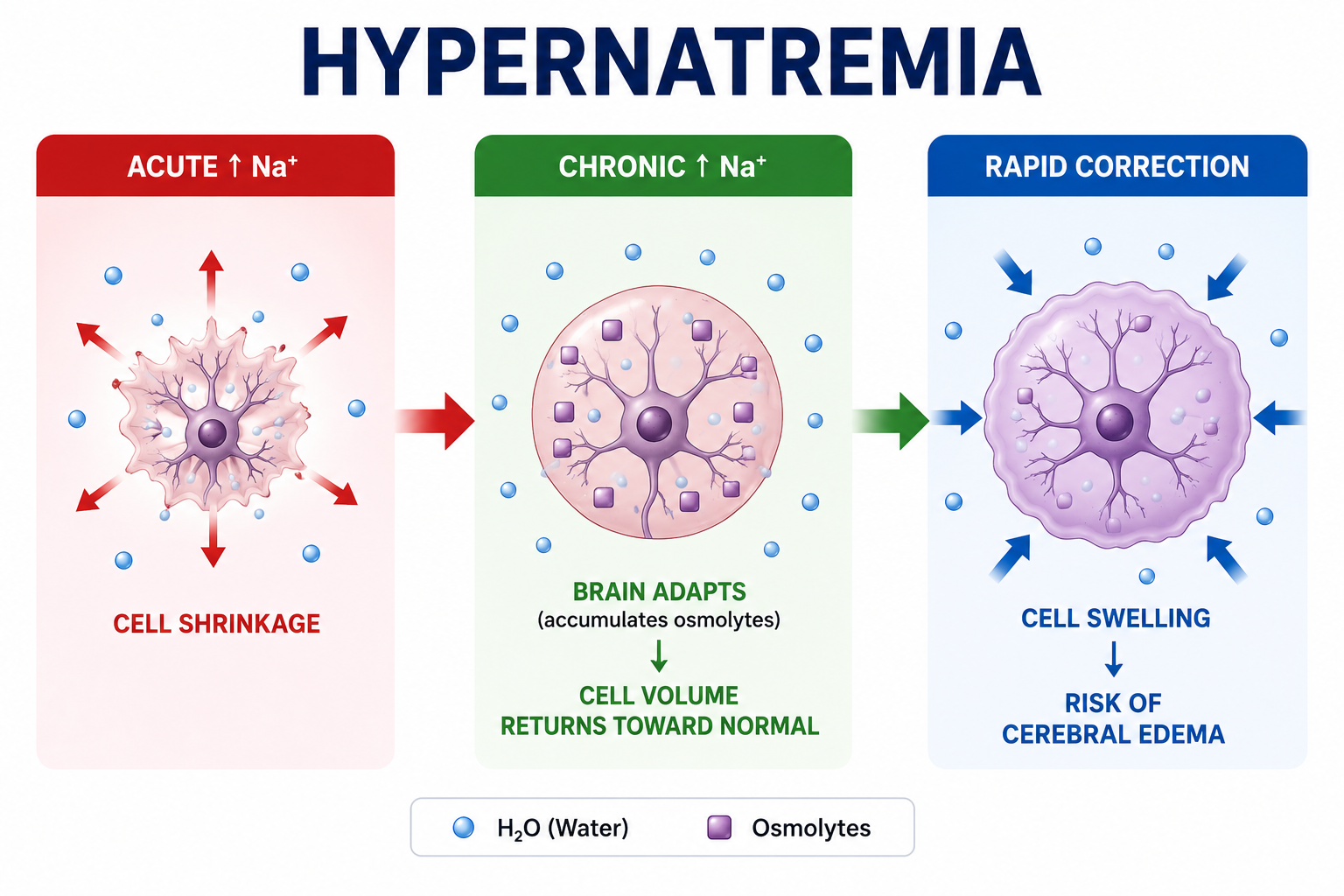
Sodium salts are the major effective extracellular osmoles. When serum sodium rises, extracellular tonicity rises and water shifts from the intracellular compartment into the extracellular compartment.
Brain cells shrink during acute hypertonicity. Over time, the brain adapts by accumulating intracellular osmoles, which helps restore cell volume but creates concern when chronic hypernatremia is corrected too quickly.
Hypernatremia is usually a problem of hypertonicity and cellular dehydration, not simply a high laboratory number.
Defences Against Hypernatremia
The body has two powerful defences against rising plasma tonicity: vasopressin and thirst. Vasopressin helps the kidney conserve water by concentrating urine. Thirst drives water intake.
Sustained hypernatremia is therefore unusual in healthy adults with intact thirst, access to water and intact renal concentrating ability. Clinically significant hypernatremia should prompt the question: why could this patient not obtain or conserve enough water?
| Defence | Normal Response to Hypertonicity | Failure Pattern |
|---|---|---|
| Vasopressin / ADH | Concentrates urine and reduces free-water loss | Diabetes insipidus or impaired renal response |
| Thirst | Increases water intake | Impaired consciousness, poor access to water, infants, older adults, critical illness |
Who Is Vulnerable?
Hypernatremia occurs particularly when normal drinking behaviour cannot match water losses. Important groups include infants, older adults, critically ill patients, patients with impaired consciousness, people with limited access to water and patients with high renal or extrarenal water losses.
Patients with diabetes insipidus physiology are especially vulnerable when thirst or access to water is impaired, because they can lose large volumes of dilute urine.
Causes of Hypernatremia
Hypernatremia can result from pure water loss, hypotonic fluid loss or sodium gain. Most clinical hypernatremia is related to water deficit rather than sodium gain.
| Mechanism | Examples | Volume Pattern |
|---|---|---|
| Pure water loss | Diabetes insipidus, inadequate water intake | Often euvolemic or mildly hypovolemic |
| Hypotonic fluid loss | Diarrhoea, vomiting, sweating, fever, burns, osmotic diuresis | Often hypovolemic |
| Sodium gain | Hypertonic saline, sodium bicarbonate, salt ingestion, mineralocorticoid excess contexts | Often hypervolemic |
Hypovolemic, Euvolemic and Hypervolemic Hypernatremia
Hypovolemic Hypernatremia
Water and sodium are both lost, but proportionally more water is lost. Examples include gastrointestinal losses, sweating, burns and osmotic diuresis. The patient may have tachycardia, hypotension, dry mucous membranes or reduced perfusion.
Euvolemic Hypernatremia
Predominant water loss occurs with relatively little sodium loss. Diabetes insipidus is the classic pattern. Clinical volume depletion may be subtle unless water intake cannot keep up.
Hypervolemic Hypernatremia
Net sodium gain exceeds water gain. This is less common than water-deficit hypernatremia and may occur after hypertonic sodium administration or other sodium-loading states.
Symptoms of Hypernatremia
Symptoms are mainly neurological because hypertonicity affects brain-cell volume. Thirst may be prominent when the patient can perceive and express it.
| Pattern | Possible Features |
|---|---|
| Mild or early | Thirst, weakness, irritability, lethargy |
| Moderate | Confusion, agitation, neuromuscular irritability |
| Severe | Seizures, coma, intracranial bleeding risk in severe acute shifts, shock if major volume depletion is present |
The speed of onset matters. Acute hypernatremia may produce marked symptoms, while chronic hypernatremia may look less dramatic because the brain adapts.
How to Diagnose Hypernatremia
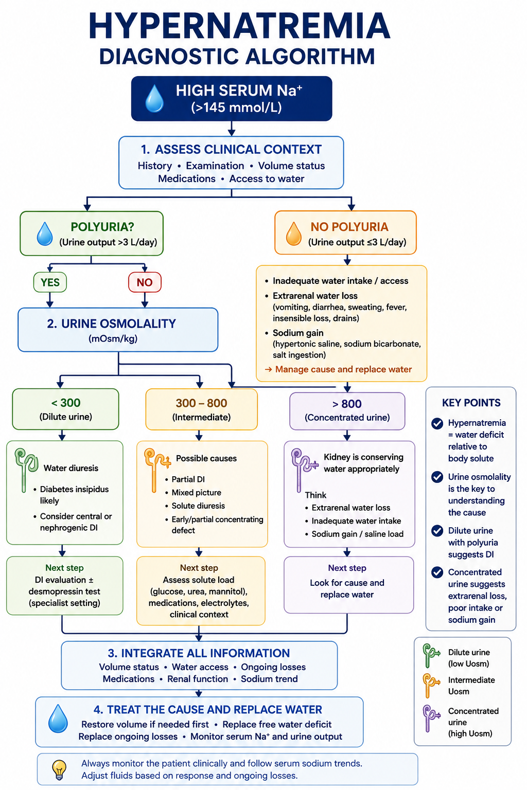
The diagnosis begins with serum sodium above about 145 mmol/L. The next step is to identify the mechanism using history, examination, volume status, urine volume and urine osmolality.
Urine Volume and Urine Osmolality
In hypernatremia, an appropriate renal response is to conserve water and produce concentrated urine. Urine osmolality therefore becomes central to diagnosis, and the physiology is explained in Urine Osmolality Explained.
| Urine Osmolality | Interpretation | Think About |
|---|---|---|
| Less than 300 mOsm/kg | Inappropriately dilute urine in hypernatremia | Diabetes-insipidus pattern if polyuria is present |
| 300 to 800 mOsm/kg | Indeterminate or mixed concentrating response | Partial DI, osmotic diuresis, kidney disease, mixed losses |
| Greater than 800 mOsm/kg | Appropriate renal water conservation | Extrarenal water loss, inadequate intake, sodium gain with intact response |
Dilute urine during hypernatremia is abnormal and suggests renal free-water loss. Concentrated urine argues that the kidney is responding appropriately.
Diabetes Insipidus Pattern
Diabetes insipidus causes excessive free-water loss because vasopressin secretion is deficient or the kidney does not respond to vasopressin. Hypernatremia develops when water losses exceed intake.
| Pattern | Mechanism | Typical Clue |
|---|---|---|
| Central DI | Reduced vasopressin secretion | Polyuria with dilute urine; response to vasopressin analogue may occur |
| Nephrogenic DI | Renal resistance to vasopressin | Polyuria with dilute urine; limited response to vasopressin analogue |
Hypernatremia alone does not diagnose diabetes insipidus. The diagnostic pattern requires polyuria, inappropriately dilute urine and the correct clinical context. Detailed water-deprivation testing and copeptin-based diagnosis are covered in Diabetes Insipidus Explained.
Osmotic Diuresis
Osmotic diuresis causes high urine output because solute in the tubular fluid drags water with it. Causes include severe hyperglycemia, mannitol and high urea generation in selected settings.
This differs from diabetes insipidus. In osmotic diuresis, urine osmolality is often not maximally dilute because solute excretion is high. The patient may lose both water and electrolytes, producing a mixed picture.
Free-Water Deficit
The free-water deficit estimates how much water is needed to return serum sodium toward a chosen target. It is an estimate, not a complete prescription, because it does not automatically include ongoing losses, sodium gains, potassium shifts or changes in renal water handling.
Total body water is commonly estimated as a fraction of body weight, with lower fractions used in older adults and women. The target sodium is selected clinically rather than assumed blindly.
Worked Example
If estimated total body water is 42 L, serum sodium is 160 mmol/L and the target sodium is 145 mmol/L:
This estimate helps planning but must be adjusted for ongoing losses, volume status, urine output, clinical trajectory and repeated sodium measurements.
Treatment of Hypernatremia
Hypernatremia is treated by correcting the underlying cause, restoring circulation when necessary, replacing the free-water deficit and replacing ongoing water losses. Monitoring is essential because the sodium can change unpredictably as water losses, kidney function and intake change.
| Clinical Situation | General Principle |
|---|---|
| Shock or major hypovolemia | Restore effective circulation first with appropriate resuscitation fluid, then correct free-water deficit |
| Water-deficit hypernatremia | Provide enteral or intravenous free water according to clinical context and monitoring |
| Diabetes-insipidus pattern | Replace water losses and address central or nephrogenic mechanism with specialist-guided therapy |
| Osmotic diuresis | Treat the solute-driven diuresis and replace water/electrolyte losses |
| Sodium-gain hypernatremia | Stop sodium source and remove excess sodium/water according to volume status and renal function |
This article teaches principles rather than patient-specific fluid prescriptions. Severe hypernatremia, neurological symptoms, major volume depletion, sodium loading or suspected diabetes insipidus require individualized monitored clinical management.
How Quickly Should Hypernatremia Be Corrected?
Correction strategy depends on whether hypernatremia is acute or chronic, symptom severity, volume status and ongoing losses. Acute hypernatremia is generally corrected more rapidly than chronic hypernatremia because the brain has not fully adapted.
For chronic or unknown-duration hypernatremia, traditional teaching favours slower correction to reduce concern for cerebral edema. However, adult evidence defining the exact safe correction rate is limited, so the article should not present one number as an absolute biological rule.
Traditional correction-rate limits are a safety framework. Robust adult evidence defining a precise universal safe rate is limited, so local protocols and specialist judgement matter.
Worked Clinical Examples
Case 1: Inadequate Water Intake
An older adult with impaired access to water has serum sodium 154 mmol/L and concentrated urine above 800 mOsm/kg. The kidney is conserving water appropriately; the problem is inadequate intake relative to losses.
Case 2: Diabetes-Insipidus Pattern
A patient has hypernatremia, very high urine volume and urine osmolality below 300 mOsm/kg. This is inappropriate dilute urine in the face of hypertonicity and suggests renal free-water loss from a DI pattern.
Case 3: Osmotic Diuresis
A patient with severe hyperglycemia has high urine output and intermediate urine osmolality. Solute-driven diuresis causes water loss and electrolyte shifts, so this should not be labelled simple DI.
Case 4: Sodium Gain
A patient receives a large hypertonic sodium load and develops hypernatremia with volume expansion. This is sodium-gain hypernatremia, a different mechanism from most water-deficit cases.
Common Mistakes
- Misconception: Hypernatremia means sodium overload. Reality: most cases reflect relative water deficit.
- Misconception: All polyuria in hypernatremia is diabetes insipidus. Reality: osmotic diuresis and mixed disorders can also cause polyuria.
- Misconception: Concentrated urine indicates DI. Reality: concentrated urine is usually an appropriate kidney response to hypernatremia.
- Misconception: Water-deficit formulas produce the full treatment plan. Reality: formulas estimate a deficit and do not include ongoing losses or clinical priorities.
- Misconception: One correction-rate number is an absolute biological rule for every adult. Reality: traditional limits are safety guidance, but adult evidence is imperfect.
One Minute Revision
- Hypernatremia is serum sodium above the reference range, usually above about 145 mmol/L.
- It usually reflects too little water relative to body solute, not sodium overload.
- Sodium salts are effective extracellular osmoles, so hypernatremia usually causes hypertonicity.
- Healthy adults are protected by thirst and vasopressin-mediated renal water conservation.
- Urine volume and urine osmolality are central to diagnosis.
- Urine osmolality below 300 mOsm/kg during hypernatremia suggests a DI pattern if polyuria is present.
- Urine osmolality above 800 mOsm/kg suggests appropriate renal water conservation.
- Free-water deficit formulas are estimates and must be adjusted for ongoing losses.
- Treatment addresses cause, circulation, water deficit and ongoing losses with monitoring.
Frequently Asked Questions
Conclusion
The correct approach to hypernatremia is not "high sodium, calculate water, give fluid." It is a structured physiological sequence: confirm high serum sodium, recognise hypertonicity, distinguish relative water deficit from sodium gain, ask why the patient could not drink or conserve enough water, use urine volume and urine osmolality to identify renal versus extrarenal water loss, estimate water deficit cautiously and correct with monitored attention to volume status, ongoing losses and acuity.
This article is intended for medical education only. Severe hypernatremia or hypernatremia associated with neurological symptoms, major volume depletion, sodium loading or suspected diabetes insipidus requires individualized monitored clinical management.