Electrolytes are electrically charged minerals that regulate fluid balance, acid-base homeostasis, nerve conduction, muscle contraction and cardiac rhythm. Every electrolyte abnormality reflects an underlying disturbance of water balance, hormonal regulation, renal function or cellular transport — not simply a number outside a reference range.
This article serves as the cornerstone of the Electrolytes cluster on this site. Rather than covering every individual disorder in depth, it provides the physiological framework needed to understand sodium, potassium, calcium, magnesium and phosphate, and complements the acid-base series — see Acid-Base Disorders Explained, Metabolic Acidosis Explained, Metabolic Alkalosis Explained, Anion Gap Explained and ABG Interpretation Explained. Dedicated articles will subsequently explore each electrolyte and its disorders in depth.
Electrolytes are minerals that dissolve in body fluids and separate into charged ions. The body regulates their concentrations tightly because even small changes can significantly affect cellular function.
Learning Objectives
- Define electrolytes and understand their physiological importance
- Describe body fluid compartments and electrolyte distribution
- Explain the major physiological functions of sodium, potassium, calcium, magnesium and phosphate
- Understand hormonal regulation of electrolyte homeostasis
- Apply a systematic approach to interpreting electrolyte abnormalities
- Recognise common electrolyte patterns and clinical emergencies
What Are Electrolytes?
Electrolytes are minerals that dissolve in body fluids and separate into positively or negatively charged particles called ions. Because they carry electrical charge, electrolytes allow cells to generate the electrical gradients essential for nerve conduction, muscle contraction and many cellular transport mechanisms. The body carefully regulates electrolyte concentrations because even small changes may significantly affect cellular function.
Electrolytes are divided into cations (positively charged: sodium, potassium, calcium, magnesium) and anions (negatively charged: chloride, bicarbonate, phosphate). The body maintains electrical neutrality by balancing positive and negative charges.

Why Electrolytes Are Important
Every cell depends on electrolyte gradients to function normally. Electrolytes participate in the regulation of body water, blood pressure control, plasma osmolality, nerve impulse transmission, skeletal muscle contraction, cardiac conduction, acid-base balance, cellular metabolism, enzyme activation and bone mineralisation. Because electrolytes influence multiple organ systems simultaneously, disturbances often present with neurological, cardiovascular and muscular manifestations.
| Disturbance | Mechanism | Consequence |
|---|---|---|
| Low sodium | Brain swelling | Confusion, seizures |
| High potassium | Reduced cardiac membrane potential | Cardiac arrhythmias |
| Low calcium | Increased neuromuscular excitability | Tetany |
| Low magnesium | Neuromuscular dysfunction | Arrhythmias |
| Low phosphate | Reduced ATP production | Muscle weakness |
Body Fluid Compartments
Understanding electrolyte disorders begins with understanding body water, since electrolytes are dissolved in water rather than existing independently — changes in water balance often produce changes in electrolyte concentrations. Total body water is approximately 60% of body weight in healthy adult males, roughly 50–55% in adult females, lower in elderly individuals due to reduced lean body mass, and substantially higher in infants. These are approximations that vary with age, sex and body composition.
Intracellular fluid (ICF) makes up approximately two-thirds of total body water and is located inside cells. Extracellular fluid (ECF) makes up approximately one-third and comprises plasma, interstitial fluid and small transcellular compartments. Different electrolytes predominate in different compartments, and the body expends considerable energy maintaining these concentration gradients because they are essential for normal cellular function.

Distribution of Major Electrolytes
Each electrolyte has a characteristic distribution between intracellular and extracellular compartments, and understanding this distribution helps explain many clinical disorders.
- Extracellular: sodium is the major cation; chloride and bicarbonate are the major anions.
- Intracellular: potassium is the major cation; proteins and organic phosphates are the major intracellular anions.
- Calcium: most is stored in bone, with only a very small proportion circulating in extracellular fluid. Even small changes in circulating calcium may have major physiological effects.
- Magnesium: mostly intracellular or stored in bone, with only a small fraction circulating in plasma.
- Phosphate: the majority is stored in bone, much of the remainder is intracellular, and only a small proportion is present in extracellular fluid.
Major Body Electrolytes
Although many ions exist within the body, five electrolytes are particularly important in routine clinical medicine.
Sodium (Na+)
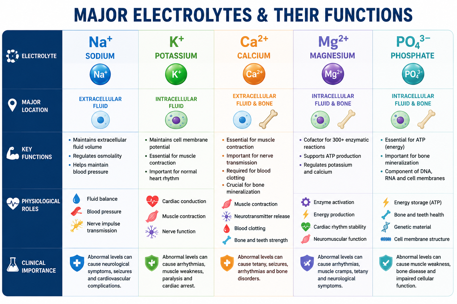
The primary extracellular cation. It determines extracellular fluid volume, maintains plasma osmolality, influences blood pressure and supports nerve conduction. Common disorders: hyponatremia and hypernatremia. For the full physiology, see Sodium Homeostasis Explained.
Potassium (K+)
The primary intracellular cation. It governs resting membrane potential, muscle contraction, cardiac conduction and nerve impulse transmission. Common disorders: hypokalemia and hyperkalemia.
Calcium (Ca2+)
Essential for bone mineralisation, muscle contraction, neurotransmitter release, blood coagulation and cellular signalling. Common disorders: hypocalcemia and hypercalcemia.
Magnesium (Mg2+)
Involved in ATP-dependent enzyme reactions, neuromuscular stability, potassium regulation and cardiac rhythm. Common disorders: hypomagnesemia and hypermagnesemia. For the full physiology, see Magnesium Homeostasis Explained. For the low-magnesium clinical disorder, see Hypomagnesemia Explained.
Phosphate (PO4 3-)
Required for ATP production, bone formation, cell membrane structure, DNA/RNA synthesis and intracellular buffering. Common disorders: hypophosphatemia and hyperphosphatemia. For normal regulation, see Phosphate Homeostasis Explained. For low and high phosphate disorders, see Hypophosphatemia Explained and Hyperphosphatemia Explained.
| Electrolyte | Major Location | Major Function |
|---|---|---|
| Sodium | Extracellular | Water balance |
| Potassium | Intracellular | Membrane potential |
| Calcium | Bone / ECF | Muscle, nerves, coagulation |
| Magnesium | Intracellular | Enzymes, ATP, potassium regulation |
| Phosphate | Bone / Intracellular | ATP, DNA, bone |
Electrolyte Homeostasis
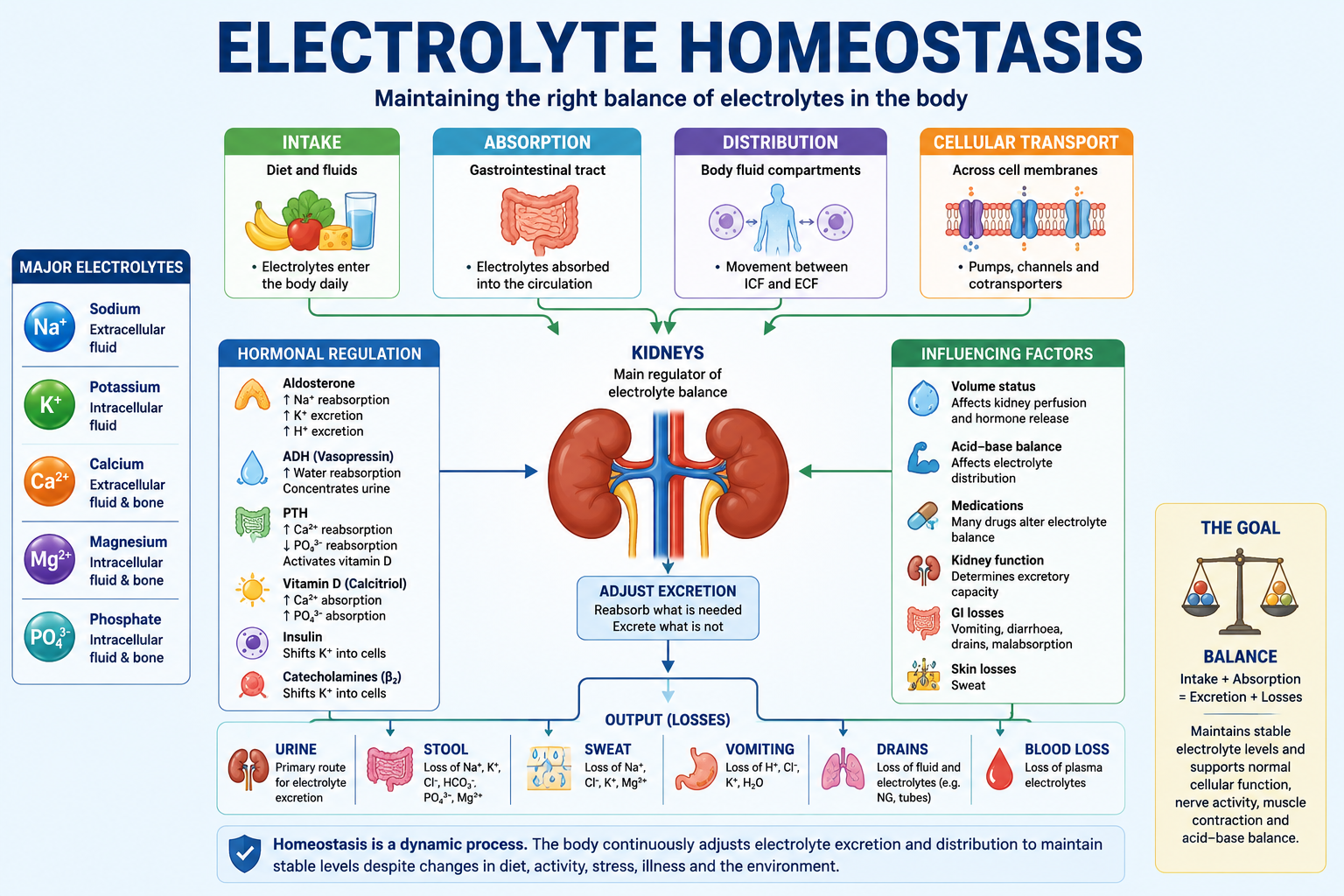
Despite daily variations in dietary intake and ongoing losses through urine, stool and sweat, plasma electrolyte concentrations remain within remarkably narrow limits. This stability depends on hormonal regulation, renal function, gastrointestinal absorption and the movement of electrolytes between intracellular and extracellular compartments — the kidneys are the principal organs responsible for long-term balance, while several hormones fine-tune the process.
Aldosterone
Produced by the adrenal cortex, acting on the distal nephron to increase sodium reabsorption (and therefore water retention), potassium excretion and hydrogen ion excretion. Excess aldosterone may cause mild hypernatremia, hypokalemia, metabolic alkalosis and hypertension; deficiency may cause hyponatremia, hyperkalemia, metabolic acidosis and hypotension.
Antidiuretic Hormone (ADH)
Regulates water rather than sodium directly, increasing water reabsorption in the collecting ducts and diluting plasma sodium. Excess ADH causes SIADH and dilutional hyponatremia; deficiency causes diabetes insipidus, hypernatremia and hyperosmolality.
Parathyroid Hormone (PTH)
Regulates calcium and phosphate by increasing bone resorption, renal calcium reabsorption, phosphate excretion and calcitriol production — the net effect is a rise in calcium and a fall in phosphate.
Vitamin D (Calcitriol)
Primarily increases intestinal absorption of calcium and phosphate, and contributes to bone mineralisation.
Calcitonin
Has a relatively minor physiological role in adults, reducing bone resorption and lowering circulating calcium, but contributing considerably less to day-to-day regulation than PTH and vitamin D.
Insulin and Catecholamines
Insulin shifts potassium into cells, lowering serum potassium — this is why insulin is used therapeutically in severe hyperkalemia. Beta2-adrenergic stimulation has the same effect, which is why inhaled beta2 agonists may be used as adjunctive treatment for hyperkalemia.
Daily Intake and Losses
Electrolytes are obtained primarily through food and beverages, and most healthy individuals meet their daily requirements through a balanced diet. They are lost through urine, stool, sweat, vomiting, gastrointestinal drainage and, to a smaller extent, bleeding. Among these routes, the kidneys provide the greatest capacity for adjusting excretion according to physiological needs.
Different gastrointestinal secretions contain different electrolyte compositions, which is why different losses produce different patterns. Vomiting commonly causes hydrogen ion loss, chloride loss and potassium depletion. Diarrhoea commonly causes sodium loss, potassium loss and bicarbonate loss.
Normal Laboratory Reference Ranges
Electrolyte interpretation always begins with the laboratory reference range, though clinicians should remember that ranges vary between laboratories because of differences in analytical methods and reference populations. The values below are commonly used approximate adult reference intervals.
| Electrolyte | Typical Adult Reference Range |
|---|---|
| Sodium | 135–145 mmol/L |
| Potassium | 3.5–5.0 mmol/L |
| Total Calcium | 2.1–2.6 mmol/L |
| Ionized Calcium | 1.10–1.30 mmol/L |
| Magnesium | 0.70–1.00 mmol/L |
| Phosphate | 0.80–1.50 mmol/L |
| Chloride | 98–106 mmol/L |
| Bicarbonate | 22–28 mmol/L |
Always interpret laboratory values using the reference intervals provided by the reporting laboratory. Clinical decisions should not rely solely on memorised numbers.
Laboratory Assessment of Electrolytes
Routine electrolyte testing usually includes sodium, potassium, chloride and bicarbonate, with calcium, magnesium and phosphate added depending on the clinical situation. Further investigations may include serum osmolality, urine sodium and potassium, urine osmolality, urinary chloride, blood glucose, renal and liver function, arterial blood gas, albumin, parathyroid hormone, vitamin D, cortisol and thyroid function — these help determine the underlying cause rather than merely confirming an abnormal concentration.
Albumin is particularly important when interpreting total serum calcium. Because a substantial proportion of circulating calcium is protein-bound, hypoalbuminaemia lowers the measured total calcium even when the physiologically active ionized calcium remains normal. Whenever clinically appropriate, correct total calcium for albumin or measure ionized calcium directly.
A Systematic Approach to Electrolyte Interpretation
Electrolyte abnormalities should never be interpreted in isolation. A structured approach improves diagnostic accuracy.
- Confirm the result is genuine. Consider haemolysis, sampling error, delayed processing or laboratory error; repeat unexpected results when clinically appropriate.
- Assess the severity — mild, moderate or severe. Urgency of treatment depends on both the biochemical abnormality and the patient's clinical condition.
- Assess symptoms — neurological (confusion, seizures, reduced consciousness), cardiovascular (palpitations, syncope, arrhythmias) and neuromuscular (weakness, tetany, cramps, paralysis). Many severe abnormalities require urgent treatment regardless of the underlying cause.
- Review the clinical context — history, examination, medications, fluid status, kidney and liver function, endocrine disorders, recent surgery and gastrointestinal losses.
- Determine the mechanism — reduced intake, excessive losses, increased intake, water imbalance, cellular redistribution, hormonal disturbance or kidney disease. Identifying the mechanism is often more important than simply naming the abnormality.
- Plan further investigation — urine electrolytes, hormonal studies, blood gas analysis, imaging, ECG or endocrine investigations as indicated.
Water Balance and Electrolytes
Water and electrolyte balance are closely related. Changes in body water often alter electrolyte concentrations without changing the total body electrolyte content. Loss of pure water produces hypernatremia despite no increase in total body sodium; excess water retention produces hyponatremia despite normal total body sodium. This distinction is particularly important for sodium disorders — in many patients, an abnormal sodium concentration reflects a disturbance of water balance rather than sodium balance.
Electrolytes and Acid-Base Balance
Electrolytes interact closely with acid-base physiology (see Acid-Base Disorders Explained for the full framework). Metabolic acidosis often shifts potassium out of cells, while metabolic alkalosis often shifts it back in. Chloride is closely related to bicarbonate, and chloride depletion contributes to many forms of metabolic alkalosis. Bicarbonate is the principal extracellular buffer, and phosphate acts as an intracellular and urinary buffer. These relationships explain why electrolyte interpretation frequently overlaps with arterial blood gas interpretation.
Clinical Importance of Electrolyte Disorders
Electrolyte disturbances range from asymptomatic laboratory abnormalities to life-threatening medical emergencies. Neurological effects include headache, confusion, delirium, seizures and coma. Cardiovascular effects include bradycardia, tachycardia, conduction abnormalities, ventricular arrhythmias and cardiac arrest. Muscular effects include cramps, weakness, paralysis and respiratory muscle weakness. Renal effects can worsen kidney injury, result from kidney disease, impair concentrating ability, or promote nephrolithiasis in selected disorders. Several endocrine disorders — SIADH, diabetes insipidus, Addison disease, hyperaldosteronism and hyperparathyroidism — primarily present with electrolyte abnormalities.
Overview of Electrolyte Disorders
Although each electrolyte has unique physiological functions, disorders generally arise through a limited number of mechanisms: reduced intake, excessive gastrointestinal or renal losses, excessive sweating, cellular redistribution, hormonal disorders, kidney disease, excessive administration, or water imbalance. Understanding these mechanisms helps identify the underlying cause rather than simply treating the laboratory abnormality.
Sodium Disorders
Sodium is the principal extracellular cation and the primary determinant of extracellular fluid osmolality. Importantly, abnormalities in plasma sodium usually reflect disturbances in water balance rather than total body sodium.
Hyponatremia (<135 mmol/L)
Causes: SIADH, heart failure, liver cirrhosis, chronic kidney disease, excess water intake, diuretic therapy, gastrointestinal losses, adrenal insufficiency. Features: nausea, headache, confusion, lethargy, seizures, coma. Severity of symptoms depends not only on the sodium level but also on how rapidly it develops.
Hypernatremia (>145 mmol/L)
Causes: water deprivation, diabetes insipidus, osmotic diuresis, excessive sweating, severe diarrhoea, burns, hypertonic saline administration. Features: intense thirst, irritability, confusion, muscle twitching, seizures, coma. Hypernatremia almost always represents water deficiency.
Potassium Disorders
Potassium is the major intracellular cation; only about 2% of total body potassium is extracellular, yet this small fraction is critical for neuromuscular and cardiac function. Even minor changes in serum potassium can significantly alter membrane excitability.
Hypokalemia (<3.5 mmol/L)
Causes: diuretics, vomiting, diarrhoea, hyperaldosteronism, magnesium deficiency, insulin therapy, beta2 agonists. Features: muscle weakness, constipation, paralysis, cardiac arrhythmias, ECG abnormalities. Hypomagnesemia often prevents correction of hypokalemia, so persistent hypokalemia should prompt evaluation of magnesium.
Hyperkalemia (>5.0 mmol/L)
Causes: acute kidney injury, chronic kidney disease, ACE inhibitors, ARBs, potassium-sparing diuretics, Addison disease, tissue breakdown, metabolic acidosis. Features: muscle weakness, paraesthesia, bradycardia, ventricular arrhythmias, cardiac arrest. ECG changes may occur before symptoms develop, making severe hyperkalemia a medical emergency.
Calcium Disorders
Approximately 99% of total body calcium is stored in bone, with only a small fraction circulating in plasma. Despite this, plasma calcium is tightly regulated because it is essential for neuromuscular function and coagulation. For the full regulatory framework, see Calcium Homeostasis Explained.
Hypocalcemia
Causes: hypoparathyroidism, vitamin D deficiency, chronic kidney disease, acute pancreatitis, massive blood transfusion, hypomagnesemia. Features: perioral numbness, muscle cramps, tetany, carpopedal spasm, seizures, QT prolongation.
Hypercalcemia
Causes: primary hyperparathyroidism, malignancy, vitamin D excess, granulomatous disease, thiazide diuretics. Features are classically remembered as "stones, bones, groans, thrones and psychiatric overtones" — kidney stones, bone pain, abdominal symptoms, polyuria and neuropsychiatric symptoms.
Magnesium Disorders
Magnesium is an essential intracellular cation involved in hundreds of enzyme reactions and plays a crucial role in potassium and calcium regulation.
Hypomagnesemia
Causes: alcohol misuse, diuretics, proton pump inhibitors, diarrhoea, malabsorption. Features: tremor, muscle cramps, tetany, ventricular arrhythmias, refractory hypokalemia, hypocalcemia.
Hypermagnesemia
Usually occurs in advanced kidney failure or with excess magnesium-containing medications. Features: loss of deep tendon reflexes, hypotension, bradycardia, respiratory depression, cardiac arrest.
Phosphate Disorders
Phosphate is essential for ATP production, intracellular signalling and bone mineralisation.
Hypophosphatemia
Causes: refeeding syndrome, alcohol misuse, respiratory alkalosis, hyperparathyroidism, antacid overuse. Features: muscle weakness, respiratory failure, rhabdomyolysis, reduced cardiac contractility, neurological dysfunction.
Hyperphosphatemia
Commonly occurs in chronic kidney disease, tumour lysis syndrome, rhabdomyolysis and hypoparathyroidism. Usually asymptomatic; complications result primarily from secondary hypocalcemia and soft tissue calcification.
Common Electrolyte Patterns
Many electrolyte abnormalities occur together, and recognising these patterns improves diagnostic accuracy.
| Clinical Situation | Typical Pattern |
|---|---|
| Vomiting | Hypokalemia, hypochloraemia, metabolic alkalosis |
| Diarrhoea | Hypokalemia, metabolic acidosis, volume depletion |
| SIADH | Hyponatremia, low serum osmolality, concentrated urine |
| Addison disease | Hyponatremia, hyperkalemia, hypotension, metabolic acidosis |
| Chronic kidney disease | Hyperkalemia, hyperphosphatemia, hypocalcemia, metabolic acidosis |
| Refeeding syndrome | Hypophosphatemia, hypokalemia, hypomagnesemia |
Electrolyte Emergencies
Some electrolyte abnormalities require immediate treatment: severe hyperkalemia, severe symptomatic hyponatremia or hypernatremia, severe hypocalcemia with tetany, severe hypercalcemia, severe hypomagnesemia with arrhythmias, and severe hypophosphatemia causing respiratory failure.
Emergency management should focus on stabilising the patient, preventing life-threatening complications, identifying the underlying cause, correcting the abnormality safely, and monitoring for treatment-related complications.
A Practical Clinical Approach
When reviewing electrolyte results, ask the following in sequence:
This structured approach can be applied to almost every electrolyte abnormality encountered in clinical practice.
Common Pitfalls
- Assuming every abnormal result requires immediate correction — some abnormalities are chronic and should be corrected gradually.
- Ignoring clinical symptoms — treat the patient, not the laboratory value alone.
- Forgetting water balance — especially in sodium disorders.
- Overlooking medication effects — many commonly prescribed drugs alter electrolyte balance.
- Forgetting magnesium — persistent hypokalemia often reflects concurrent magnesium deficiency.
- Ignoring acid-base disorders — electrolyte abnormalities frequently coexist with acid-base disturbances.
- Failing to repeat unexpected results — haemolysis and sampling errors are common causes of spurious abnormalities.
One Minute Revision
- Electrolytes are charged minerals essential for cellular function: sodium, potassium, calcium, magnesium and phosphate.
- Sodium governs water balance; potassium governs membrane potential; calcium governs muscle contraction; magnesium governs enzyme activity; phosphate governs ATP production.
- Major regulators: the kidneys, aldosterone, ADH, PTH, vitamin D and insulin.
- Sodium abnormalities usually reflect water balance, not total body sodium.
- Major emergencies: severe hyperkalemia, severe symptomatic hyponatremia, severe hypocalcemia and severe hypercalcemia.
- Approach: confirm the result → assess severity and symptoms → review clinical context → determine the mechanism → investigate → treat.
Frequently Asked Questions
This article is intended for medical education only. Electrolyte disturbances range from incidental findings to medical emergencies. Clinical management requires proper assessment, local protocols and senior or specialist input.