Hyperphosphatemia at a Glance
Hyperphosphatemia means serum phosphate is above the laboratory reference interval. The diagnostic reflex is: high phosphate, check kidney function first.
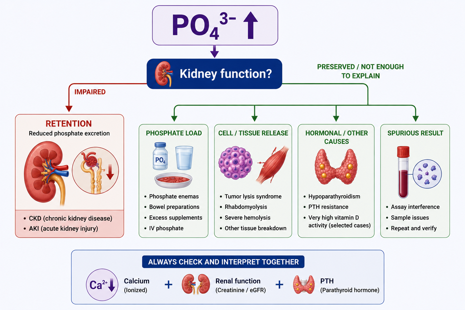
If kidney function is impaired, renal phosphate retention is likely central. If renal function does not explain it, look for phosphate load, cell breakdown, reduced PTH effect or pseudohyperphosphatemia.
CKD pattern: nephron mass falls, FGF23 rises, calcitriol falls, PTH rises, and later phosphate retention develops.

What Is Hyperphosphatemia?
Hyperphosphatemia is a serum phosphate concentration above the laboratory reference interval. Major mechanisms include reduced renal phosphate excretion, increased phosphate load and release of intracellular phosphate.
The exact reference range varies with age, laboratory methodology and clinical context, so use the local laboratory reference interval rather than one universal value.
The clinical significance depends on degree of elevation, speed of development, kidney function, calcium level, underlying cause, associated cell or tissue breakdown and chronicity.
For the baseline physiology, see how PTH, FGF23 and the kidneys normally regulate phosphate.
Causes of Hyperphosphatemia
Do not learn hyperphosphatemia as a disconnected list. Organize the causes by mechanism.
| Mechanism | Examples | Core Question |
|---|---|---|
| Reduced renal excretion | Advanced CKD, severe AKI | Can the kidney eliminate excess phosphate? |
| Increased phosphate load | Phosphate enemas, bowel preparations, excessive phosphate products, IV phosphate over-replacement | Has phosphate input exceeded elimination? |
| Cell or tissue release | Tumor lysis, rhabdomyolysis, severe hemolysis or extensive cellular destruction | Is intracellular phosphate being released? |
| Reduced phosphaturic hormonal effect | Hypoparathyroidism, PTH resistance in appropriate contexts | Is renal phosphate excretion hormonally reduced? |
| Spurious elevation | Laboratory or sample interference | Does the result fit the patient? |
Why Kidney Failure Raises Phosphate
The kidneys are the major route for eliminating excess phosphate. Persistent hyperphosphatemia should immediately prompt assessment of renal phosphate excretion, especially kidney function.
Reduced renal phosphate excretion is one of the most important causes of persistent hyperphosphatemia, especially in advanced CKD and severe AKI.
Hyperphosphatemia in CKD
CKD reduces renal phosphate excretory capacity. FGF23 and PTH initially increase phosphate excretion per remaining nephron, but in advanced kidney disease these compensatory mechanisms become insufficient and phosphate retention develops.
Serum phosphate may remain within the laboratory reference interval during earlier CKD despite declining nephron mass. Normal serum phosphate does not prove normal phosphate physiology in CKD.
Later, functional nephron mass falls further, compensation becomes insufficient, phosphate excretion becomes inadequate and phosphate retention produces hyperphosphatemia.
FGF23 and Phosphate in CKD
FGF23 is an important early compensatory hormone in CKD. As renal phosphate excretory capacity declines, FGF23 rises and reduces proximal tubular phosphate reabsorption in remaining nephrons.
FGF23 also suppresses calcitriol production. Reduced functioning renal mass can also limit normal calcitriol production.
Hyperphosphatemia and Calcium
Acute severe hyperphosphatemia can reduce ionized calcium through calcium-phosphate interactions and altered mineral equilibrium, potentially producing hypocalcemia and secondary PTH stimulation.
Phosphate interacts with calcium in extracellular fluid and tissues. Acute major phosphate elevation can reduce biologically available calcium through calcium-phosphate complex formation, altered mineral equilibrium and precipitation when conditions become sufficiently unfavorable.
For the calcium side of this physiology, see how PTH and vitamin D regulate calcium. For clinical low-calcium effects, see clinical effects and diagnosis of hypocalcemia.

Hyperphosphatemia and PTH
If ionized calcium falls, PTH rises. PTH attempts to restore calcium and increases renal phosphate excretion when the kidneys are capable of responding.
In severe kidney dysfunction, the phosphaturic effect of PTH becomes limited by reduced renal excretory capacity.
In CKD, phosphate retention, calcitriol deficiency and impaired calcium availability converge to stimulate secondary hyperparathyroidism.
Vascular and Soft-Tissue Calcification
Calcium and phosphate are essential for normal bone mineralization. The problem is not that calcium and phosphate should never combine; they should combine appropriately in the skeleton.
When mineral metabolism is chronically abnormal, especially in CKD, elevated phosphate contributes to conditions favoring vascular calcification, soft-tissue calcification and abnormal mineral deposition.
This is not a simple consequence of one isolated phosphate value. It reflects a broader disturbed environment involving phosphate, calcium balance, PTH, FGF23, vitamin D, kidney function and bone turnover.
Persistent phosphate excess in CKD contributes to a mineral environment associated with vascular and soft-tissue calcification.
Increased Phosphate Load
Hyperphosphatemia can occur if phosphate intake or administration exceeds the body's ability to eliminate it.
Potential settings include phosphate-containing bowel preparations, phosphate enemas, excessive phosphate-containing products and IV phosphate over-replacement. Risk rises substantially when kidney function is impaired.
Phosphate Enemas and Bowel Preparations
Some bowel preparations and enemas contain substantial phosphate. Excess absorption can produce acute hyperphosphatemia, especially in patients with impaired renal function, dehydration, impaired bowel motility, excessive or repeated exposure and vulnerable age groups.
Severe phosphate loading can also contribute to hypocalcemia. This article does not provide product-specific dosing.
IV Phosphate Over-Replacement
Excessive IV phosphate replacement can produce iatrogenic hyperphosphatemia. This can be accompanied by hypocalcemia, calcium-phosphate precipitation and renal complications.
This reinforces why IV phosphate replacement in hypophosphatemia must be monitored carefully. For contrast, see how low phosphate and renal phosphate wasting are evaluated.
Tumor Lysis Syndrome
Tumor lysis releases intracellular phosphate from rapidly destroyed tumor cells, and associated acute kidney injury can further increase phosphate by reducing renal excretion.
High phosphate can interact with calcium, so hyperphosphatemia and hypocalcemia may occur together. This section does not become a complete tumor lysis management guideline.
Rhabdomyolysis
Skeletal muscle contains potassium, phosphate, intracellular enzymes and proteins. During rhabdomyolysis, muscle cells break down and intracellular phosphate is released.
Rhabdomyolysis can also cause acute kidney injury. Then phosphate release and reduced phosphate excretion combine, and hyperphosphatemia can become more severe. Interpret phosphate with potassium, creatinine, CK, calcium and the full clinical context.
Hypoparathyroidism and High Phosphate
PTH normally promotes renal phosphate excretion. When PTH is deficient, proximal tubular phosphate reabsorption increases and serum phosphate rises.
Reduced PTH effect therefore reduces phosphaturia. PTH resistance can produce a related high-phosphate pattern in appropriate clinical contexts.
Pseudohyperphosphatemia
Pseudohyperphosphatemia should be considered when the laboratory result does not fit the clinical context. Assay or sample-related interference can create an apparently elevated phosphate result.
Do not ignore the patient. Recheck the result, review sample quality and interpret phosphate with renal function, calcium, PTH context and associated laboratory findings.
Symptoms of Hyperphosphatemia
Hyperphosphatemia may be asymptomatic, especially when chronic. Symptoms often reflect hypocalcemia, kidney failure, cell breakdown or the underlying illness rather than phosphate alone.
| Clinical Context | What May Dominate |
|---|---|
| Acute severe phosphate rise | Hypocalcemia-related manifestations may become clinically important. |
| Advanced CKD | Chronic mineral metabolism disturbance and CKD complications. |
| Tumor lysis | Cell breakdown, potassium, uric acid, calcium, kidney injury and phosphate abnormalities. |
| Rhabdomyolysis | Muscle injury, CK rise, potassium, calcium, phosphate and AKI context. |
How to Diagnose Hyperphosphatemia
Begin by confirming the result and assessing the patient. Then check renal function because impaired renal phosphate excretion is central to persistent hyperphosphatemia.
- Confirm the phosphate result and decide whether it fits the patient.
- Assess kidney function and urine output in the clinical context.
- Check calcium and PTH context because high phosphate directly links to calcium-PTH physiology.
- Look for phosphate load: enemas, bowel preparations, supplements, products or IV phosphate.
- Look for cell or tissue breakdown: tumor lysis, rhabdomyolysis or severe hemolysis/extensive cellular destruction.
- Consider reduced PTH effect or pseudohyperphosphatemia when the pattern suggests it.
Treatment of Hyperphosphatemia
Treatment must match the mechanism. Do not treat every high phosphate result as the same problem.
Acute phosphate load requires stopping the source. Tumor lysis and rhabdomyolysis require treatment of the underlying emergency. Chronic CKD management reduces phosphate burden through appropriate dietary management, phosphate binders, dialysis contribution and broader CKD-mineral management.
Dietary Phosphate
Dietary management can reduce phosphate burden in chronic CKD-associated hyperphosphatemia. This article does not provide universal dietary phosphate limits because targets must be individualized and handled within the broader clinical context.
Phosphate Binders
Phosphate binders reduce intestinal absorption of dietary phosphate by binding phosphate within the gastrointestinal tract.
They do not remove phosphate directly from the blood. Their role is to reduce ongoing intestinal phosphate absorption as part of chronic phosphate-burden management in appropriate patients.
This article does not create binder comparison tables, binder selection algorithms or dosing protocols.
Dialysis and Hyperphosphatemia
Dialysis contributes to phosphate removal when kidney excretory function is inadequate. It may be required in severe accumulation with poor renal clearance or when broader renal replacement therapy indications exist.
Do not use a single universal phosphate threshold for dialysis. The decision depends on the full clinical context, kidney function, associated electrolyte abnormalities, volume status, symptoms and underlying emergency.
Worked Clinical Cases
Case 1: Advanced CKD
A patient with advanced CKD develops persistent hyperphosphatemia. Nephron mass is reduced, compensation is insufficient and renal phosphate excretion cannot match phosphate burden.
Case 2: Early CKD With Normal Phosphate
A patient with earlier CKD has serum phosphate within the reference interval. This does not prove normal phosphate physiology because FGF23 and PTH may be increasing phosphate excretion per remaining nephron.
Case 3: Phosphate Enema Exposure
A vulnerable patient receives phosphate-containing bowel therapy and develops acute hyperphosphatemia. The mechanism is phosphate load, with risk increased if renal elimination is impaired.
Case 4: Tumor Lysis
A patient with rapid tumor-cell destruction develops high phosphate and low calcium. Cell lysis releases phosphate, and AKI may worsen phosphate retention.
Case 5: Hypoparathyroidism
A patient has low PTH effect and high phosphate. PTH normally promotes renal phosphate excretion, so PTH deficiency permits increased phosphate reabsorption and phosphate rise.
Common Mistakes
- Misconception: All hyperphosphatemia is caused by CKD. Reality: phosphate load, cell breakdown, reduced PTH effect and spurious elevation also matter.
- Misconception: Normal phosphate in CKD means normal phosphate physiology. Reality: early FGF23 and PTH compensation may keep serum phosphate normal.
- Misconception: PTH retains phosphate. Reality: PTH is phosphaturic.
- Misconception: Hypoparathyroidism causes low phosphate. Reality: reduced PTH effect can raise phosphate.
- Misconception: Hyperphosphatemia always causes symptoms. Reality: many patients are asymptomatic or symptoms reflect the underlying condition.
- Misconception: Phosphate binders remove phosphate from blood. Reality: they reduce intestinal phosphate absorption.
- Misconception: All severe hyperphosphatemia requires dialysis. Reality: dialysis decisions depend on the full clinical context, not one universal phosphate value.
- Misconception: Vascular calcification is determined solely by calcium-phosphate product. Reality: it reflects broader disturbed mineral metabolism.
One-Minute Revision
- Hyperphosphatemia is serum phosphate above the laboratory reference interval.
- High phosphate should first prompt assessment of kidney function.
- The kidneys are the major route for eliminating excess phosphate.
- Major mechanisms include reduced renal excretion, phosphate load, cell release, reduced PTH effect and spurious elevation.
- In CKD, FGF23 rises early, calcitriol falls, PTH rises and later phosphate retention develops.
- Acute severe phosphate elevation may lower ionized calcium and stimulate PTH.
- Tumor lysis and rhabdomyolysis release intracellular phosphate.
- PTH deficiency reduces phosphaturia and can raise phosphate.
- Phosphate binders reduce intestinal absorption; they do not remove phosphate directly from blood.
- Treatment depends on mechanism: stop phosphate source, treat cell-breakdown emergencies, reduce CKD phosphate burden and use dialysis when clinically required.
Frequently Asked Questions
Key Take-Home Messages
High phosphate means you should check kidney function first. Reduced renal phosphate excretion is central to persistent hyperphosphatemia.
If kidney failure does not explain the result, look for phosphate load, cell or tissue breakdown, reduced PTH effect or pseudohyperphosphatemia.
Hyperphosphatemia directly connects kidney function, calcium, PTH, FGF23, vitamin D and bone-mineral physiology. In CKD, early compensation may hide abnormal phosphate physiology until advanced disease produces phosphate retention.
Treatment matches the mechanism: stop the source, treat the underlying emergency, reduce phosphate burden in CKD and consider dialysis when severe accumulation occurs with poor renal clearance or broader clinical indications.
Next in the Cluster
See how phosphate retention integrates with calcium, PTH, vitamin D and bone disease in CKD-Mineral and Bone Disorder Explained.
This article is intended for medical education only. It explains hyperphosphatemia physiology, diagnosis and treatment principles, not patient-specific medical advice, phosphate targets, binder doses or dialysis thresholds.