Low calcium is the starting point — not the diagnosis
Hypocalcemia means that the concentration of biologically available calcium in the extracellular fluid is below normal.
LOW Ca → CONFIRM IT IS REAL → PTH → PO₄ → Mg → RENAL FUNCTION → VITAMIN D → CAUSE
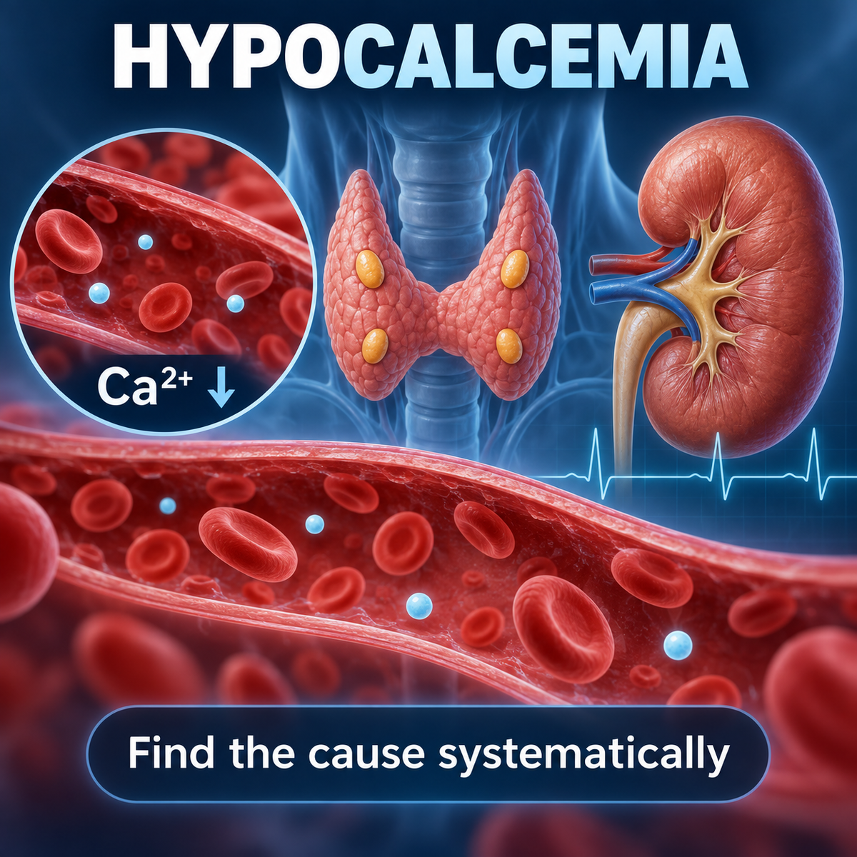
What Is Hypocalcemia?
Opening Article Content
Hypocalcemia means that the concentration of biologically available calcium in the extracellular fluid is below normal.
A low calcium result, however, is not itself a diagnosis.
The first questions should be:
- Is the calcium genuinely low?
- Is the patient symptomatic or unstable?
- Is PTH responding appropriately?
- What do phosphate, magnesium, renal function and vitamin D show?
This produces a much more useful approach than memorizing a long list of causes.
The diagnostic framework is:
Ca²⁺ ↓ CONFIRM TRUE HYPOCALCEMIA PTHPTH low or inappropriately normal
Think:
PTH DEFICIENCY / IMPAIRED PTH SECRETIONPTH appropriately high
Think:
THE PARATHYROIDS ARE RESPONDINGThen determine why the compensatory response is failing to restore calcium.
Endotext identifies phosphate, magnesium, intact PTH, creatinine/renal function and vitamin D measurements as central investigations in the evaluation of hypocalcemia. (NCBI)
Why Calcium Matters
Calcium participates in:
- neuromuscular excitability;
- muscle contraction;
- cardiac electrophysiology;
- intracellular signalling;
- coagulation;
- enzyme activity;
- bone mineralization.
Serum calcium is tightly regulated because relatively small changes in extracellular ionized calcium can substantially alter cellular excitability.
The physiologically active fraction is:
IONIZED CALCIUMTotal serum calcium also includes calcium:
- bound mainly to albumin;
- complexed with anions.
Therefore:
TOTAL Ca ≠ IONIZED Cain every clinical situation.
Calcium Homeostasis
Total Calcium vs Ionized Calcium
First Question: Is The Calcium Really Low?
A low total calcium concentration does not always mean the biologically active ionized calcium is low.
One important example is:
HYPOALBUMINEMIAWhen albumin falls:
albumin-bound calcium ↓therefore:
TOTAL Ca ↓while:
IONIZED Ca MAY REMAIN NORMALThis is sometimes called:
PSEUDOHYPOCALCEMIAor factitious hypocalcemia.
Endotext specifically recommends confirming hypocalcemia and excluding an artifactually low total calcium caused by hypoalbuminemia. (NCBI)
Ionized Calcium
Ionized calcium is the biologically active fraction.
Direct ionized calcium measurement is especially useful when:
- albumin is substantially abnormal;
- total calcium does not fit the clinical picture;
- critical illness is present;
- significant acid-base disturbance exists;
- rapid physiological changes are occurring.
In critically ill patients with acid-base abnormalities, direct ionized calcium is preferable because pH changes alter calcium-albumin binding. (NCBI)
Albumin-Corrected Calcium
Traditional correction equations attempt to estimate what total calcium might have been at a normal albumin concentration.
A commonly taught formula is:
Corrected Ca (mg/dL) = measured Ca + 0.8 × [4.0 − albumin (g/dL)]However, this must not be presented as a perfect substitute for ionized calcium.
Albumin correction is an estimate. When accurate determination matters, especially in critical illness or major protein/pH abnormalities, measure ionized calcium directly.
Why Ph Matters
Hydrogen ions compete with calcium for albumin-binding sites.
Therefore:
Alkalosis
Albumin binds more calcium.
IONIZED Ca ↓even when total calcium changes little.
Acidosis
Albumin binds less calcium.
IONIZED Ca ↑This explains why acute respiratory alkalosis can cause symptoms of hypocalcemia even without a major change in total calcium.
Hyperventilation Example
A patient with acute anxiety hyperventilates.
CO₂ ↓ RESPIRATORY ALKALOSIS Ca binding to albumin ↑ IONIZED Ca ↓Possible:
- perioral tingling;
- paresthesia;
- carpopedal spasm.
The lesson is:
SYMPTOMS FOLLOW IONIZED CALCIUM MORE CLOSELY THAN TOTAL CALCIUMSymptoms and Signs of Hypocalcemia
Symptoms Of Hypocalcemia
Symptoms depend on:
- absolute calcium concentration;
- ionized calcium;
- rate of fall;
- chronicity;
- associated electrolyte disturbances.
A rapidly developing fall may produce marked symptoms at a calcium concentration that a chronically hypocalcemic patient may tolerate better.
Neuromuscular Manifestations
Possible manifestations include:
- perioral numbness;
- tingling;
- distal paresthesia;
- muscle cramps;
- muscle twitching;
- carpopedal spasm;
- tetany;
- hyperreflexia.
The underlying concept is:
HYPOCALCEMIA → NEUROMUSCULAR EXCITABILITY ↑Severe Manifestations
Severe hypocalcemia may cause:
- seizures;
- laryngospasm;
- bronchospasm;
- altered neurological function;
- cardiac dysfunction;
- arrhythmias.
Current ESE guidance recognizes seizures, cardiac failure, bronchospasm and laryngospasm among potentially life-threatening manifestations of severe hypocalcemia. (OUP Academic)
Trousseau Sign
Trousseau sign is:
CARPAL SPASM PROVOKED BY PROLONGED BP-CUFF INFLATIONIt reflects increased neuromuscular excitability.
It can be a useful clinical clue but should not replace biochemical confirmation.
Chvostek Sign
Chvostek sign is contraction of facial muscles after tapping over the facial nerve.
It may occur in hypocalcemia but:
IT IS NOT SUFFICIENTLY SPECIFIC TO DIAGNOSE HYPOCALCEMIA ALONEUse it as a bedside clue, not a diagnostic test.
Cardiac Effects
Hypocalcemia can produce:
QT INTERVAL PROLONGATIONand, in severe cases:
- dysrhythmias;
- impaired myocardial function.
Therefore symptomatic or severe acute hypocalcemia may require:
ECG MONITORINGparticularly during IV calcium treatment.
The PTH-Based Diagnostic Approach
The First Major Diagnostic Test: Pth
Once genuine hypocalcemia has been established:
MEASURE PTHBut never ask only:
“Is the PTH inside the laboratory reference range?”
Ask:
IS THE PTH APPROPRIATE FOR THE CALCIUM?When calcium is low:
PTH SHOULD BE HIGHEndotext specifically emphasizes that PTH must be interpreted in the context of serum calcium. (NCBI)
Low Pth
If:
Ca ↓and:
PTH ↓the parathyroid response is inadequate.
Think:
HYPOPARATHYROIDISMPossible causes include:
- neck surgery;
- autoimmune destruction;
- genetic disease;
- infiltrative/destructive disease;
- severe magnesium disturbance.
Internal link prominently:
Hypoparathyroidism Explained
“Normal” Pth Can Be Abnormal
Suppose:
Ca = LOWbut:
PTH = NORMAL RANGEA common error is:
“PTH is normal.”
Physiologically:
IT IS NOT NORMALbecause hypocalcemia should stimulate PTH above its usual resting level.
Therefore:
LOW Ca + NORMAL-RANGE PTHcan represent:
INAPPROPRIATELY NORMAL PTHand support hypoparathyroidism.
The 2025 ESE guideline defines chronic hypoparathyroidism using persistent hypocalcemia with PTH that is low or inappropriately normal. (OUP Academic)
High Pth
If:
Ca ↓and:
PTH ↑the parathyroid glands are responding.
This tells us:
THE PROBLEM IS NOT SIMPLE PTH DEFICIENCYNow investigate why the elevated PTH is unable to normalize calcium.
Major possibilities include:
- vitamin D deficiency;
- CKD;
- malabsorption;
- PTH resistance;
- phosphate-related disorders;
- high skeletal calcium uptake;
- selected acute/critical illnesses.
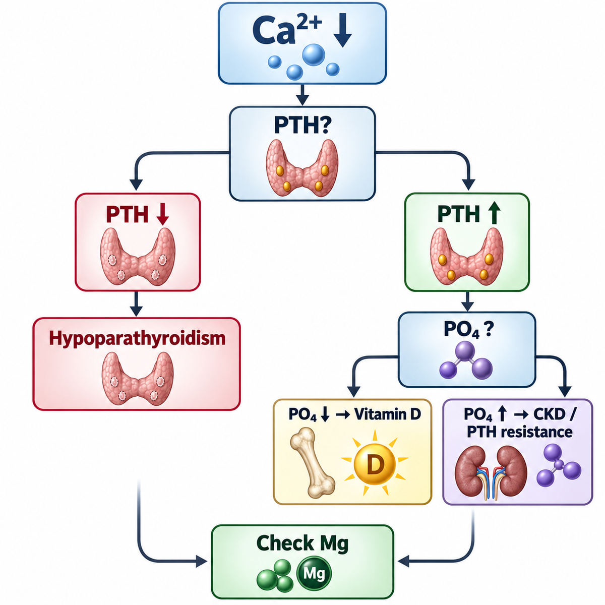
Why Phosphate Is the Next Major Clue
Phosphate Is The Second Major Discriminator
After PTH, phosphate is extremely useful.
Use:
Ca ↓ + PTH ↑ WHAT IS PO₄?PO₄ low / low-normal
Think particularly:
- vitamin D deficiency;
- malabsorption;
- secondary hyperparathyroidism.
PO₄ high
Think particularly:
- CKD;
- PTH resistance;
- excessive phosphate load;
- impaired phosphate excretion.
Why Vitamin D Deficiency Tends To Lower Phosphate
Vitamin D deficiency reduces intestinal calcium absorption.
Ca tendency ↓ PTH ↑PTH increases:
RENAL PHOSPHATE EXCRETION PO₄ ↓Therefore the classic advanced pattern is:
Ca ↓/low-normal PO₄ ↓ PTH ↑ 25-OH D ↓Vitamin D Deficiency Explained
Pth Resistance
In pseudohypoparathyroidism:
PTH ↑but renal tissues do not respond normally.
Therefore:
PHOSPHATURIA FAILS PO₄ ↑while:
Ca ↓Thus:
Ca ↓ + PO₄ ↑ + PTH ↑should raise suspicion for:
PTH RESISTANCEafter important alternatives, especially CKD, have been considered.
Pseudohypoparathyroidism Explained
Ckd
CKD can produce:
- phosphate retention;
- reduced calcitriol production;
- altered calcium balance;
- secondary hyperparathyroidism.
Therefore a common CKD-MBD pattern is:
PO₄ ↑ PTH ↑with:
Ca normal or ↓This can superficially resemble PTH resistance.
Therefore:
CHECK CREATININE / eGFRbefore diagnosing pseudohypoparathyroidism.
CKD-MBD Explained
Magnesium and Refractory Hypocalcemia
Magnesium: Never Forget It
Magnesium is required for:
- normal PTH secretion;
- normal PTH action.
Severe:
HYPOMAGNESEMIAcan produce:
PTH SECRETION ↓and:
PTH RESISTANCEtherefore causing hypocalcemia.
Endotext identifies hypomagnesemia as a key cause to investigate in hypocalcemia. (NCBI)
Refractory Hypocalcemia
HYPOCALCEMIA NOT CORRECTING? CHECK MAGNESIUMBecause:
Ca replacement alone may failuntil magnesium deficiency is corrected.
Hypomagnesemia Explained
Hypermagnesemia
Severe hypermagnesemia can also interfere with PTH secretion and calcium physiology.
This is much less common than hypomagnesemia.
Clinical context may include:
- severe renal impairment;
- excessive magnesium administration.
Internal link if existing:
Hypermagnesemia Explained
Vitamin D and Malabsorption
Vitamin D Testing
The primary test for nutritional vitamin D status is:
25-HYDROXYVITAMIN Dnot:
1,25-DIHYDROXYVITAMIN DA normal or even elevated calcitriol concentration does not exclude nutritional vitamin D deficiency because secondary hyperparathyroidism can stimulate renal calcitriol production when renal function permits.
Internal links:
- Vitamin D Metabolism
- Vitamin D Deficiency
When 1,25-(Oh)₂D Can Help
Calcitriol measurement is not the routine nutritional vitamin D test.
It can be useful in selected physiological questions involving:
- renal activation of vitamin D;
- PTH-related calcitriol regulation;
- rare vitamin D metabolism disorders.
Malabsorption
Reduced intestinal calcium and/or vitamin D absorption can produce hypocalcemia.
Possible clinical contexts include:
- celiac disease;
- inflammatory intestinal disease;
- pancreatic/biliary disorders affecting fat absorption;
- bariatric surgery;
- extensive intestinal disease/resection.
Expected physiology often resembles:
Ca ↓ PTH ↑with vitamin D abnormalities depending on cause.
Testing should be guided by clinical context.
Drug-Induced Hypocalcemia
Drugs That Can Cause Or Worsen Hypocalcemia
Include a concise table.
| Drug/category | Important mechanism/context |
|---|---|
| Bisphosphonates | Suppress bone resorption |
| Denosumab | Potent antiresorptive effect |
| Calcimimetics | Suppress PTH signalling/secretion |
| Some anticonvulsants | Can alter vitamin D metabolism |
| Massive citrate exposure | Binds ionized calcium |
| Selected chemotherapy/critical-care therapies | Mechanism varies |
Risk of antiresorptive-related hypocalcemia is greater in susceptible patients, including those with:
- CKD;
- vitamin D deficiency;
- high bone turnover.
Denosumab And Ckd
Denosumab does not require renal clearance in the same way as bisphosphonates, but:
SEVERE HYPOCALCEMIA RISK CAN BE GREATER IN ADVANCED CKDespecially when mineral metabolism is already abnormal.
Teach the principle only.
Hungry Bone Syndrome
Hungry Bone Syndrome
After correction of severe hyperparathyroidism, particularly following parathyroid surgery:
PTH FALLS RAPIDLYwhile highly active bone takes up:
- calcium;
- phosphate;
- magnesium.
may develop.
This is:
HUNGRY BONE SYNDROMETypical clues:
- recent parathyroidectomy;
- severe preoperative hyperparathyroidism/high bone turnover;
- prolonged postoperative hypocalcemia;
- often phosphate falling rather than rising.
This should link eventually to the planned dedicated:
Hungry Bone Syndrome Explained
Hungry Bone Vs Postsurgical Hypoparathyroidism
This is a high-value comparison.
| Feature | Hungry bone syndrome | Postsurgical HypoPT |
|---|---|---|
| Calcium | ↓ | ↓ |
| PTH | Variable according to underlying surgery/recovery | ↓ / inappropriate normal |
| Phosphate | Often ↓ | Often ↑ |
| Mechanism | Skeletal mineral uptake | PTH deficiency |
| Context | High-turnover bone after correction | Parathyroid injury/removal |
The phosphate difference is especially useful.
Acute and Critical Illness Causes
Acute Pancreatitis
Hypocalcemia may occur in acute pancreatitis.
Mechanisms can be multifactorial and include:
- fat necrosis and calcium sequestration;
- critical illness physiology;
- magnesium abnormalities;
- altered PTH/vitamin D responses.
Use it only as a recognized clinical association.
Massive Transfusion
Stored blood products contain:
CITRATECitrate binds:
IONIZED CaTherefore rapid or massive transfusion can produce:
ACUTE IONIZED HYPOCALCEMIARisk is greater when citrate metabolism is impaired or citrate delivery is very rapid.
This is particularly important in:
- massive hemorrhage;
- major trauma;
- major surgery.
Critical Illness
Hypocalcemia is common in critical illness and may involve multiple mechanisms.
Potential contributors include:
- inflammation;
- renal dysfunction;
- magnesium disturbance;
- altered PTH response;
- vitamin D abnormalities;
- citrate exposure;
- medications.
Ionized calcium is especially useful in this context. (NCBI)
Sepsis
Hypocalcemia can accompany severe sepsis.
LOW Ca → GIVE CALCIUM ROUTINELY TO EVERY SEPTIC PATIENTTreatment depends on severity, symptoms and clinical circumstances.
Keep the focus on diagnosis rather than ICU treatment controversy.
Hyperphosphatemia Causing Hypocalcemia
A substantial acute phosphate load can lower calcium through:
- calcium-phosphate complex formation;
- precipitation;
- altered mineral equilibrium.
Possible settings include:
- severe renal failure;
- tissue breakdown;
- excessive phosphate administration;
- selected bowel-preparation/toxic exposures.
The clinical context is essential.
Tumor Lysis Syndrome
Rapid cell breakdown can cause:
PO₄ ↑ Ca ↓Other abnormalities may include:
- K ↑;
- uric acid ↑;
- AKI.
This is a medical emergency with a broader metabolic management strategy.
Rhabdomyolysis
During acute rhabdomyolysis:
phosphate release ↑and calcium can shift into injured tissues.
Therefore early:
HYPOCALCEMIAmay occur.
Later calcium may rebound as tissue injury resolves.
Osteoblastic Metastases
Extensive osteoblastic skeletal disease can rarely lower serum calcium because calcium is incorporated into newly forming bone.
Think especially when there is:
- extensive skeletal tumor burden;
- high bone formation;
- elevated alkaline phosphatase;
- appropriate secondary PTH response.
Keep concise.
Genetic Causes
Genetic Causes
Genetic hypocalcemia can arise through abnormalities involving:
- parathyroid development;
- PTH production;
- calcium sensing;
- magnesium handling;
- vitamin D metabolism;
- PTH signalling.
Use:
Genetic testing becomes particularly important in unexplained, early-onset, familial or syndromic hypocalcemia.
Activating Casr Disorders
Activating calcium-sensing receptor abnormalities can shift the calcium set point downward.
This can produce:
Ca ↓with:
PTH low / inappropriately normaland relatively:
URINARY Ca HIGHfor the degree of hypocalcemia.
This differs from typical acquired hypoparathyroidism and is important because aggressive calcium treatment can worsen hypercalciuria.
Internal link to Hypoparathyroidism article where the concept is already developed.
Complete Hypocalcemia Diagnostic Algorithm
The Complete Initial Laboratory Set
For persistent/unexplained hypocalcemia, the core initial laboratory assessment should include:
Ca / IONIZED Ca PTH PO₄ Mg CREATININE / eGFR 25-OH VITAMIN DAdditional investigations depend on the emerging pattern and clinical context.
Endotext identifies essentially this biochemical framework as central to determining the cause. (NCBI)
Complete Diagnostic Algorithm
Implement this as a prominent responsive HTML algorithm.
LOW TOTAL Ca CONFIRMAlbumin / ionized Ca / clinical context
Ionized Ca normal
APPARENT / ALBUMIN-RELATED LOW TOTAL CaInvestigate underlying albumin disorder as appropriate.
True Ca low
SYMPTOMATIC / UNSTABLE?YES
URGENT MANAGEMENT + DIAGNOSIS IN PARALLELNO / STABLE
PTHBranch A — PTH LOW / INAPPROPRIATELY NORMAL
CHECK MgMg severely low
→ functional impairment of PTH secretion/action.
Mg not explanatory
Think:
HYPOPARATHYROIDISMAsk:
- neck surgery?
- autoimmune?
- genetic?
- infiltrative/destructive?
- activating CaSR?
Branch B — PTH HIGH
CHECK PO₄PO₄ LOW
Think:
VITAMIN D DEFICIENCY / MALABSORPTIONCheck:
25-OH Dand clinical context.
PO₄ HIGH
CHECK eGFRCKD
→ CKD-MBD
Kidney function not explanatory
→ PTH RESISTANCE
or:
→ phosphate load / acute tissue breakdown depending context.
Master Biochemical Comparison
Implement prominently.
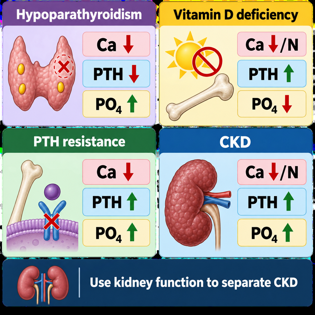
| Cause | Ca | PTH | PO₄ | Key clue |
|---|---|---|---|---|
| Hypoparathyroidism | ↓ | ↓ / inappropriate normal | ↑ | PTH deficiency |
| Pseudohypoparathyroidism | ↓ | ↑ | ↑ | PTH resistance |
| Vitamin D deficiency | ↓/N | ↑ | ↓/N | 25-OH D ↓ |
| CKD-MBD | ↓/N | ↑ | ↑ often | eGFR ↓ |
| Severe hypomagnesemia | ↓ | ↓/inappropriate ± resistance | Variable | Mg markedly ↓ |
| Hungry bone syndrome | ↓ | Variable | ↓ often | Post-parathyroid surgery/high bone uptake |
| Acute phosphate load | ↓ | ↑ expected | ↑ | Acute context |
| Citrate exposure | ionized ↓ | Compensatory | Variable | Massive transfusion |
| Antiresorptive therapy | ↓ | ↑ expected | Variable | Medication history |
Use `N` only if the site's table conventions already define it as normal; otherwise write normal.
Pattern-Recognition Box
Use a high-visibility revision box:
Pattern 1
Ca ↓ + PTH ↓ + PO₄ ↑→ Hypoparathyroidism
Pattern 2
Ca ↓ + PTH ↑ + PO₄ ↑→ CKD or PTH resistance
Pattern 3
Ca ↓ + PTH ↑ + PO₄ ↓→ Vitamin D deficiency / malabsorption
Pattern 4
Ca ↓ + Mg ↓→ Correct magnesium and reassess
Pattern 5
Ca ↓ after parathyroid surgery + PO₄ ↓→ Consider hungry bone syndrome
When Is Hypocalcemia an Emergency?
When Is Hypocalcemia An Emergency?
Assess:
- symptoms;
- ionized calcium;
- absolute calcium;
- speed of fall;
- ECG;
- airway manifestations;
- seizures;
- underlying cause.
Urgent treatment is particularly important with:
- tetany;
- seizures;
- laryngospasm;
- bronchospasm;
- significant arrhythmia/QT abnormality;
- severe acute symptomatic hypocalcemia.
Current endocrine guidance treats severe symptomatic hypocalcemia as an endocrine emergency requiring IV calcium. (Society for Endocrinology)
Acute Treatment Principle
The article is primarily diagnostic, so treatment should remain concise.
Use:
SEVERE / SYMPTOMATIC HYPOCALCEMIA IV CALCIUMusually:
CALCIUM GLUCONATE ECG / CLINICAL MONITORING CORRECT Mg IDENTIFY AND TREAT THE CAUSEtransition to appropriate oral/chronic therapy when stable.
Current ESE guidance uses IV calcium gluconate for life-threatening hypocalcemia. (OUP Academic)
Do not turn this hub article into an IV-calcium prescribing protocol.
Why Calcium Gluconate?
Calcium gluconate is generally preferred for peripheral IV administration because it causes less severe tissue injury if extravasation occurs than calcium chloride.
Calcium chloride delivers more elemental calcium but is more irritating and is generally reserved for selected emergency/monitored settings.
No dosing table is needed here.
Correct Magnesium
Display:
Ca LOW + Mg LOW CORRECT BOTHBecause calcium can remain refractory when severe magnesium deficiency continues to impair PTH physiology.
Endotext emphasizes magnesium deficiency as an important contributor to persistent hypocalcemia. (NCBI)
Treatment Principles
Cause-Specific Treatment
Once the patient is stabilized:
Hypoparathyroidism
→ active vitamin D + adequate calcium ± modern PTH replacement in selected chronic inadequately controlled disease.
Vitamin D deficiency
→ correct vitamin D deficiency and contributing calcium deficiency.
Hypomagnesemia
→ magnesium replacement.
CKD-MBD
→ CKD-specific mineral management.
PTH resistance
→ active vitamin D ± calcium; do not routinely use PTH replacement.
Hungry bone syndrome
→ calcium + active vitamin D with monitoring according to severity.
Medication-related
→ reassess contributing drug and correct relevant mineral deficiencies.
Acute phosphate load / tumor lysis
→ treat underlying emergency.
Do Not Treat The Number Alone
TREAT THE PATIENT + THE MECHANISMnot simply:
THE TOTAL CALCIUM NUMBERWhy?
Because:
- low total calcium may occur with normal ionized calcium;
- chronic hypocalcemia may be better tolerated than an acute fall;
- treatment differs radically by mechanism;
- excessive calcium treatment can cause harm in some disorders.
Chronic Hypocalcemia
Chronic management depends entirely on cause.
Examples:
- HypoPT → hormone-deficiency physiology.
- PHP → hormone-resistance physiology.
- vitamin D deficiency → substrate deficiency.
- CKD → altered renal mineral metabolism.
- magnesium deficiency → impaired PTH physiology.
The same calcium concentration therefore does not imply the same long-term treatment.
Worked Clinical Cases
Use all 12 cases exactly in substance.
Case 1 — Low total calcium, normal ionized calcium
Patient:
- total Ca low;
- albumin markedly low;
- ionized Ca normal;
- no tetany.
Interpretation
NOT TRUE IONIZED HYPOCALCEMIAThe low total calcium reflects reduced protein-bound calcium.
Lesson
CONFIRM FIRSTCase 2 — Postsurgical hypoparathyroidism
Two days after thyroidectomy:
- tingling;
- Ca ↓;
- PO₄ ↑;
- PTH ↓.
Interpretation
POSTSURGICAL HYPOPARATHYROIDISMLesson
Ca ↓ + PO₄ ↑ + PTH ↓Case 3 — “Normal” PTH that is inappropriate
Patient:
- Ca clearly low;
- PTH mid-reference range.
Wrong interpretation
“PTH normal.”
Correct interpretation
PTH should be elevated during hypocalcemia.
Therefore:
PTH IS INAPPROPRIATELY NORMALThink hypoparathyroidism.
Case 4 — Vitamin D deficiency
Patient:
- Ca low-normal;
- phosphate low;
- PTH high;
- 25-OH D markedly low.
Interpretation
VITAMIN D DEFICIENCY WITH SECONDARY HYPERPARATHYROIDISMCase 5 — PTH resistance
Patient:
- Ca ↓;
- PO₄ ↑;
- PTH markedly ↑;
- renal function normal.
Interpretation
PTH RESISTANCEafter appropriate exclusion of other causes.
Pseudohypoparathyroidism.
Case 6 — CKD
Patient:
- Ca low;
- PO₄ high;
- PTH high;
- eGFR 19.
Interpretation
CKD-MBDrather than assuming pseudohypoparathyroidism.
Case 7 — Severe hypomagnesemia
Patient:
- Ca ↓;
- Mg profoundly ↓;
- PTH low-normal;
- calcium remains low despite supplementation.
Interpretation
MAGNESIUM-RELATED FUNCTIONAL HYPOPARATHYROIDISMLesson
REFRACTORY Ca ↓ → CHECK MgCase 8 — Hungry bone syndrome
Patient after parathyroidectomy for severe hyperparathyroidism:
- prolonged Ca ↓;
- PO₄ ↓;
- high preoperative bone turnover.
Interpretation
HUNGRY BONE SYNDROMErather than simple postoperative HypoPT.
Case 9 — Massive transfusion
Trauma patient receiving rapid massive transfusion develops:
- hypotension;
- prolonged QT;
- low ionized Ca.
Mechanism
CITRATE BINDS IONIZED CALCIUMLesson
Ionized calcium is particularly relevant during rapid transfusion.
Case 10 — Antiresorptive-associated hypocalcemia
Patient with CKD and vitamin D deficiency receives potent antiresorptive therapy.
Later:
- Ca ↓;
- PTH ↑.
Interpretation
Medication-associated hypocalcemia in a high-risk physiological setting.
Lesson
Medication history matters.
Case 11 — Acute pancreatitis
Patient with severe pancreatitis develops low calcium.
PTH appropriately elevated.
Interpretation
Hypocalcemia is secondary to the acute illness rather than primary parathyroid failure.
Lesson
Clinical context matters.
Case 12 — Hypocalcemic emergency
Patient:
- severe acute hypocalcemia;
- carpopedal spasm;
- seizure;
- QT prolongation.
Immediate principle
URGENT IV CALCIUM ECG MONITORING CHECK/CORRECT Mgwhile determining the underlying cause.
Common Mistakes
Include all.
Mistake 1
Every low total calcium means true hypocalcemia. Wrong.
Mistake 2
Albumin correction is always perfectly accurate. Wrong.
Mistake 3
Ionized calcium is never needed. Wrong.
Mistake 4
Normal-range PTH is normal during hypocalcemia. Wrong.
Mistake 5
PTH should be interpreted without calcium. Wrong.
Mistake 6
High PTH means hyperparathyroidism is the primary disease. Wrong. It may be an appropriate response to hypocalcemia.
Mistake 7
High PTH + low calcium always means vitamin D deficiency. Wrong.
Mistake 8
Phosphate is unimportant. Wrong.
Mistake 9
High PTH + high phosphate automatically means pseudohypoparathyroidism. Wrong. Check renal function.
Mistake 10
Magnesium is optional in the workup. Wrong.
Mistake 11
Calcium replacement will always correct severe magnesium-related hypocalcemia. Wrong.
Mistake 12
25-OH D and calcitriol are interchangeable tests. Wrong.
Mistake 13
Hypocalcemia after parathyroid surgery is always HypoPT. Wrong. Consider hungry bone syndrome.
Mistake 14
Hungry bone syndrome usually causes high phosphate. Wrong; phosphate is commonly low because mineral is entering bone.
Mistake 15
Every ICU low total calcium represents an endocrine disease. Wrong.
Mistake 16
Massive transfusion lowers calcium because PTH suddenly disappears. Wrong. Citrate binding is important.
Mistake 17
QT shortening is typical of hypocalcemia. Wrong. Hypocalcemia tends to prolong QT.
Mistake 18
Symptoms depend only on the absolute calcium value. Wrong.
Mistake 19
All chronic hypocalcemia has the same treatment. Wrong.
Mistake 20
Diagnosis should wait until emergency treatment is finished. Wrong. In severe disease, stabilization and diagnostic evaluation proceed in parallel.
Hypocalcemia in One Minute
One-Minute Revision
HYPOCALCEMIA IN ONE MINUTE
Ca ↓ REAL?Check:
albumin / ionized Ca
SYMPTOMATIC?If severe:
urgent treatment
PTHPTH ↓ / inappropriate normal
→ Hypoparathyroidism / Mg problem
PTH ↑
PO₄PO₄ ↓
→ Vitamin D deficiency / malabsorption
PO₄ ↑
eGFReGFR ↓
→ CKD-MBD
eGFR preserved
→ PTH resistance / phosphate-related cause
Always check:
Mg + 25-OH D + RENAL FUNCTION + HISTORYGolden Rules
A low total calcium result must first be confirmed as physiologically meaningful.
Ionized calcium is the biologically active calcium fraction.
PTH must always be interpreted relative to calcium.
Low calcium should stimulate PTH.
Low calcium with low or inappropriately normal PTH suggests hypoparathyroid physiology.
Low calcium with high PTH means the parathyroids are responding.
Phosphate helps determine why the response is failing.
Low Ca + high PTH + low phosphate strongly suggests vitamin D deficiency or another secondary hyperparathyroid state.
Low Ca + high PTH + high phosphate suggests CKD or PTH resistance.
Check renal function before diagnosing pseudohypoparathyroidism.
Magnesium deficiency can impair both PTH secretion and PTH action.
Refractory hypocalcemia should trigger a magnesium check.
Hypocalcemia after parathyroid surgery is not always hypoparathyroidism—consider hungry bone syndrome.
Rapid massive transfusion can lower ionized calcium through citrate binding.
Severe symptomatic hypocalcemia is an emergency.
Treat the cause, not merely the calcium number.
Frequently Asked Questions
What is hypocalcemia?
Hypocalcemia is a reduction in blood calcium, particularly the biologically active ionized calcium concentration. A low total calcium result should be interpreted with albumin and clinical context.
What is the first step when calcium is low?
Confirm that the hypocalcemia is genuine by considering albumin and, when appropriate, directly measuring ionized calcium.
What tests are important in unexplained hypocalcemia?
Important initial tests include PTH, phosphate, magnesium, renal function and 25-hydroxyvitamin D, together with confirmation of calcium status.
Why is ionized calcium important?
Ionized calcium is the physiologically active fraction and can differ from total calcium when albumin or blood pH is abnormal.
Can low albumin cause low calcium?
Low albumin can lower total calcium without lowering ionized calcium, producing an apparently low total calcium concentration.
What should PTH do when calcium is low?
PTH should rise. Therefore a low or even normal-range PTH can be inappropriate during genuine hypocalcemia.
What does low calcium with low PTH suggest?
It suggests hypoparathyroid physiology, after considering magnesium disturbance and the clinical context.
What does low calcium with high PTH mean?
It means the parathyroid glands are responding to hypocalcemia; vitamin D deficiency, CKD, malabsorption, PTH resistance and other causes should then be considered.
Why measure phosphate?
Phosphate helps distinguish causes: hypoparathyroidism and PTH resistance commonly raise phosphate, whereas secondary hyperparathyroidism from vitamin D deficiency often lowers it.
Can magnesium deficiency cause hypocalcemia?
Yes. Severe hypomagnesemia can impair both PTH secretion and PTH action, sometimes making hypocalcemia refractory until magnesium is corrected.
How do hypoparathyroidism and pseudohypoparathyroidism differ?
Both can cause low calcium and high phosphate, but PTH is low or inappropriately normal in hypoparathyroidism and elevated in pseudohypoparathyroidism.
What is hungry bone syndrome?
Hungry bone syndrome is prolonged hypocalcemia caused by rapid skeletal mineral uptake after correction of severe hyperparathyroidism, commonly after parathyroid surgery.
Why can massive transfusion cause hypocalcemia?
Citrate in transfused blood products binds ionized calcium, so rapid large-volume transfusion can acutely lower biologically active calcium.
What ECG change occurs in hypocalcemia?
Hypocalcemia typically prolongs the QT interval and severe cases may produce clinically important cardiac dysfunction or arrhythmias.
When is hypocalcemia an emergency?
Severe acute hypocalcemia with manifestations such as tetany, seizures, laryngospasm, bronchospasm or significant cardiac abnormalities requires urgent treatment, generally including intravenous calcium.
Key Take-Home Messages
Hypocalcemia is not a diagnosis.
It is a biochemical finding that should trigger a structured physiological investigation.
Start with:
IS THE CALCIUM REALLY LOW?A low total calcium can simply reflect low albumin, while ionized calcium remains normal.
If true hypocalcemia is present, ask:
IS THE PATIENT STABLE?Severe neurological, airway or cardiac manifestations require urgent treatment while the cause is investigated.
Then ask the most important diagnostic question:
WHAT IS THE PTH DOING?Because:
LOW CALCIUM SHOULD STIMULATE PTHTherefore:
Ca ↓ + PTH ↓ / inappropriate normalpoints toward:
HYPOPARATHYROID PHYSIOLOGYwhile:
Ca ↓ + PTH ↑means:
THE PARATHYROIDS ARE RESPONDINGThe next major discriminator is:
PHOSPHATEIf phosphate is low, consider particularly:
VITAMIN D DEFICIENCY / MALABSORPTIONIf phosphate is high, consider:
CKDor:
PTH RESISTANCEThen always remember:
MAGNESIUMbecause severe magnesium deficiency can impair both PTH secretion and action.
The complete reasoning pathway is therefore:
Ca ↓ → CONFIRM → URGENCY → PTH → PO₄ → Mg → eGFR → 25-OH D → CAUSEThis approach converts a long differential diagnosis into a small number of physiological branches.
End the article with:
The most useful question in hypocalcemia is not simply “Why is calcium low?” but “Is the calcium truly low, and is the physiological response appropriate?” Once PTH, phosphate, magnesium, renal function and vitamin D are interpreted together, most causes of hypocalcemia fall into a logical diagnostic pathway.