Magnesium homeostasis becomes much easier when viewed as gut entry, body storage and renal conservation or loss.
Magnesium homeostasis is the regulation of magnesium absorption, distribution and excretion. The gastrointestinal tract controls magnesium entry, while the kidneys provide the major adaptive control of magnesium excretion.
Magnesium is one of the major intracellular cations and is essential for normal cellular function. Although serum magnesium is routinely measured, circulating extracellular magnesium represents only a small fraction of total-body magnesium.
Gut absorbs Mg2+. Blood exchanges with tissues and bone. Kidney filters Mg2+, the TAL performs major reabsorption, the DCT provides fine control, and urine carries the final excreted magnesium. When Mg2+ falls, the kidney should conserve Mg2+.

Learning Objectives
- Explain why magnesium is physiologically important and where it is stored
- Understand why serum magnesium represents only a small fraction of total-body magnesium
- Describe intestinal magnesium absorption through paracellular and transcellular pathways
- Follow filtered magnesium through the nephron
- Explain TAL magnesium reabsorption through NKCC2, ROMK and lumen-positive voltage
- Explain DCT fine control through TRPM6
- Distinguish renal from nonrenal magnesium loss conceptually
- Explain magnesium links with potassium, calcium and PTH
Why Magnesium Is Important
Magnesium participates in enzyme reactions, ATP-dependent processes, nucleic acid and protein function, membrane stability, neuromuscular function, cardiac electrophysiology, potassium homeostasis, and calcium and PTH physiology.
A large number of ATP-dependent reactions use ATP in association with magnesium. Conceptually, ATP plus Mg2+ forms a biologically useful complex for many enzymatic processes involving energy metabolism, ion transport, phosphorylation, cellular signalling and protein synthesis.
Magnesium contributes to neuronal function, skeletal muscle function, cardiac electrical stability and membrane ion transport. Magnesium disorders therefore rarely behave as isolated laboratory abnormalities and may present with potassium and calcium abnormalities.
Where Magnesium Is Stored
Magnesium is distributed mainly in bone and intracellular tissues. Only a small proportion of total body magnesium is present in extracellular fluid. Therefore serum magnesium represents only a small fraction of total-body magnesium.
A substantial proportion of total body magnesium is associated with bone, which acts as an important reservoir. Much of the remaining magnesium is intracellular, especially within skeletal muscle and other soft tissues.
Serum Magnesium and Body Stores
Serum magnesium is clinically useful because it is readily measurable, relevant to acute physiology and helpful for detecting many clinically important abnormalities.
However, serum magnesium is not a direct measurement of total-body magnesium stores. A patient may have significant magnesium depletion before serum magnesium becomes profoundly abnormal. Conversely, a clearly abnormal serum concentration must still be taken seriously.
Circulating magnesium exists as free ionized magnesium, protein-bound magnesium and magnesium complexed with circulating anions. The physiologically active fraction is ionized Mg2+, but routine clinical laboratories generally report total serum magnesium.
Intestinal Magnesium Absorption
Magnesium enters the body through the gastrointestinal tract. Dietary magnesium is absorbed mainly through the small intestine, with additional absorption in more distal gastrointestinal segments.
Absorption occurs through both paracellular and transcellular pathways. Paracellular absorption occurs between intestinal epithelial cells and is particularly important when luminal magnesium availability is relatively high. It is influenced by concentration gradients, intestinal permeability and electrochemical conditions.
When magnesium availability is lower, regulated transcellular transport becomes particularly important. Magnesium enters epithelial cells through magnesium-permeable channels, including members of the TRPM family. TRPM6 contributes to regulated intestinal magnesium absorption.
Reduced intestinal magnesium absorption or excessive gastrointestinal loss can produce magnesium depletion. When the loss is primarily gastrointestinal, healthy kidneys should respond by conserving magnesium.
How the Kidney Regulates Magnesium
The kidney filters circulating magnesium and then reabsorbs most filtered magnesium along the nephron. The gastrointestinal tract controls entry into the body, while the kidney provides the major adaptive control of magnesium excretion.
If magnesium intake decreases or gastrointestinal loss increases, the kidney can reduce urinary magnesium loss. When magnesium availability is greater, renal magnesium excretion can increase. This adaptive response is central to magnesium disorders.
Magnesium Handling Along the Nephron
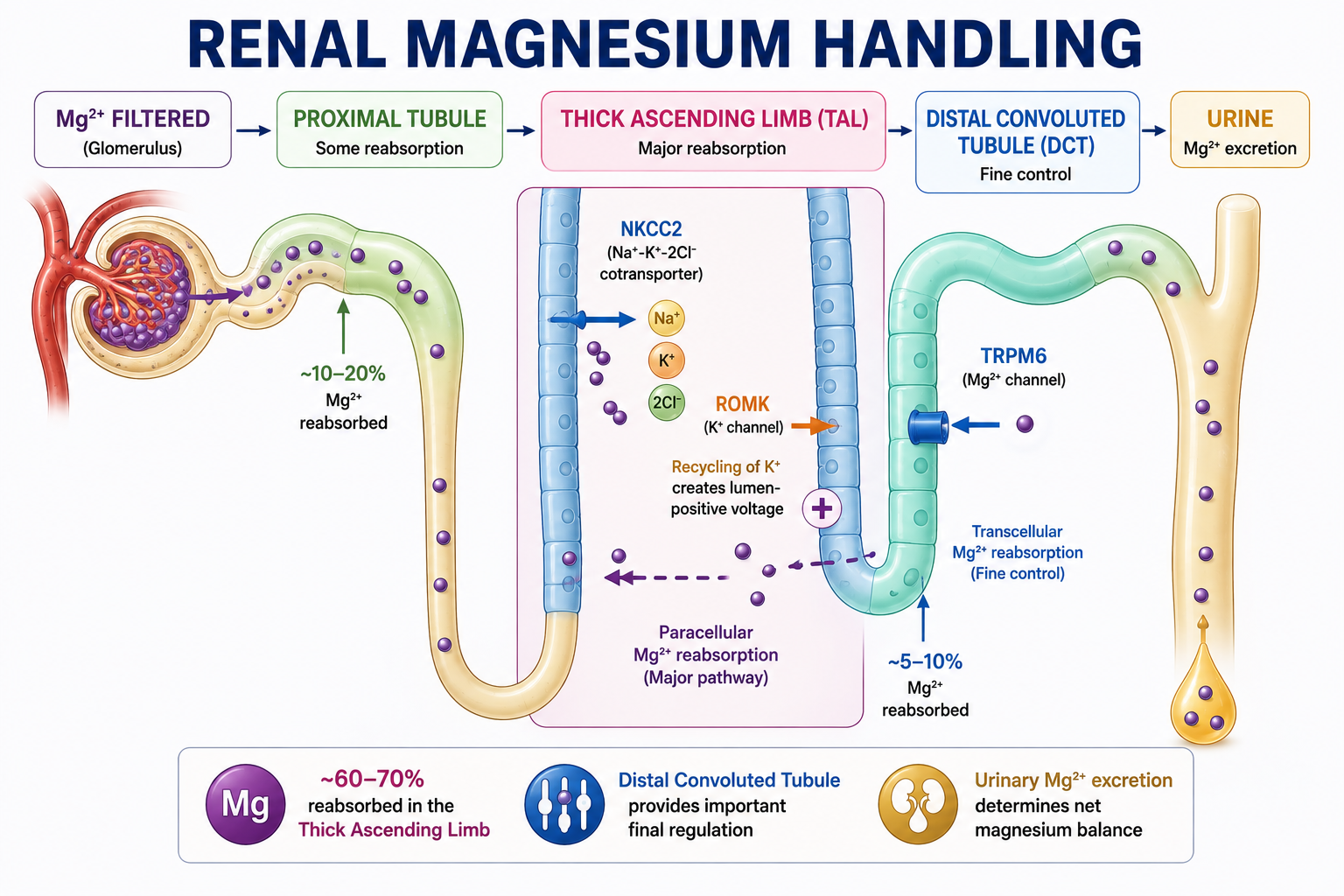
The broad nephron sequence is glomerulus, proximal tubule, thick ascending limb, distal convoluted tubule and urine. Only the ultrafilterable fraction of circulating magnesium passes freely through the glomerular filtration barrier; protein-bound magnesium is not freely filtered.
The proximal tubule reabsorbs a portion of filtered magnesium, but it is not the major site of magnesium reabsorption. The largest fraction is reabsorbed later in the thick ascending limb.
| Nephron Segment | Main Role in Mg2+ Handling | Key Concept |
|---|---|---|
| Proximal tubule | Some reabsorption | Not the major site |
| Thick ascending limb | Largest fraction reabsorbed | Paracellular, voltage-driven |
| Distal convoluted tubule | Smaller fraction, final regulation | TRPM6-mediated transcellular transport |
| Beyond DCT | Limited recovery | Distal loss strongly affects final excretion |
Magnesium Reabsorption in the Thick Ascending Limb
The thick ascending limb reabsorbs the largest fraction of filtered magnesium, mainly through a paracellular pathway driven by the lumen-positive voltage, while the distal convoluted tubule provides important final regulation through transport including TRPM6.
Much TAL magnesium reabsorption occurs between tubular cells. The driving force is closely related to the lumen-positive transepithelial voltage.
On the apical membrane of TAL cells, NKCC2 transports sodium, potassium and chloride from the tubular lumen into the cell. Potassium entering through NKCC2 is partly recycled back into the lumen through ROMK channels. This potassium recycling helps create a lumen-positive electrical potential.
The lumen-positive voltage promotes paracellular movement of positively charged ions from tubular lumen toward interstitium and blood. This includes Mg2+ and Ca2+.
Loop Diuretics and Magnesium Loss
Loop diuretics inhibit NKCC2 in the thick ascending limb, reducing the lumen-positive voltage that normally promotes paracellular magnesium reabsorption and thereby increasing urinary magnesium loss.
NKCC2 inhibition reduces potassium recycling and the lumen-positive voltage. Paracellular Mg2+ reabsorption falls, so urinary Mg2+ loss increases. This explains why loop diuretics can contribute to hypomagnesemia.
The same lumen-positive voltage also contributes to paracellular calcium reabsorption, so loop diuretics can increase urinary loss of Mg2+ and Ca2+.
Magnesium Reabsorption in the DCT
The distal convoluted tubule reabsorbs a smaller absolute fraction of filtered magnesium than the TAL, but it is crucial because it provides important final regulation of magnesium excretion.
By the time tubular fluid reaches the DCT, much of the filtered magnesium has already been reabsorbed. Small changes in distal magnesium transport can significantly affect final urinary magnesium excretion.
TRPM6 and Magnesium
Magnesium enters DCT cells from the tubular lumen through TRPM6, an important apical magnesium channel. TRPM6 plays an important role in regulated distal magnesium reabsorption.
Abnormalities affecting distal magnesium transport can produce substantial renal magnesium wasting even though the DCT handles a smaller fraction of filtered magnesium than the TAL. There is little opportunity to recover magnesium after the DCT.
TAL = major bulk Mg2+ reabsorption. DCT = important final fine control.
How the Kidney Responds to Low Magnesium
Suppose body magnesium begins to fall. A healthy kidney should respond by increasing magnesium conservation. Magnesium depletion should increase renal magnesium reabsorption and reduce urinary magnesium excretion.
If a patient has low serum magnesium, ask: is the kidney appropriately conserving magnesium? If yes, urinary magnesium should be low. If the kidney continues losing an inappropriate amount of magnesium despite hypomagnesemia, renal magnesium wasting is present.
Low Mg2+ with kidney conserving Mg2+ suggests nonrenal loss or reduced availability. Low Mg2+ with persistent inappropriate urinary Mg2+ loss suggests renal magnesium wasting.
Urinary Magnesium and FEMg
Urinary magnesium helps answer whether the kidney is appropriately conserving magnesium. Assessment can involve urinary magnesium concentration in context, 24-hour urinary magnesium or fractional excretion of magnesium. The exact method depends on the clinical situation.
Fractional excretion of magnesium, or FEMg, estimates the fraction of filtered magnesium ultimately excreted in urine. During magnesium depletion, a normal kidney should reduce FEMg because it is conserving magnesium. In renal magnesium wasting, FEMg remains inappropriately elevated for the degree of hypomagnesemia.
For the clinical diagnostic approach to low magnesium, see Hypomagnesemia Explained. This physiology article does not include a FEMg calculator.
Magnesium and Potassium
Magnesium deficiency can increase renal potassium secretion through effects involving ROMK, causing ongoing urinary potassium loss and making hypokalemia difficult to correct until magnesium deficiency is treated.
Magnesium normally influences renal potassium handling. When intracellular magnesium is deficient, inhibitory regulation of ROMK is reduced. This allows increased potassium secretion into the tubular lumen, increasing urinary potassium loss and lowering potassium.
Persistent or refractory hypokalemia should prompt assessment of magnesium. This relationship connects directly with renal potassium handling and hypokalemia.
Magnesium, Calcium and PTH
Severe magnesium deficiency can cause hypocalcemia by impairing both PTH secretion and tissue responsiveness to PTH.
Severe magnesium deficiency can impair PTH secretion. This is physiologically unusual because hypocalcemia would normally stimulate PTH secretion. Instead, severe magnesium deficiency can produce inappropriately low or normal PTH despite low calcium.
Severe magnesium deficiency can also impair tissue responsiveness to PTH. Hypocalcemia associated with significant magnesium deficiency may therefore not correct appropriately until magnesium is corrected.
For the normal PTH framework, see how PTH normally regulates calcium homeostasis. For clinical low-calcium diagnosis, see how low calcium is evaluated using PTH, magnesium and phosphate.

Magnesium, Potassium and Calcium Together
Magnesium deficiency can produce two important secondary abnormalities. First, low magnesium can increase renal potassium loss and contribute to hypokalemia. Second, severe low magnesium can impair PTH secretion and action, contributing to hypocalcemia.
A patient may therefore present with low magnesium, low potassium and low calcium from a common magnesium-related mechanism. When hypokalemia or hypocalcemia is difficult to correct, check magnesium.
Magnesium Excess and Kidney Failure
When magnesium availability increases and kidney function is normal, renal magnesium excretion can increase. This protects against major magnesium accumulation, so clinically important hypermagnesemia is uncommon with normal renal excretory capacity unless magnesium exposure is substantial.
When renal function is markedly reduced, magnesium filtration and excretion capacity fall, making magnesium retention more likely. If exogenous magnesium is also administered or consumed, accumulation risk rises further. Renal failure plus magnesium exposure is an important setting for hypermagnesemia.
Clinical Assessment of Magnesium Balance
Serum magnesium is the standard starting test. Interpret it alongside potassium, calcium, renal function, medication history, gastrointestinal history and clinical context.
When magnesium is low, actively look for low potassium and low calcium. Likewise, unexplained or refractory abnormalities of potassium or calcium should prompt magnesium assessment. Renal function is central because the kidney determines magnesium conservation versus excretion.
If hypomagnesemia is present, ask whether the kidney is conserving magnesium. Use urinary magnesium assessment when needed. Consider diarrhea, malabsorption, poor nutritional intake, medications, diuretics, renal tubular disorders, renal function and other electrolyte abnormalities. Investigation should follow the physiology.
Worked Clinical Cases
Case 1: Gastrointestinal Loss
A patient has prolonged diarrhea, low serum magnesium and appropriately low urinary magnesium. The gastrointestinal tract is losing magnesium and the kidneys are responding appropriately by conserving Mg2+.
Case 2: Loop Diuretic
A patient receiving a loop diuretic develops hypomagnesemia. NKCC2 is inhibited, the TAL lumen-positive voltage falls, paracellular Mg2+ reabsorption falls and urinary Mg2+ loss rises. This is renal magnesium wasting.
Case 3: Refractory Hypokalemia
A patient has low potassium and low magnesium. Potassium remains low despite repeated replacement because magnesium deficiency maintains renal ROMK-mediated potassium secretion and urinary potassium loss.
Case 4: Hypocalcemia
A patient has markedly low magnesium, low calcium and PTH that is not appropriately elevated. Severe magnesium deficiency can impair PTH secretion and PTH responsiveness, so hypocalcemia may persist until magnesium deficiency is corrected.
Case 5: Kidney Failure
A patient with markedly impaired renal function is exposed to a substantial amount of magnesium-containing medication. Serum magnesium rises because renal magnesium excretion is reduced while magnesium exposure increases.
Case 6: Multiple Electrolyte Abnormalities
A patient has low magnesium, low potassium and low calcium. Do not automatically assume three unrelated diseases. Magnesium deficiency can connect renal potassium wasting and impaired PTH secretion/action.
Common Mistakes
- Misconception: Magnesium is mainly extracellular. Reality: most magnesium is in bone and intracellular tissues.
- Misconception: Serum magnesium perfectly reflects total-body stores. Reality: serum magnesium is useful but incomplete.
- Misconception: The proximal tubule reabsorbs most magnesium. Reality: the thick ascending limb handles the largest fraction.
- Misconception: The DCT is unimportant. Reality: it provides final regulation with little recovery afterward.
- Misconception: TAL magnesium transport is mainly transcellular. Reality: a large proportion is paracellular and voltage-driven.
- Misconception: ROMK only matters for potassium. Reality: ROMK helps create the TAL voltage that facilitates Mg2+ and Ca2+ reabsorption.
- Misconception: Low magnesium with low urinary magnesium means renal wasting. Reality: it usually indicates appropriate renal conservation.
- Misconception: Hypomagnesemia only affects magnesium. Reality: it can contribute to hypokalemia and hypocalcemia.
- Misconception: Potassium replacement always corrects hypokalemia even if magnesium is low. Reality: magnesium deficiency can maintain renal potassium wasting.
- Misconception: Magnesium deficiency always increases PTH. Reality: severe deficiency can impair PTH secretion and action.
One-Minute Revision
- Magnesium is mostly in bone and intracellular tissues.
- Serum magnesium is useful but represents only a small fraction of body magnesium.
- The gut controls magnesium entry; the kidney controls final magnesium excretion.
- The proximal tubule reabsorbs some magnesium but is not the major site.
- The TAL performs major magnesium reabsorption through paracellular, voltage-driven movement.
- NKCC2 and ROMK help generate the lumen-positive voltage.
- The DCT provides final fine control through transport including TRPM6.
- When Mg2+ falls, healthy kidneys should conserve Mg2+ and urinary Mg2+ should fall.
- Persistent urinary Mg2+ loss during hypomagnesemia suggests renal magnesium wasting.
- Low magnesium can contribute to low potassium and low calcium.
Key Clinical Pearls
The kidney is the final regulator of magnesium balance: when magnesium falls, urinary magnesium should fall. If the kidney continues losing magnesium despite hypomagnesemia, think renal magnesium wasting.
- Magnesium is important for ATP-dependent cellular processes.
- The kidney provides the major adaptive control of magnesium excretion.
- Loop diuretics reduce TAL lumen-positive voltage and can increase magnesium loss.
- TRPM6 is important in distal magnesium reabsorption.
- Refractory hypokalemia should prompt magnesium assessment.
- Refractory hypocalcemia should prompt magnesium assessment.
- Renal failure reduces the ability to excrete magnesium.
Frequently Asked Questions
Key Take-Home Messages
Magnesium homeostasis is a balance between gut entry and renal excretion. Magnesium enters through the gastrointestinal tract, circulates in a small extracellular fraction, exchanges with bone and intracellular stores, and is then filtered and reabsorbed by the kidney.
The central diagnostic rule is that when magnesium falls, the kidney should conserve magnesium. Low magnesium with low urinary magnesium suggests an appropriate renal response and favors nonrenal loss or reduced availability. Low magnesium with inappropriate urinary magnesium loss suggests renal magnesium wasting.
Magnesium cannot be viewed in isolation. Low magnesium can drive renal potassium wasting and impair PTH secretion/action, producing both low potassium and low calcium.
This article is intended for medical education only. It explains physiology and clinical interpretation principles, not magnesium replacement protocols or patient-specific treatment.