Potassium balance depends not only on total body potassium but on its distribution between intracellular and extracellular compartments, dietary intake, renal excretion, hormonal regulation and acid-base status. Rather than memorising lists of causes of hypokalemia and hyperkalemia, this article builds the physiological foundation needed to understand why potassium disorders develop in the first place.
This article follows on from Electrolytes Explained and complements Acid-Base Disorders Explained, Metabolic Acidosis Explained, Metabolic Alkalosis Explained and ABG Interpretation Explained. It provides the physiological foundation for the potassium cluster; later articles will explore hypokalemia, hyperkalemia and their ECG changes and management in greater depth.
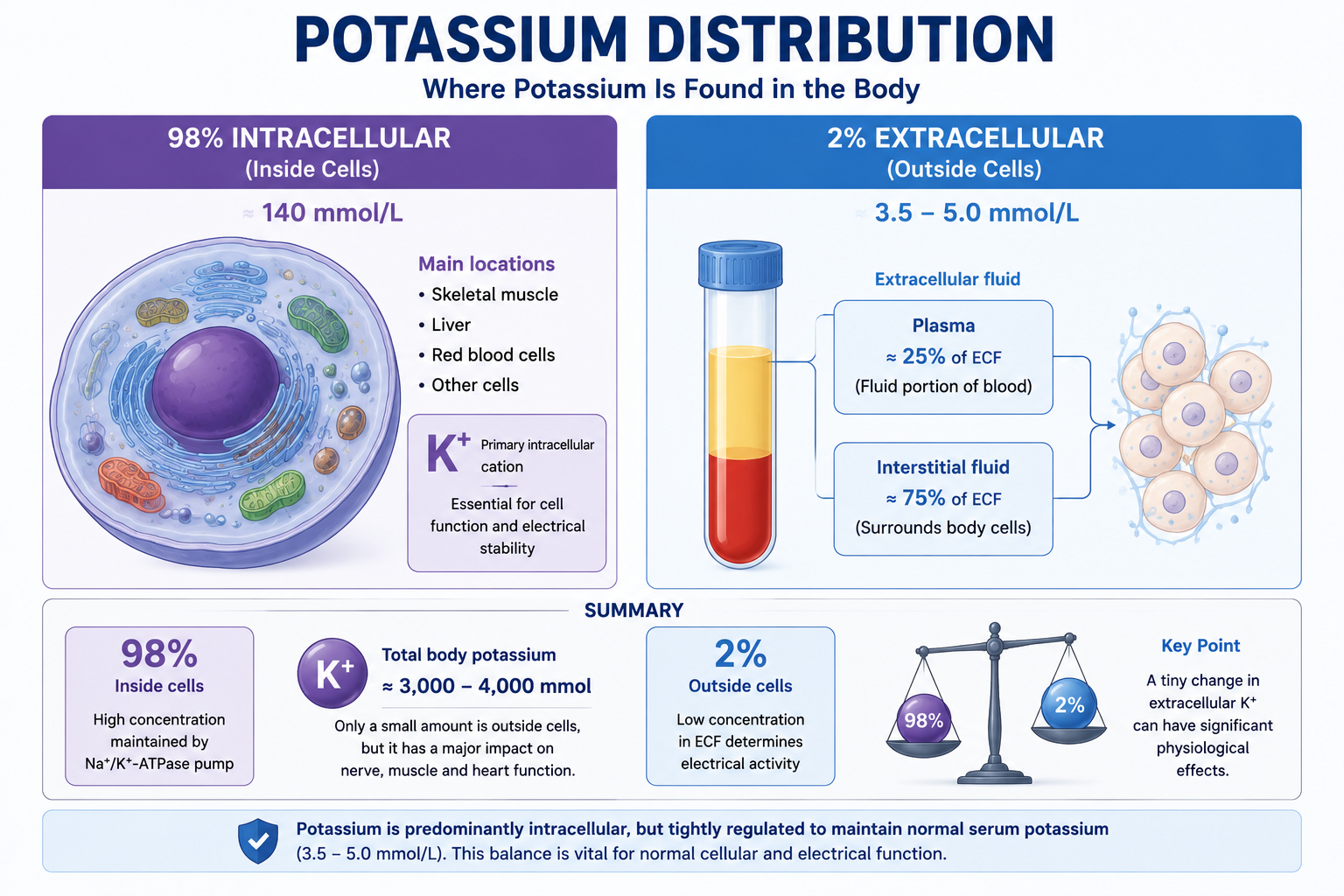
Potassium is the major intracellular cation. Roughly 98% of total body potassium sits inside cells and only about 2% circulates in the extracellular fluid — the fraction that laboratory tests actually measure.
Learning Objectives
- Explain why potassium is one of the body's most tightly regulated electrolytes
- Describe the normal distribution of potassium and why it is mainly intracellular
- Explain the role of the Na+/K+-ATPase pump and resting membrane potential
- Describe renal potassium handling and the role of aldosterone
- Understand rapid redistribution mechanisms (insulin, beta2 stimulation, acid-base status)
- Apply potassium physiology to common clinical scenarios and ECG changes
Why Potassium Matters
Potassium participates in virtually every electrically active tissue in the body. Its concentration determines how easily cells can generate electrical impulses, so potassium is essential for resting membrane potential, action potential generation, skeletal muscle contraction, cardiac conduction, smooth muscle function, nerve impulse transmission, cellular enzyme activity, protein synthesis and glycogen synthesis.
Abnormal potassium concentrations may produce muscle weakness, paralysis, cardiac arrhythmias, respiratory failure and sudden cardiac death — complications that arise because potassium directly influences the electrical properties of excitable cells.

Total Body Potassium
An average healthy adult contains approximately 3,000–4,000 mmol of total body potassium, depending on body size, muscle mass, age and sex. Because skeletal muscle contains most intracellular potassium, individuals with greater muscle mass generally have a higher total body potassium content. Most adults require approximately 2–3 g (≈50–75 mmol) of potassium daily, though requirements vary with diet, activity, renal function and clinical conditions.
Potassium Distribution
One of the most important concepts in potassium physiology is that serum potassium represents only a very small proportion of total body potassium.
Approximately 98% of total body potassium is found within cells, with the highest concentrations in skeletal muscle, liver and red blood cells. Only about 2% is present in the extracellular fluid — this small compartment is what laboratory tests measure as the serum potassium concentration.
A patient may have severe total body potassium depletion with only a mildly reduced serum concentration, or a markedly elevated serum concentration without a large increase in total body potassium due to redistribution. Understanding this distinction is fundamental to clinical potassium interpretation.
The Na+/K+-ATPase Pump
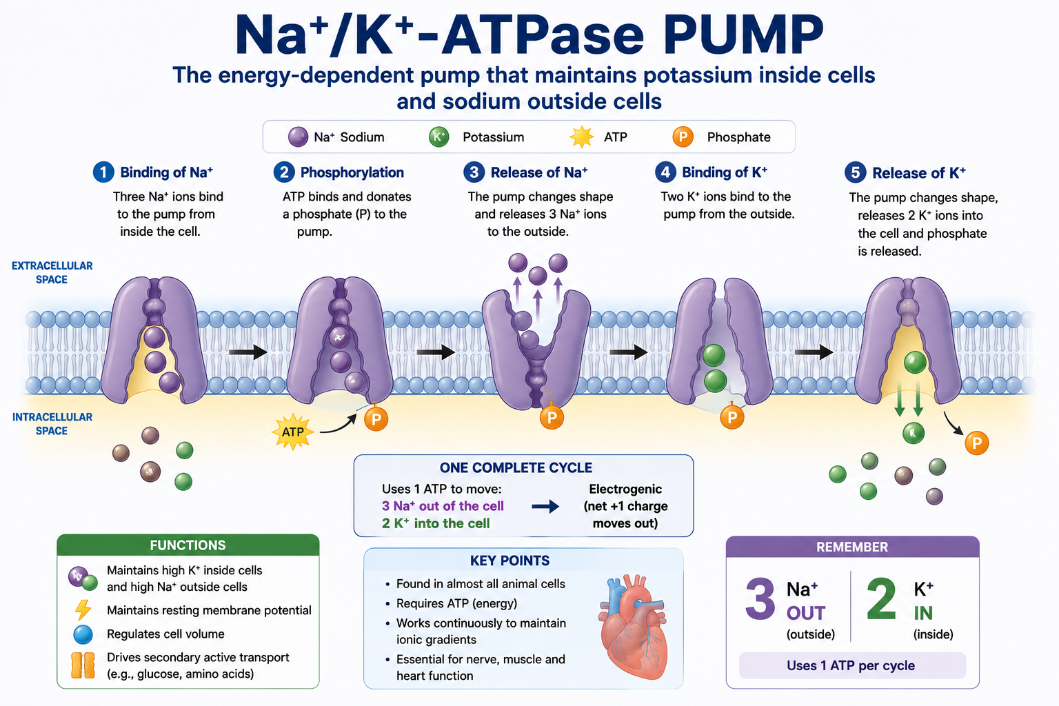
Cells actively maintain a high intracellular potassium concentration. This distribution is not passive — it requires continuous energy expenditure through the Na+/K+-ATPase pump, an energy-dependent membrane protein found in virtually all human cells. Without this active transport system, potassium would gradually diffuse out of cells, destroying the electrical gradients essential for life.
For every cycle, the pump moves 3 sodium ions out of the cell and 2 potassium ions into the cell, consuming one molecule of ATP. Because both ions are transported against their concentration gradients, this active transport requires energy from ATP hydrolysis.
| Compartment | Potassium Concentration |
|---|---|
| Intracellular fluid | ≈140 mmol/L |
| Extracellular fluid | ≈3.5–5.0 mmol/L |
This steep concentration gradient underlies the resting membrane potential of most excitable cells. Beyond maintaining potassium and sodium gradients, the pump also preserves resting membrane potential, regulates cell volume, supports secondary active transport of glucose and amino acids, and enables normal nerve and muscle function.
Resting Membrane Potential
Every excitable cell maintains a difference in electrical charge across its membrane, known as the resting membrane potential. The unequal distribution of potassium between intracellular and extracellular compartments is the principal factor responsible for this electrical gradient. Because potassium tends to diffuse out of the cell through potassium channels, leaving behind negatively charged proteins, the interior of the cell becomes electrically negative relative to the exterior.
| Cell Type | Typical Resting Membrane Potential |
|---|---|
| Skeletal muscle | −90 mV |
| Neurons | −70 mV |
| Cardiac ventricular myocytes | −90 mV |
Small changes in extracellular potassium alter the resting membrane potential, changing the excitability of nerves and muscles — this explains why even modest potassium abnormalities can produce profound clinical effects.

Potassium Intake and Absorption
Potassium is obtained almost entirely from the diet, and under normal conditions dietary intake greatly exceeds daily requirements. Healthy kidneys efficiently excrete excess potassium, allowing a wide range of dietary intake without significant changes in serum potassium. Potassium-rich foods include bananas, oranges, avocados, tomatoes, potatoes, leafy green vegetables, beans, lentils, nuts, milk, yoghurt, fish and meat. Most adults consume approximately 70–120 mmol (≈2.7–4.7 g) of potassium daily.
Approximately 90–95% of ingested potassium is absorbed under normal conditions, largely passively, primarily in the small intestine with a minor contribution from the colon. Inadequate absorption is a relatively uncommon cause of hypokalemia; gastrointestinal potassium disorders more commonly result from excessive losses such as persistent vomiting, chronic diarrhoea, high-output ileostomy or enterocutaneous fistulae.
Renal Potassium Handling
Although potassium is lost through urine (90–95%), stool (5–10%) and a small variable amount in sweat, the kidneys are responsible for maintaining long-term potassium balance — increasing excretion after a potassium-rich meal and reducing it during depletion. Unlike sodium, potassium regulation depends primarily on variable distal secretion rather than variable filtration.
Glomerulus and Proximal Tubule
Potassium is freely filtered at the glomerulus. Approximately 65–70% of filtered potassium is reabsorbed in the proximal tubule, predominantly through passive paracellular transport driven by solvent drag from water and sodium reabsorption. This reabsorption is relatively constant and undergoes little physiological regulation.
Thick Ascending Limb
An additional 20–25% of filtered potassium is reabsorbed here via the Na+-K+-2Cl- cotransporter (NKCC2). Loop diuretics inhibit NKCC2, reducing sodium reabsorption and increasing potassium losses, which may cause hypokalemia.
Distal Convoluted Tubule and Collecting Duct
The distal nephron is the major regulatory site for potassium balance. Unlike earlier segments, transport here is highly variable — the kidneys can increase potassium secretion or reduce it to almost zero, allowing precise regulation of serum potassium.
Principal cells are responsible for most potassium secretion: sodium enters the cell, the Na+/K+-ATPase pump brings potassium in, and potassium is then secreted into the urine. Secretion depends on aldosterone, distal sodium delivery, tubular flow rate, plasma potassium concentration and acid-base status. Intercalated cells contribute to potassium conservation during depletion — secretion decreases, reabsorption increases, and urinary excretion falls to very low levels.
| Nephron Segment | Main Potassium Handling |
|---|---|
| Glomerulus | Free filtration |
| Proximal tubule | ~65–70% reabsorption |
| Thick ascending limb | ~20–25% reabsorption |
| Distal convoluted tubule | Variable secretion |
| Collecting duct | Final regulation of excretion |
Aldosterone
Aldosterone is the most important hormonal regulator of renal potassium excretion, released in response to increased plasma potassium, renin-angiotensin activation or reduced circulating volume. It increases sodium reabsorption and potassium secretion, thereby increasing potassium excretion. Excess aldosterone causes persistent hypokalemia, metabolic alkalosis and hypertension; deficiency (as in Addison disease) causes hyperkalemia and mild metabolic acidosis.
Distal Sodium Delivery, Tubular Flow and Feedback
When more sodium reaches the distal nephron, more potassium is secreted — this is why several diuretics increase potassium loss. Higher tubular flow removes secreted potassium from the lumen, maintaining a favourable gradient for continued secretion, so high urine flow increases potassium excretion while low flow reduces it. Plasma potassium itself provides negative feedback: high plasma potassium increases aldosterone and secretion, while low plasma potassium reduces both, conserving potassium. Healthy kidneys can reduce urinary potassium losses to less than 10 mmol/day when stores are depleted.
Hyperkalemia rarely develops solely from increased dietary potassium intake if renal function is normal. Impaired kidney function reduces the capacity to excrete potassium, explaining why hyperkalemia is common in acute kidney injury, chronic kidney disease, advanced diabetic nephropathy and patients on potassium-retaining medications.
Potassium Redistribution Between Cells
Maintaining potassium homeostasis involves more than balancing intake and excretion. The body can rapidly alter serum potassium by shifting potassium between intracellular and extracellular compartments without changing total body potassium — these mechanisms act within minutes, buffering meals, exercise and acute illness while the kidneys make slower adjustments over hours.
Insulin
Insulin stimulates the Na+/K+-ATPase pump, driving potassium into cells and lowering serum potassium. After a potassium-rich meal, insulin secretion rises and potassium rapidly moves into cells, preventing a marked rise in serum potassium. This is why intravenous insulin (with glucose) is routinely used in the emergency treatment of severe hyperkalemia — it does not remove potassium from the body, only redistributes it until definitive removal occurs.
Beta2-Adrenergic Stimulation
Beta2 stimulation also activates the Na+/K+-ATPase pump, moving potassium into cells during exercise, emotional stress, acute illness or with inhaled beta2 agonists such as salbutamol. Nebulized or intravenous beta2 agonists may be used as adjunctive therapy for severe hyperkalemia for the same reason.
Acid-Base Status
The relationship is more nuanced than the traditional teaching that "acidosis causes hyperkalemia, alkalosis causes hypokalemia." In selected forms of metabolic acidosis, hydrogen ions move into cells and potassium moves out, raising serum potassium — this effect is more pronounced in mineral (non-organic) acidosis than in organic acidoses such as lactic acidosis and ketoacidosis, which involve additional mechanisms. In metabolic alkalosis, hydrogen ions leave cells and potassium shifts in, lowering serum potassium, with increased renal excretion often contributing further. Acute respiratory acid-base disorders generally have smaller effects on potassium than metabolic ones.
Hyperosmolality
Hyperosmolality draws water out of cells; as intracellular potassium concentration rises, potassium moves into the extracellular fluid. Common causes include severe hyperglycaemia, hyperosmolar hyperglycaemic state and mannitol administration. Patients with uncontrolled diabetes may present with hyperkalemia despite significant total body potassium depletion — an apparent paradox from redistribution and urinary losses occurring simultaneously.
Exercise and Cell Lysis
Exercise causes transient potassium release from contracting muscle, raising serum potassium slightly; recovery involves beta2 stimulation, insulin secretion and Na+/K+-ATPase activation returning potassium into muscle. Cell lysis — from rhabdomyolysis, tumour lysis syndrome, massive trauma, burns, haemolysis or crush injury — releases large amounts of intracellular potassium and may rapidly produce severe hyperkalemia requiring emergency treatment.
Potassium in Common Diseases
| Condition | Typical Findings |
|---|---|
| Diabetic ketoacidosis | Total body potassium markedly depleted, but serum potassium normal or initially raised due to insulin deficiency, acidosis and osmotic diuresis; potassium replacement is often needed during treatment |
| Hyperosmolar hyperglycaemic state | Potassium depletion, variable serum potassium, severe dehydration |
| Chronic kidney disease | Reduced potassium excretion, increased hyperkalemia risk, higher susceptibility to medication-induced elevation |
| Primary hyperaldosteronism | Hypokalemia, metabolic alkalosis, hypertension |
| Addison disease | Hyperkalemia, mild metabolic acidosis, hyponatremia, hypotension |
Potassium and ECG Physiology
Potassium strongly influences cardiac electrical activity; changes in serum potassium alter myocardial depolarisation and repolarisation.
Hypokalemia may produce flattened T waves, ST depression, prominent U waves, apparent QT prolongation (actually QU prolongation) and ventricular arrhythmias.
Hyperkalemia produces a progressive sequence: tall peaked T waves, PR interval prolongation, loss of P waves, QRS widening, a sine-wave pattern, and ultimately ventricular fibrillation or asystole.
The ECG patterns of low and high potassium are compared in detail in ECG Changes in Potassium Disorders.
ECG changes do not always correlate perfectly with serum potassium concentration. Some patients develop life-threatening ECG abnormalities at relatively modest potassium elevations, whereas others tolerate higher concentrations. Clinical assessment should integrate both laboratory values and ECG findings.
Practical Interpretation of Potassium Results
- Confirm the result. Exclude haemolysis, sample contamination or delayed processing; consider pseudohyperkalemia when the clinical picture is inconsistent.
- Assess severity — mild, moderate or severe.
- Assess symptoms — muscle weakness, paralysis, palpitations, syncope, arrhythmias.
- Review the ECG. Hyperkalemia may need immediate treatment even before laboratory confirmation if characteristic changes are present.
- Determine the mechanism — redistribution, reduced renal excretion, increased intake, gastrointestinal or renal loss, hormonal disorder or medication effect.
Common Misconceptions
- "Serum potassium reflects total body potassium." False — serum potassium represents only about 2% of total body potassium.
- "Hyperkalemia always means excess total body potassium." False — redistribution alone may produce severe hyperkalemia.
- "Normal serum potassium excludes potassium deficiency." False — patients may have substantial depletion despite a normal serum concentration.
- "Acidosis always causes hyperkalemia." False — the effect varies by the type of acidosis and accompanying mechanisms.
- "Insulin removes potassium from the body." False — insulin shifts potassium into cells but does not eliminate it; definitive removal requires renal excretion, gastrointestinal potassium binders or dialysis.
Clinical Cases
Insulin Treatment in DKA
A patient receives intravenous insulin for DKA. Why does serum potassium fall after treatment?
Rhabdomyolysis
A patient with rhabdomyolysis develops severe hyperkalemia. What is the mechanism?
Loop Diuretics
A patient taking loop diuretics develops persistent hypokalemia. Why?
Addison Disease
A patient with Addison disease has hyperkalemia. Why?
One Minute Revision
- Potassium distribution: 98% intracellular, 2% extracellular.
- Main long-term regulator: the kidneys, chiefly through distal nephron secretion.
- Main hormone: aldosterone.
- Rapid regulators: insulin, beta2 stimulation, acid-base status, exercise, hyperosmolality.
- Serum potassium does not equal total body potassium.
- Hyperkalemia: think kidney failure, cell lysis, aldosterone deficiency, redistribution.
- Hypokalemia: think diuretics, GI losses, hyperaldosteronism, magnesium deficiency.
Frequently Asked Questions
This article is intended for medical education only. Both hyperkalemia and hypokalemia can be life-threatening. Clinical management requires proper assessment, ECG review, local protocols and senior or specialist input.