Hypomagnesemia at a Glance
Hypomagnesemia is a serum magnesium concentration below the laboratory reference interval. It may cause neuromuscular and cardiac abnormalities and can contribute to hypokalemia and hypocalcemia.

What Is Hypomagnesemia?
Hypomagnesemia means that serum magnesium is below the appropriate laboratory reference interval.
Its clinical significance depends on the degree of deficiency, symptoms, associated electrolyte abnormalities and underlying cause.
A low magnesium result should not be interpreted in isolation. It should be interpreted with symptoms, ECG context when indicated, potassium, calcium, renal function, medication exposure and gastrointestinal history.
For the physiology behind normal magnesium balance, review Magnesium Homeostasis Explained.
Serum Magnesium and Total-Body Magnesium
Most body magnesium is located in bone and intracellular tissues. Only a small fraction is present in the extracellular fluid.
Serum magnesium is clinically useful because it is accessible and reflects the measured circulating compartment. However, it does not perfectly represent total-body magnesium stores.
A serum magnesium result should be interpreted alongside the clinical picture, associated potassium and calcium abnormalities, renal function and possible ongoing loss.
Symptoms of Hypomagnesemia
Mild hypomagnesemia may be asymptomatic. When symptoms occur, they often reflect neuromuscular irritability, cardiac electrical effects, or associated potassium and calcium abnormalities.
| System | Possible Manifestations | Clinical Meaning |
|---|---|---|
| Neuromuscular | Tremor, muscle cramps, weakness, paresthesia, neuromuscular irritability | Low magnesium can disturb membrane and neuromuscular stability |
| Neurological | Seizures in severe deficiency | Severe deficiency can produce major neurological manifestations |
| Cardiac | Electrical instability and arrhythmias | Risk is higher when hypokalemia or QT-prolonging medication exposure coexists |
| Electrolyte | Hypokalemia and hypocalcemia | Low magnesium can make potassium and calcium abnormalities difficult to correct |
Neuromuscular Effects
Magnesium contributes to membrane and neuromuscular stability. When magnesium is low, neuromuscular irritability may occur.
Clinical manifestations may include tremor, muscle cramps, weakness, paresthesia and neuromuscular irritability. Severe magnesium deficiency can produce seizures.
Neuromuscular symptoms should prompt assessment of magnesium in context, especially when potassium or calcium is also abnormal.
ECG and Cardiac Effects
Magnesium contributes to cardiac electrical stability. Hypomagnesemia can increase susceptibility to cardiac electrical instability, especially when other risk factors are present.
There is no single ECG abnormality that uniquely diagnoses hypomagnesemia. ECG changes are often influenced by associated hypokalemia, medications and the wider clinical setting.
Hypomagnesemia can contribute to abnormal ventricular repolarization and may be associated with QT prolongation in relevant clinical settings. Significant magnesium deficiency can increase susceptibility to ventricular arrhythmias including torsades de pointes, particularly when other risk factors are present.
For ECG patterns driven by potassium disorders, see ECG Changes in Potassium Disorders.
Causes of Hypomagnesemia
Do not begin by memorizing a long list. First ask why magnesium is low.
The major mechanisms are reduced magnesium availability, gastrointestinal loss, renal magnesium wasting and other clinical mechanisms.
| Mechanism | Examples From the Guide | Diagnostic Clue |
|---|---|---|
| Reduced availability | Reduced intake, impaired intestinal absorption | May coexist with other nutritional or gastrointestinal problems |
| Gastrointestinal loss | Diarrhea, malabsorption | Kidney should conserve magnesium, so urinary magnesium should fall |
| Medication-associated | Long-term PPI exposure, diuretics, selected nephrotoxic medications | Medication history is essential |
| Renal wasting | Loop diuretics, thiazides, cisplatin, aminoglycosides, calcineurin inhibitors, renal tubular disorders, Gitelman syndrome | Urinary magnesium remains inappropriate for hypomagnesemia |
Gastrointestinal Magnesium Loss
Prolonged diarrhea is an important cause of magnesium loss. Diarrhea can directly increase gastrointestinal magnesium loss and can coexist with poor intake or malabsorption.
In gastrointestinal magnesium loss, the expected renal response is conservation. Urinary Mg2+ excretion should fall.
Disorders that reduce intestinal magnesium absorption can also cause magnesium deficiency. When the cause appears gastrointestinal, urinary magnesium helps confirm that the kidney is responding appropriately.
Low Mg2+ with appropriately low urinary Mg2+ supports nonrenal magnesium loss or reduced availability.
PPI-Associated Hypomagnesemia
Long-term proton pump inhibitor exposure is associated with hypomagnesemia in susceptible patients.
The key mechanism to teach is reduced intestinal magnesium absorption. In unexplained hypomagnesemia, medication history should include long-term PPI exposure.
This is especially important when hypomagnesemia is persistent, recurrent or associated with hypokalemia or hypocalcemia.
Renal Magnesium Wasting
When serum magnesium is low, healthy kidneys should conserve magnesium. Low magnesium should normally cause urinary Mg2+ excretion to fall.
If the kidneys continue excreting an inappropriate amount of magnesium despite hypomagnesemia, renal magnesium wasting is present.
Important causes include loop diuretics, thiazide diuretics, selected nephrotoxic medications, cisplatin, aminoglycosides, calcineurin inhibitors, renal tubular disorders and inherited tubulopathies such as Gitelman syndrome.
Loop and Thiazide Diuretics
Loop diuretics inhibit NKCC2 in the thick ascending limb, reducing the lumen-positive voltage that normally promotes paracellular magnesium reabsorption and thereby increasing urinary magnesium loss.
The thick ascending limb is the major site of renal magnesium reabsorption. NKCC2 inhibition reduces potassium recycling and the lumen-positive voltage. Paracellular Mg2+ reabsorption falls, so urinary Mg2+ loss increases.
Thiazide diuretics can also contribute to renal magnesium wasting. Their clinical importance is that a medication history can reveal an ongoing renal cause of hypomagnesemia.
For the nephron physiology, see TAL magnesium handling.
Medication-Related Renal Magnesium Loss
Cisplatin can damage renal tubular magnesium handling and produce renal magnesium wasting.
Aminoglycosides can interfere with renal tubular electrolyte handling and contribute to renal magnesium wasting.
Calcineurin inhibitors can promote renal magnesium wasting through effects on distal tubular magnesium handling.
The article should teach the mechanism rather than attempt an exhaustive rare-disease list. When hypomagnesemia is unexplained, the medication history should be deliberate and specific.
Gitelman Syndrome
Gitelman syndrome is an inherited salt-losing tubulopathy involving the distal convoluted tubule.
The DCT is important for final magnesium regulation. Distal tubular dysfunction can therefore cause renal magnesium wasting.
The high-yield clinical pattern is hypomagnesemia with renal magnesium wasting in a distal tubular disorder context.
How to Diagnose Hypomagnesemia
When magnesium is low, first confirm and assess the patient. Evaluate symptoms, ECG when indicated, potassium, calcium and renal function.
Next, look for an obvious cause: gastrointestinal loss, reduced intake, medication exposure or renal context. If the cause remains uncertain, assess renal magnesium handling.
The central question is simple: is the kidney conserving magnesium?
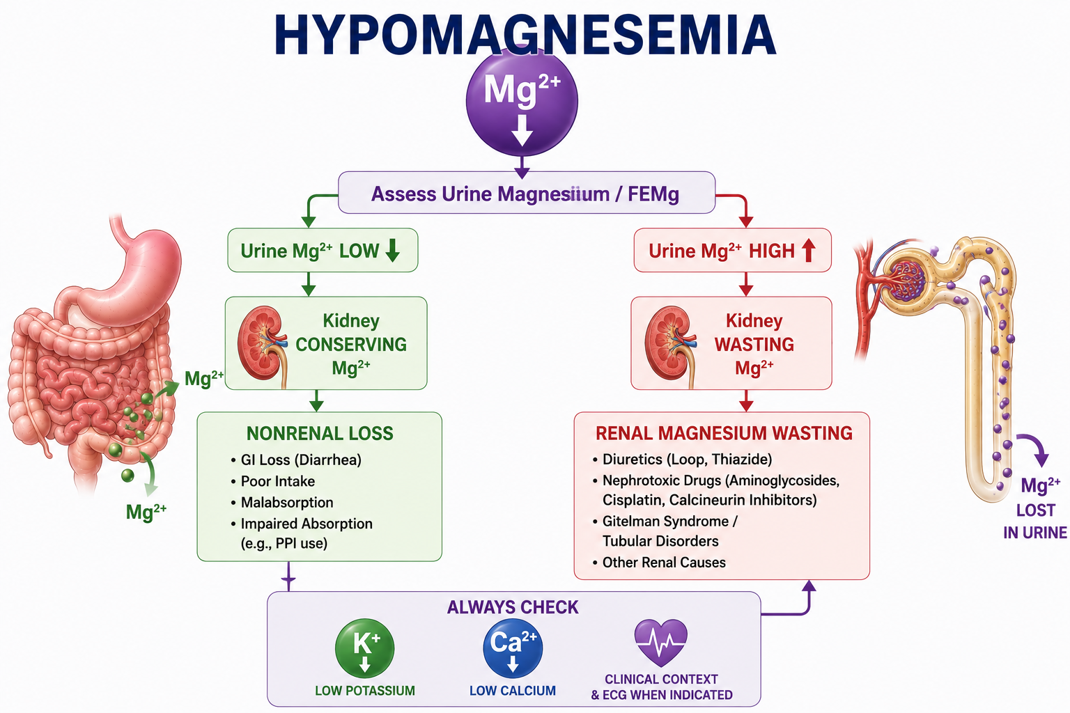
Renal vs Nonrenal Magnesium Loss
During hypomagnesemia, healthy kidneys should conserve magnesium. Appropriately low urinary magnesium suggests a nonrenal source such as gastrointestinal loss, while inappropriate urinary magnesium loss suggests renal magnesium wasting.
Urinary magnesium appropriately low
This pattern suggests that the kidney is conserving magnesium. The problem is more likely reduced magnesium availability or nonrenal loss, such as gastrointestinal loss.
Urinary magnesium inappropriately high
This pattern suggests renal magnesium wasting. The kidney is not reducing magnesium excretion appropriately despite hypomagnesemia.
Urinary Magnesium and FEMg
Urinary magnesium can be assessed using urinary magnesium in context, 24-hour urinary magnesium or fractional excretion of magnesium.
FEMg is the fractional excretion of magnesium. It estimates the fraction of filtered magnesium that is excreted in urine.
In appropriate renal conservation, urinary magnesium and FEMg should be low for the clinical context. In renal magnesium wasting, urinary magnesium loss or FEMg is inappropriate for the degree of hypomagnesemia.
FEMg must be interpreted with clinical context and renal function. This article does not provide diagnostic thresholds or a FEMg calculator.
Hypomagnesemia and Hypokalemia
Magnesium deficiency increases renal potassium secretion through effects involving ROMK, causing urinary potassium loss and making hypokalemia difficult to correct until magnesium deficiency is addressed.
This is one of the most clinically important consequences of magnesium deficiency. Hypomagnesemia frequently coexists with hypokalemia.
When intracellular magnesium is low, inhibitory regulation of ROMK is reduced. Renal potassium secretion can increase, urinary potassium loss rises and serum potassium falls.
If magnesium deficiency persists, renal potassium wasting can continue despite potassium replacement. Difficult-to-correct hypokalemia should prompt assessment of magnesium. See Hypokalemia Explained for the broader low-potassium approach.
Hypomagnesemia and Hypocalcemia
Severe magnesium deficiency can cause hypocalcemia by impairing both PTH secretion and tissue responsiveness to PTH.
Normally, hypocalcemia should stimulate PTH secretion. Severe magnesium deficiency can create a paradoxical pattern where PTH secretion is impaired despite low calcium.
Magnesium deficiency can also impair target tissue responsiveness to PTH. Therefore hypocalcemia associated with significant magnesium deficiency may be difficult to correct until magnesium is addressed.
For the calcium diagnostic framework, see Hypocalcemia Explained and Calcium Homeostasis Explained.

Mg2+, K+ and Ca2+ Together
A patient may present with low magnesium, low potassium and low calcium. Do not automatically assume three unrelated diseases.
Magnesium deficiency can connect the pattern through renal potassium wasting and impaired PTH secretion or action.
| Finding | Magnesium-Linked Mechanism | Clinical Lesson |
|---|---|---|
| Mg2+ low | Reduced availability, GI loss or renal wasting | Ask why Mg2+ is low |
| K+ low | Increased renal potassium loss through ROMK-related effects | Refractory hypokalemia should prompt magnesium assessment |
| Ca2+ low | Impaired PTH secretion and tissue responsiveness to PTH | Refractory hypocalcemia should prompt magnesium assessment |
Treatment of Hypomagnesemia
Management should address three goals: the magnesium deficit, associated complications and the underlying cause.
The choice of replacement depends on severity, symptoms, urgency, gastrointestinal function and renal function. This article does not provide magnesium replacement doses or infusion rates.
Mild, stable or asymptomatic
Oral magnesium may be appropriate when hypomagnesemia is mild, the patient is stable, gastrointestinal absorption is adequate and urgent correction is not required.
Severe, symptomatic or urgent setting
IV magnesium may be appropriate when hypomagnesemia is severe, symptomatic, associated with seizures, associated with significant arrhythmia, associated with an urgent cardiac situation or when oral therapy is not suitable.
Treat the magnesium deficit, correct associated potassium and calcium problems when present, and address the cause of ongoing magnesium loss.
Oral Magnesium, IV Magnesium and Renal Impairment
Oral magnesium preparations can cause gastrointestinal adverse effects, particularly diarrhea. This can complicate treatment because diarrhea itself can increase magnesium loss. Therefore tolerability matters.
IV replacement requires monitoring because magnesium affects cardiac and neuromuscular physiology, and because the kidney is responsible for magnesium excretion.
Kidney function changes treatment because reduced renal excretion increases the risk of magnesium accumulation. Magnesium replacement should be interpreted in the context of renal function and local clinical protocols.
Treat the Cause
If gastrointestinal loss is driving magnesium depletion, ongoing diarrhea or malabsorption must be addressed.
If medication-associated hypomagnesemia is suspected, the relevant medication exposure should be identified and reviewed in the clinical context.
If renal magnesium wasting is present, the cause of renal wasting should be sought, such as diuretic effect, nephrotoxic medication exposure, renal tubular disorder or Gitelman-type pattern.
Worked Clinical Cases
Case 1: Diarrhea
A patient has hypomagnesemia after prolonged diarrhea. Urinary magnesium is appropriately low. Interpretation: nonrenal magnesium loss. Lesson: the kidney is conserving magnesium appropriately.
Case 2: Loop Diuretic
A patient receiving a loop diuretic develops hypomagnesemia. NKCC2 inhibition reduces the TAL lumen-positive voltage, paracellular magnesium reabsorption falls and urinary magnesium loss increases. Interpretation: renal magnesium wasting.
Case 3: Gitelman Pattern
A patient has a distal tubular disorder pattern with hypomagnesemia. The DCT is important for final magnesium handling. Interpretation: Gitelman-type distal tubular disorder with renal magnesium wasting.
Case 4: Refractory Hypokalemia
A patient has low potassium and low magnesium. Potassium remains difficult to correct because magnesium deficiency maintains renal potassium wasting. Lesson: correct magnesium deficiency as part of correcting hypokalemia.
Case 5: Hypocalcemia
A patient has low calcium, low magnesium and PTH that is not appropriately elevated. Severe magnesium deficiency can impair PTH secretion and PTH action. Interpretation: hypocalcemia may be secondary to severe magnesium deficiency.
Case 6: Cardiac Risk
A patient has significant hypomagnesemia, hypokalemia, QT-prolonging medication exposure and prolonged QT. Interpretation: combined risk factors increase concern for cardiac electrical instability.
Case 7: PPI Exposure
A patient has unexplained recurrent hypomagnesemia and long-term PPI exposure. Interpretation: PPI-associated impaired intestinal magnesium absorption should be considered.
Common Mistakes
- Misconception: Serum magnesium perfectly reflects total-body magnesium. Reality: serum magnesium is useful but incomplete.
- Misconception: Hypomagnesemia only matters when it is symptomatic. Reality: low magnesium can contribute to potassium, calcium and cardiac risk patterns.
- Misconception: Low magnesium always means renal wasting. Reality: low urinary magnesium may indicate appropriate renal conservation.
- Misconception: FEMg alone diagnoses the cause. Reality: FEMg must be interpreted with clinical context and renal function.
- Misconception: All hypomagnesemia requires IV magnesium. Reality: treatment depends on severity, symptoms, urgency and route suitability.
- Misconception: Magnesium can be replaced aggressively without considering kidney function. Reality: renal function changes accumulation risk.
- Misconception: Potassium replacement alone always corrects hypokalemia. Reality: magnesium deficiency can maintain renal potassium wasting.
- Misconception: Calcium replacement alone always corrects hypocalcemia. Reality: severe magnesium deficiency can impair PTH secretion and action.
One-Minute Revision
- Hypomagnesemia is a serum magnesium below the appropriate laboratory reference interval.
- First assess symptoms, ECG when indicated, K+, Ca2+ and renal function.
- Then ask why Mg2+ is low.
- Look for GI loss, poor intake, medication exposure and renal tubular loss.
- If uncertain, assess urinary Mg or FEMg.
- Low Mg2+ with low urinary Mg suggests kidney conservation and nonrenal loss or reduced availability.
- Low Mg2+ with inappropriate urinary Mg loss suggests renal magnesium wasting.
- Low Mg2+ can cause K+ loss through ROMK-related renal potassium secretion.
- Severe low Mg2+ can cause low Ca2+ by impairing PTH secretion and action.
- Treatment addresses magnesium deficit, K+/Ca2+ consequences and the underlying cause.
Key Clinical Pearls
- Serum magnesium represents only a small fraction of total-body magnesium.
- Hypomagnesemia may be asymptomatic or may present with neuromuscular, cardiac or electrolyte patterns.
- There is no single ECG abnormality that uniquely diagnoses hypomagnesemia.
- During hypomagnesemia, the kidney should conserve magnesium.
- Inappropriate urinary magnesium loss during hypomagnesemia suggests renal magnesium wasting.
- Loop diuretics cause magnesium loss by disrupting TAL voltage-driven paracellular magnesium reabsorption.
- Difficult-to-correct hypokalemia should prompt magnesium assessment.
- Difficult-to-correct hypocalcemia should prompt magnesium assessment.
- Kidney function matters when treating hypomagnesemia because renal excretion prevents magnesium accumulation.
Frequently Asked Questions
Key Take-Home Messages
Hypomagnesemia becomes easier to understand when approached physiologically rather than as a long list of causes.
The central diagnostic question is: why is magnesium low? If the cause is unclear, ask whether the kidney is conserving magnesium. Low magnesium with appropriately low urinary magnesium suggests nonrenal loss or reduced availability. Low magnesium with inappropriate urinary magnesium loss suggests renal magnesium wasting.
Low magnesium also affects other electrolytes. It can promote renal potassium loss and make hypokalemia difficult to correct. Severe magnesium deficiency can impair PTH secretion and action, contributing to hypocalcemia.
This article is intended for medical education only. It explains diagnostic and treatment principles, not patient-specific management, magnesium replacement doses or infusion protocols.