FGF23 disorders are a major group of renal phosphate-wasting conditions. The central question is not simply whether serum phosphate is low, but whether the kidney is wasting phosphate during hypophosphatemia and whether FGF23 activity is inappropriate for that physiological context.
FGF23 Disorders at a Glance
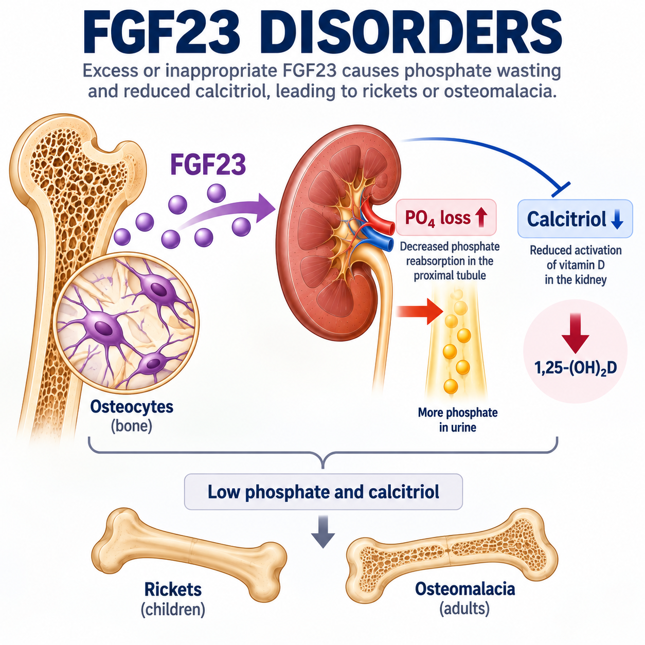
FGF23-mediated disease should not be diagnosed merely because FGF23 is detectable or because serum phosphate is low. The key abnormality is inappropriate FGF23 activity in a patient whose kidneys are wasting phosphate despite hypophosphatemia.
What Is FGF23?
Fibroblast growth factor 23 (FGF23) is a bone-derived hormone that plays a major role in phosphate and vitamin D homeostasis.
It is produced predominantly by cells of the osteoblast and osteocyte lineage. Its major physiological target is the kidney, where it regulates phosphate handling and vitamin D metabolism.
The student should think of FGF23 as a hormone that tells the kidney to waste phosphate and to reduce calcitriol physiology.
What Does FGF23 Do?
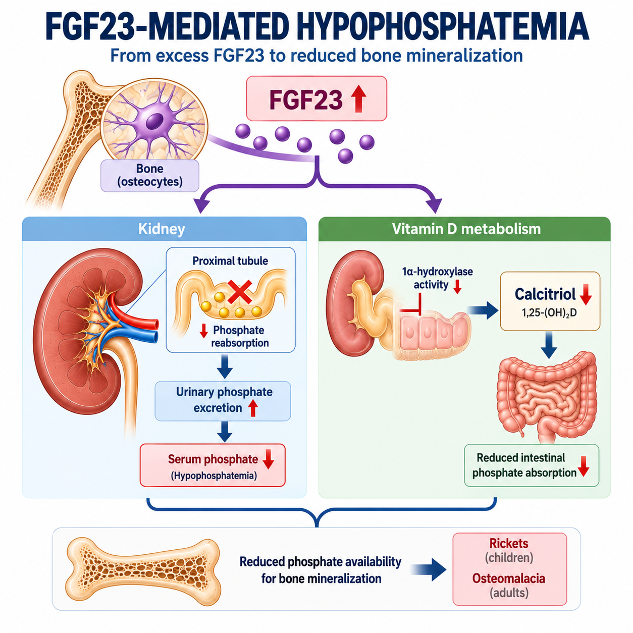
FGF23 is a bone-derived hormone that reduces renal phosphate reabsorption and suppresses calcitriol physiology. Excessive FGF23 activity therefore causes renal phosphate wasting, hypophosphatemia and impaired skeletal mineralization.
FGF23 also lowers active vitamin D physiology. It suppresses renal 1-alpha hydroxylase activity and promotes pathways that reduce calcitriol availability. The clinical consequence is that calcitriol may be low or inappropriately normal when the body should be increasing calcitriol in response to hypophosphatemia.
How FGF23 Causes Renal Phosphate Wasting
In hypophosphatemia, the normal kidney should conserve phosphate. Renal phosphate wasting means the opposite is happening: the kidney continues to lose phosphate inappropriately.
FGF23 causes renal phosphate wasting by reducing proximal tubular phosphate reabsorption. The biochemical pattern is therefore low serum phosphate with reduced renal phosphate reabsorptive capacity, often assessed using TmP/GFR.
For the calculation framework, review how TRP, FEPO₄ and TmP/GFR establish inappropriate renal phosphate loss.
How FGF23 Affects Calcitriol
Phosphate deficiency would normally support mechanisms that increase calcitriol availability, helping intestinal mineral absorption and skeletal mineralization.
Excessive FGF23 activity disrupts that response. Instead of an appropriate calcitriol rise, the patient may have low or inappropriately normal 1,25-(OH)₂D for the degree of hypophosphatemia.
This is why FGF23-mediated hypophosphatemia is not just a kidney phosphate problem. It is also a vitamin D physiology problem. See how calcitriol is produced and regulated.
What Does Inappropriately Normal FGF23 Mean?
During hypophosphatemia, FGF23 would normally be expected to decrease. Therefore, a measured FGF23 value that is not frankly elevated may still be abnormal if it is not suppressed for the low phosphate state.
Global tumor-induced osteomalacia guidance recognizes increased or inappropriately normal FGF23 as compatible with TIO in the correct biochemical context.
What Is FGF23-Mediated Hypophosphatemia?
FGF23-mediated hypophosphatemia occurs when excessive or inappropriately non-suppressed FGF23 causes renal phosphate wasting despite low serum phosphate, often with a low or inappropriately normal calcitriol response.
The diagnosis depends on physiology, not on a universal FGF23 cutoff. Use age-, sex-, laboratory- and assay-appropriate reference ranges where relevant, and interpret FGF23 in context.
Why FGF23 Excess Causes Rickets and Osteomalacia
Persistent FGF23 excess causes renal phosphate loss and abnormal calcitriol physiology. The result is inadequate mineral availability for normal bone mineralization.
In children, defective mineralization of growing bone can produce rickets. In adults, defective mineralization of osteoid produces osteomalacia. For the skeletal endpoint, see how chronic phosphate depletion causes defective bone mineralization.
What Disorders Cause FGF23 Excess?
FGF23-mediated disorders can be inherited or acquired. The key inherited prototype is X-linked hypophosphatemia. The key acquired prototype is tumor-induced osteomalacia.
| Group | Core Mechanism | Typical Clinical Direction |
|---|---|---|
| Inherited FGF23 disorders | Genetic disorders that increase FGF23 activity or impair phosphate conservation. | Often present during growth, but recognition may occur later. |
| Tumor-induced osteomalacia | Acquired FGF23 production by a usually small tumor. | Often later onset with new bone pain, weakness, fractures and osteomalacia. |
| CKD-related FGF23 elevation | FGF23 may rise in chronic kidney disease physiology. | Do not interpret FGF23 as TIO/XLH without renal and biochemical context. |
For the broader phosphate framework, review normal regulation of phosphate by the kidney, PTH and FGF23.
What Is X-Linked Hypophosphatemia?
X-linked hypophosphatemia is an inherited PHEX-related disorder characterized by excessive FGF23 activity, renal phosphate wasting, chronic hypophosphatemia and skeletal mineralization abnormalities.
Family history can be helpful, but absence of a known family history does not exclude inherited disease. Clinical history, biochemical pattern, skeletal features and genetic assessment may all contribute to diagnosis.
TODO: Link X-Linked Hypophosphatemia Explained after publication.
Clinical Features of XLH
XLH commonly manifests during growth because chronic phosphate wasting affects skeletal mineralization. Childhood rickets, longstanding skeletal abnormalities, dental problems, short stature or a family history of similar features should raise concern for inherited disease.
Recognition can still occur in adulthood, especially when childhood manifestations were missed or when persistent pain, enthesopathy, fractures or osteomalacia-like symptoms prompt reassessment.
How Is XLH Diagnosed?
XLH diagnosis begins with chronic hypophosphatemia and renal phosphate wasting. FGF23 is elevated or inappropriately non-suppressed for the low phosphate state, and PHEX-related inherited disease is considered from the clinical pattern.
Genetic confirmation can be important, but the article should not be turned into a genetic testing manual. Testing decisions depend on clinical context, available assays and specialist assessment.
Other Inherited FGF23 Disorders
XLH is the prototype, but it is not the only inherited FGF23-related hypophosphatemic disorder. Other inherited disorders can produce chronic renal phosphate wasting through FGF23 excess or related phosphate-regulatory mechanisms.
The practical student-level point is to recognize inherited clues: childhood onset, rickets, dental or skeletal history, family history where present, and a stable long-term phenotype rather than a new adult acquired syndrome.
What Is Tumor-Induced Osteomalacia?
Tumor-induced osteomalacia is a rare acquired disorder in which an FGF23-producing tumor causes renal phosphate wasting, chronic hypophosphatemia and defective bone mineralization.
The responsible tumor is often small and may be difficult to find. Many are phosphaturic mesenchymal tumors. The diagnostic sequence is therefore important: do not search for the tumor first.
Continue to Tumor-Induced Osteomalacia Explained for the acquired FGF23-mediated pathway, tumor localization sequence and treatment principles.
Clinical Features of TIO
TIO should be considered when an adult with previously normal skeletal development develops new bone pain, proximal weakness, fractures or pseudofractures with biochemical evidence of FGF23-mediated phosphate wasting.
The symptoms reflect the consequences of chronic hypophosphatemia and defective mineralization. They do not by themselves prove TIO; they must be interpreted with phosphate, renal phosphate handling, calcitriol physiology and FGF23 context.
Laboratory Findings in TIO
The typical biochemical framework is hypophosphatemia with inappropriate renal phosphate wasting, reduced TmP/GFR and increased or inappropriately normal FGF23. Calcitriol may be low or inappropriately normal for the degree of hypophosphatemia.
Do not invent or apply a universal phosphate cutoff, TmP/GFR threshold or FGF23 cutoff. Use age-, sex-, laboratory- and assay-appropriate reference ranges and specialist interpretation.
How Is TIO Diagnosed?
TIO is diagnosed by establishing an acquired FGF23-mediated renal phosphate-wasting syndrome and then identifying the causative lesion where possible.
- Confirm persistent hypophosphatemia.
- Establish renal phosphate wasting, preferably with TmP/GFR in the correct context.
- Assess whether the pattern is FGF23-mediated.
- Exclude competing explanations such as PTH-mediated phosphaturia or proximal tubular dysfunction.
- Proceed to tumor localization only after the biochemical diagnosis is established.
How Is the Tumor Located?
Do not search for a TIO tumor merely because phosphate is low. Tumor localization comes after biochemical evidence supports TIO.
Functional imaging is used to identify candidate lesions. Focused anatomical imaging then defines the lesion. Venous FGF23 sampling can have a role in selected situations, but it should not be presented as routine first-line testing.
TIO vs XLH
TIO and XLH both cause FGF23-mediated renal phosphate wasting. XLH is inherited and commonly manifests during growth, whereas TIO is acquired and usually presents later with new bone pain, weakness, fractures and osteomalacia.

| Feature | TIO | XLH |
|---|---|---|
| Nature | Acquired | Inherited |
| Driver | FGF23-producing tumor | PHEX-related excessive FGF23 activity |
| Usual timing | Later onset after previously normal skeletal development | Often manifests during growth |
| Clinical clues | New bone pain, proximal weakness, fractures, osteomalacia | Rickets, longstanding skeletal abnormalities, dental/skeletal history, possible family history |
| Localization | Tumor localization after biochemical diagnosis | No tumor search; inherited disease assessment |
FGF23 vs PTH-Mediated Phosphate Wasting
PTH is also phosphaturic. Increased PTH activity can reduce proximal tubular phosphate reabsorption and contribute to hypophosphatemia.
Calcium and PTH interpretation are therefore essential. Low phosphate with renal wasting should not automatically be called FGF23-mediated disease if the calcium-PTH pattern suggests primary or secondary hyperparathyroid physiology.
Review secondary hyperparathyroidism physiology when PTH-mediated phosphaturia is part of the differential.
FGF23 vs Fanconi Syndrome
FGF23-mediated disease mainly explains phosphate wasting and altered calcitriol physiology. Fanconi-type proximal tubular dysfunction causes a broader proximal tubular reabsorptive problem.
If phosphate wasting is accompanied by inappropriate urinary losses of other solutes normally reclaimed by the proximal tubule, the diagnostic direction moves toward proximal tubular disease rather than isolated FGF23 excess.
When Should FGF23 Be Measured?
FGF23 should not be the first test in every patient with low phosphate. It is most useful after hypophosphatemia has been confirmed and renal phosphate wasting has been demonstrated.
The reasoning sequence is:
Understanding FGF23 Assays
FGF23 assays are not interchangeable. Interpretation depends on whether intact FGF23 or C-terminal FGF23 is measured, as well as laboratory method and clinical context.
Global TIO guidance gives preference to intact FGF23 when available. Reference intervals should be laboratory- and assay-appropriate, and renal function should be considered carefully.
Do not attach a universal meaning to a single FGF23 number. Interpret the result against the phosphate state, renal phosphate handling, renal function and assay type.
CKD Warning
FGF23 may be elevated in chronic kidney disease physiology. Therefore, elevated FGF23 in a patient with CKD does not automatically mean TIO or XLH.
Interpret phosphate, kidney function, PTH, vitamin D physiology and the broader CKD-mineral bone disorder context together. See CKD-MBD Explained.
Treatment of FGF23 Disorders
Treatment depends on the cause, severity, age, skeletal disease, renal context and specialist assessment. This article explains principles, not dosing schedules.
Broad treatment concepts include correcting phosphate and calcitriol physiology where appropriate, treating the underlying lesion in TIO when feasible, and considering targeted FGF23 pathway therapy in selected FGF23-mediated disorders.
No phosphate dose, calcitriol dose, burosumab dose, pediatric schedule or treatment eligibility rule should be inferred from this article.
Treatment of TIO
When the causative tumor is localized and complete resection is feasible, complete tumor resection is the definitive treatment principle for TIO.
If curative resection is not possible or the tumor cannot be found, management becomes specialist-directed and may include medical approaches to manage phosphate and mineralization physiology. The details belong in a dedicated TIO treatment protocol, not here.
Treatment of XLH
XLH management is specialist-led and depends on age, symptoms, skeletal disease, dental disease, renal context and treatment availability. Conventional therapy and targeted FGF23 pathway therapy may be considered in appropriate settings.
This article should not be used to decide treatment eligibility, dosing or monitoring intervals.
What Is Burosumab?
Burosumab is an antibody directed against FGF23 activity. Conceptually, it reduces excessive FGF23 signaling, improving renal phosphate handling and calcitriol physiology in appropriate FGF23-mediated disorders.
It is not a general treatment for every low phosphate result. It belongs only in carefully selected FGF23-mediated disease contexts under specialist guidance.
Diagnostic Algorithm
- Confirm true hypophosphatemia using appropriate reference ranges.
- Establish whether renal phosphate wasting is present using paired serum and urine studies and TmP/GFR where appropriate.
- If there is no renal wasting, look outside the FGF23 pathway.
- If renal wasting is present, interpret FGF23, PTH, calcium, calcitriol and proximal tubular markers together.
- If FGF23 is elevated or inappropriately non-suppressed, classify inherited versus acquired disease.
- If TIO is suspected biochemically, proceed to tumor localization in the correct sequence.
Worked Clinical Cases
Case 1: Adult with new bone pain and low TmP/GFR
An adult with new bone pain, proximal weakness, fractures and renal phosphate wasting needs FGF23-mediated disease considered. If FGF23 is elevated or inappropriately non-suppressed and competing explanations are excluded, TIO becomes an important possibility. Tumor localization follows the biochemical diagnosis.
Case 2: Childhood rickets and lifelong phosphate wasting
Childhood skeletal disease with chronic renal phosphate wasting suggests inherited hypophosphatemia. XLH is the prototype, especially when PHEX-related disease is supported by clinical or genetic assessment.
Case 3: Low phosphate with high PTH pattern
Renal phosphate wasting with a calcium-PTH pattern pointing to hyperparathyroid physiology should not be mislabeled as FGF23-mediated disease without careful interpretation.
Case 4: Phosphate wasting plus other proximal tubular losses
When phosphate wasting occurs with other inappropriate proximal tubular losses, Fanconi-type physiology becomes more likely than isolated FGF23 excess.
Common Mistakes
- Diagnosing FGF23 disease just because phosphate is low.
- Ordering FGF23 before proving renal phosphate wasting.
- Assuming FGF23 must be frankly elevated.
- Missing that normal FGF23 may be inappropriate during hypophosphatemia.
- Searching for a TIO tumor before biochemical disease is established.
- Forgetting that PTH is also phosphaturic.
- Missing Fanconi-type proximal tubular dysfunction.
- Interpreting FGF23 without knowing the assay type.
- Using CKD-associated FGF23 elevation as proof of TIO or XLH.
- Inferring treatment doses from physiology articles.
FGF23 Disorders in One Minute
Low phosphate should make the kidney conserve phosphate and should normally suppress FGF23. If hypophosphatemia is accompanied by renal phosphate wasting, use TmP/GFR and the clinical context to decide whether the kidney is behaving inappropriately.
FGF23-mediated disease means excessive or inappropriately non-suppressed FGF23 activity causing renal phosphate wasting and impaired calcitriol physiology. Inherited disease points toward XLH and related disorders. Acquired adult-onset disease with osteomalacia features raises concern for TIO.
The memory rule is: low PO₄, prove renal wasting, interpret FGF23, identify the cause.
Frequently Asked Questions
Key Take-Home Messages
- FGF23 is a bone-derived hormone that acts mainly on the kidney.
- FGF23 reduces renal phosphate reabsorption and suppresses calcitriol physiology.
- FGF23-mediated disease requires inappropriate FGF23 activity in the context of renal phosphate wasting.
- Normal-range FGF23 may be abnormal if it is not suppressed during hypophosphatemia.
- XLH is the inherited PHEX-related prototype.
- TIO is the acquired tumor-related prototype.
- Do not search for a TIO tumor before establishing the biochemical disease.
- PTH-mediated and Fanconi-type phosphate wasting must be separated from FGF23-mediated disease.
- No universal FGF23 cutoff, phosphate cutoff or treatment dose belongs in this reasoning article.
This article is intended for medical education only. It explains FGF23 physiology and diagnostic reasoning, not patient-specific medical advice, treatment eligibility, medication dosing, imaging schedules or genetic testing instructions.