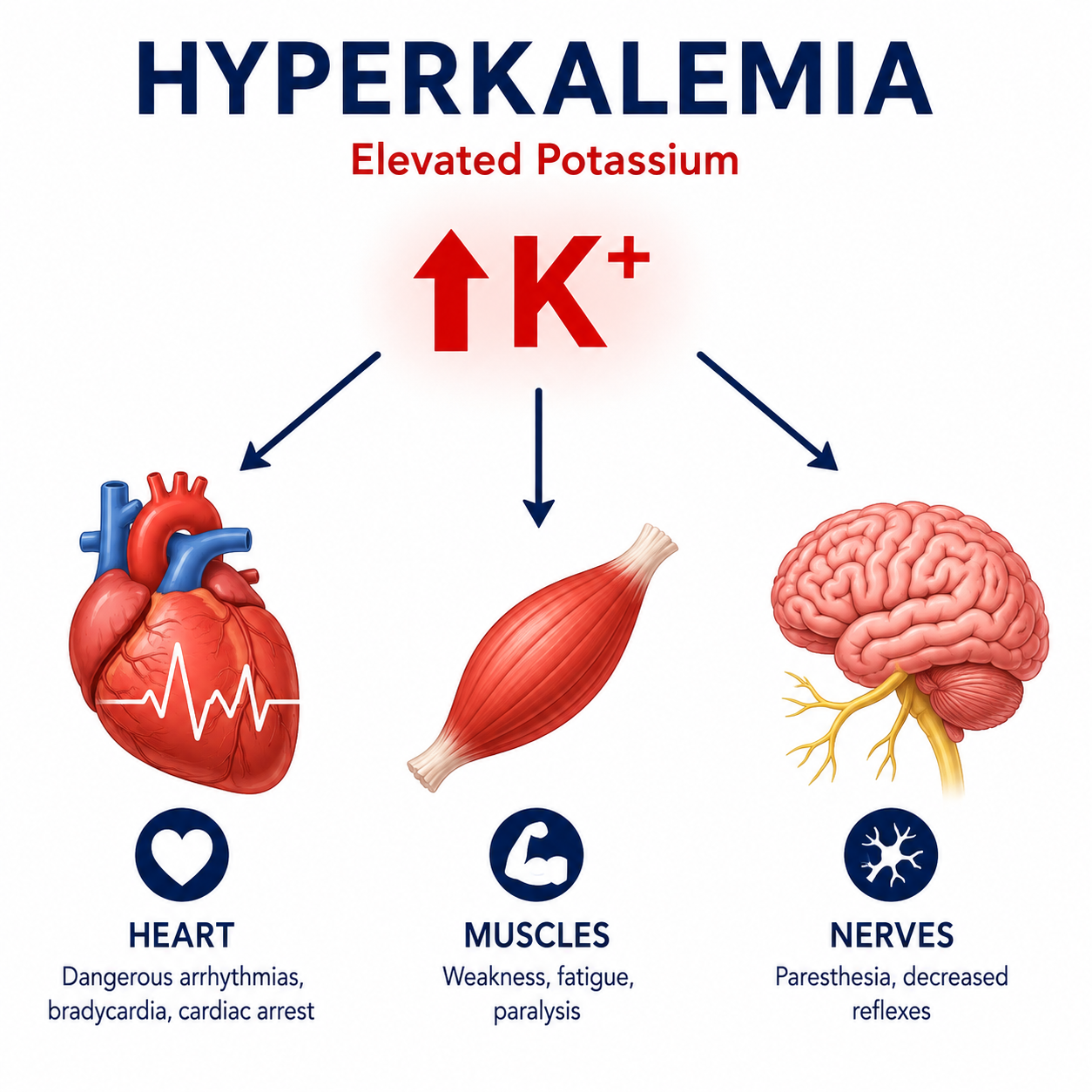
Hyperkalemia is one of the most important electrolyte disorders in emergency and inpatient medicine because elevated extracellular potassium can rapidly produce life-threatening cardiac conduction abnormalities.
This article follows Electrolytes Explained, Potassium Homeostasis Explained and Hypokalemia Explained. It also connects with Acid-Base Disorders Explained, Metabolic Acidosis Explained and ABG Interpretation Explained.
Hyperkalemia does not always mean total body potassium is increased. Some patients accumulate potassium because the kidneys cannot excrete it, while others develop a high serum potassium because potassium shifts from cells into the extracellular fluid. Distinguishing these mechanisms is central to clinical reasoning.
Hyperkalemia can cause sudden cardiovascular collapse even before prominent symptoms develop. Significant or rapidly rising hyperkalemia should be assessed with urgency.
Learning Objectives
- Define hyperkalemia and classify severity
- Explain why serum potassium rises
- Distinguish impaired renal excretion, redistribution, cell destruction and increased intake
- Recognise important symptoms and ECG changes
- Understand pseudohyperkalemia and when to repeat testing
- Apply a structured diagnostic and treatment-principles framework
Definition
Hyperkalemia refers to an elevated serum potassium concentration above the normal laboratory reference range.
Because serum potassium represents only a small fraction of total body potassium, interpretation must consider the clinical context, kidney function, acid-base status and the possibility of laboratory artefact.
Classification
Hyperkalemia is commonly classified by serum potassium concentration. Severity should never be judged by the number alone.
| Severity | Serum Potassium | Clinical Significance |
|---|---|---|
| Mild | 5.5-5.9 mmol/L | Often asymptomatic; assess cause and trend |
| Moderate | 6.0-6.4 mmol/L | Prompt clinical assessment and ECG are usually required |
| Severe | >=6.5 mmol/L | High-risk; urgent treatment and monitoring may be needed |
Risk depends on the rate of rise, ECG abnormalities, kidney function, acid-base status, medications and underlying heart disease. A rapidly rising potassium may be more dangerous than a chronically elevated value.
Pathophysiology
Potassium is the principal intracellular cation. The extracellular fraction is small, but it determines resting membrane potential and cardiac electrical stability.
Early hyperkalemia may increase excitability, but persistent depolarisation impairs conduction. The myocardium is especially vulnerable, which explains why ECG assessment is central to clinical decision-making.
For the core physiology, review Potassium Homeostasis Explained.
Causes
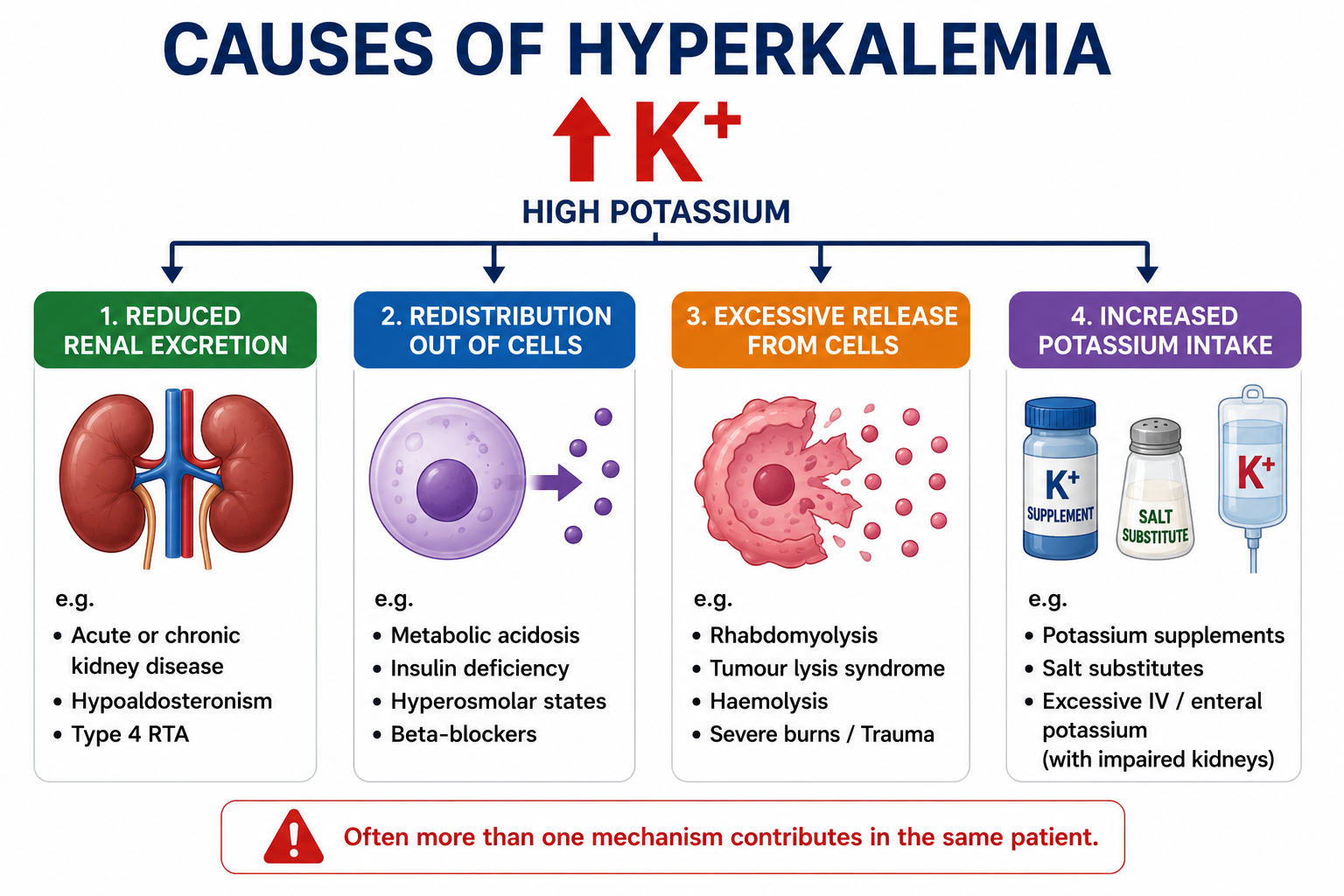
Most cases can be understood using four mechanisms: reduced renal potassium excretion, redistribution out of cells, excessive potassium release from damaged cells and increased potassium intake.
Reduced Renal Excretion
Reduced renal potassium excretion is the commonest cause of clinically significant hyperkalemia. It occurs in acute kidney injury, advanced chronic kidney disease, aldosterone deficiency, hyporeninemic hypoaldosteronism and type 4 renal tubular acidosis.
Risk is higher when kidney disease coexists with diabetes, heart failure or potassium-retaining medications.
Redistribution Out of Cells
Hyperkalemia may occur without increased total body potassium when potassium shifts from the intracellular compartment into the extracellular fluid. Important causes include mineral metabolic acidosis, insulin deficiency, hyperosmolality and beta-blockade.
In diabetic ketoacidosis, serum potassium may be normal or high initially, while total body potassium is usually depleted because osmotic diuresis causes urinary potassium loss.
Excessive Potassium Release
Large-scale cell injury releases intracellular potassium into the circulation. Examples include rhabdomyolysis, crush injury, major trauma, severe burns, tumour lysis syndrome and massive intravascular haemolysis.
Increased Potassium Intake
Dietary intake alone rarely causes hyperkalemia in patients with normal renal function. Intake becomes important with kidney impairment, potassium supplements, salt substitutes containing potassium, excessive intravenous potassium or parenteral nutrition.
Drug-Induced Hyperkalemia
Medication review is essential because many common drugs impair potassium excretion or potassium distribution.
- ACE inhibitors and ARBs reduce aldosterone activity.
- Spironolactone, eplerenone, amiloride and triamterene reduce distal potassium secretion.
- NSAIDs reduce renin release and may lower aldosterone.
- Trimethoprim acts like amiloride at the distal nephron.
- Calcineurin inhibitors such as tacrolimus and ciclosporin reduce renal potassium excretion.
- Heparin may suppress aldosterone synthesis with prolonged use.
- Non-selective beta-blockers reduce beta2-mediated cellular potassium uptake.
Pseudohyperkalemia
Pseudohyperkalemia is an artificial elevation in measured potassium caused by potassium release after blood collection. Recognising it prevents unnecessary and potentially harmful treatment.
| Cause | Mechanism |
|---|---|
| Haemolysed sample | Red cell potassium release during or after collection |
| Prolonged tourniquet or fist clenching | Local potassium shift and sample artefact |
| Delayed processing | Cellular potassium leak before analysis |
| Severe thrombocytosis or leukocytosis | Potassium release during clotting or processing |
Consider pseudohyperkalemia when the patient is well, the ECG is normal, kidney function is stable and the potassium result is unexpected. Repeat testing is usually appropriate if the patient is clinically stable.
Clinical Features
Many patients are asymptomatic until cardiac conduction becomes abnormal. Symptoms are more likely when potassium rises rapidly or reaches higher concentrations.
| System | Manifestations |
|---|---|
| General | Fatigue, malaise, weakness |
| Neuromuscular | Muscle weakness, difficulty walking, flaccid paralysis in severe cases |
| Cardiac | Palpitations, bradycardia, dizziness, syncope, cardiac arrest |
Syncope, bradycardia, hypotension, weakness with severe hyperkalemia, rapidly rising potassium or any ECG feature of hyperkalemia should prompt urgent clinical escalation.
ECG Changes
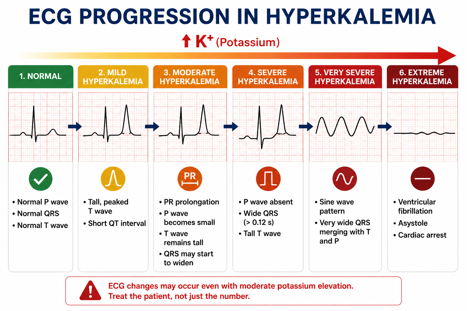
ECG changes may not appear in every patient, even with significant hyperkalemia. However, the ECG is a rapid way to assess cardiac toxicity and guide urgency.
- Tall, narrow, symmetrical peaked T waves
- Shortened QT interval early in the course
- PR prolongation
- P-wave flattening or disappearance
- QRS widening
- Sine-wave pattern
- Ventricular fibrillation or asystole
For a detailed comparison of peaked T waves, QRS widening, U waves and the limitations of ECG interpretation, see ECG Changes in Potassium Disorders.
Diagnostic Approach
A structured approach separates immediate danger from cause identification.
Laboratory Evaluation
Initial investigations usually include urea, creatinine, sodium, chloride, bicarbonate, glucose, magnesium, calcium and phosphate. Depending on the scenario, add venous or arterial blood gas, serum osmolality, creatine kinase, full blood count, cortisol, renin and aldosterone.
Clinical Scenarios
| Scenario | Likely Mechanism |
|---|---|
| Advanced CKD | Reduced renal potassium excretion |
| DKA | Redistribution despite total body depletion |
| Rhabdomyolysis | Potassium release from damaged muscle |
| ACE inhibitor plus spironolactone | Reduced aldosterone effect and reduced distal secretion |
| Unexpected isolated high potassium | Consider pseudohyperkalemia |
Treatment Principles
Hyperkalemia treatment aims to prevent fatal arrhythmias, lower serum potassium, remove potassium from the body and correct the underlying cause. Exact protocols vary by institution and are intentionally reserved for a dedicated potassium treatment article.
- Protect the myocardium when ECG changes or severe instability are present.
- Shift potassium into cells using therapies such as insulin with glucose and beta2 agonists when indicated.
- Remove potassium from the body through renal excretion, potassium binders or dialysis depending on context.
- Stop reversible contributors such as potassium supplements and potassium-retaining medications when appropriate.
- Monitor closely because rebound hyperkalemia and treatment-related hypoglycaemia can occur.
Significant ECG abnormalities, severe hyperkalemia, rapid potassium rise, symptomatic patients, severe AKI, arrhythmias or progressive weakness should be treated as urgent clinical problems.
Complications
Untreated hyperkalemia may cause bradycardia, complete heart block, ventricular tachycardia, ventricular fibrillation, asystole, sudden cardiac death, progressive weakness and flaccid paralysis. Persistent hyperkalemia may also limit the use of clinically beneficial medications such as ACE inhibitors, ARBs and mineralocorticoid receptor antagonists.
One Minute Revision
- Hyperkalemia is serum potassium above the laboratory reference range, often >5.0-5.5 mmol/L.
- Major mechanisms are reduced renal excretion, redistribution, cell destruction and increased intake.
- Common causes include AKI, CKD, ACE inhibitors, ARBs, spironolactone, DKA, rhabdomyolysis and tumour lysis syndrome.
- Always consider pseudohyperkalemia when the result is unexpected and the patient is well.
- ECG changes include peaked T waves, PR prolongation, P-wave loss, QRS widening and sine-wave pattern.
- Treatment principles are myocardium protection, intracellular shift, potassium removal, cause correction and monitoring.
Frequently Asked Questions
Conclusion
Hyperkalemia is dangerous because elevated extracellular potassium can destabilise cardiac conduction and progress rapidly to fatal arrhythmias. Most cases arise from impaired renal excretion, redistribution, tissue injury or medications rather than dietary excess alone. A systematic approach helps clinicians confirm true hyperkalemia, assess ECG risk, identify reversible causes and initiate appropriate treatment before deterioration occurs.
This article is intended for educational purposes. It should not replace clinical judgement, institutional protocols or specialist advice. Laboratory results should always be interpreted within the patient's clinical context.