Clinical Medicine • Electrolytes • Calcium and Magnesium
Hypercalcemia Explained: Causes, Symptoms, PTH-Based Diagnosis and Treatment
A clinical guide to genuine high calcium, symptoms, renal effects, ECG changes, PTH-dependent and PTH-independent causes, and treatment principles.
Dr. Seneth Gajasinghe, MBBS, MD Updated 27 Aug 2026 40 min read Reviewed educational content
Hypercalcemia becomes much easier to diagnose when the article is built around one question: is the hypercalcemia genuine, and is PTH appropriately suppressed?
Hypercalcemia is an abnormally elevated concentration of calcium in the circulation; elevation of ionized calcium is particularly important because ionized calcium is the biologically active fraction.
Calcium circulates as ionized calcium, protein-bound calcium and calcium complexed with circulating anions. Interpretation should not begin and end with a total serum calcium number. Once genuine hypercalcemia is established, the most useful next question is whether PTH is appropriately suppressed.
Hypercalcemia at a Glance
Confirm high calcium, then check PTH. If PTH is elevated or inappropriately normal, think PTH-dependent hypercalcemia. If PTH is suppressed, think PTH-independent hypercalcemia. Severe symptoms or dehydration require treatment while investigation continues.
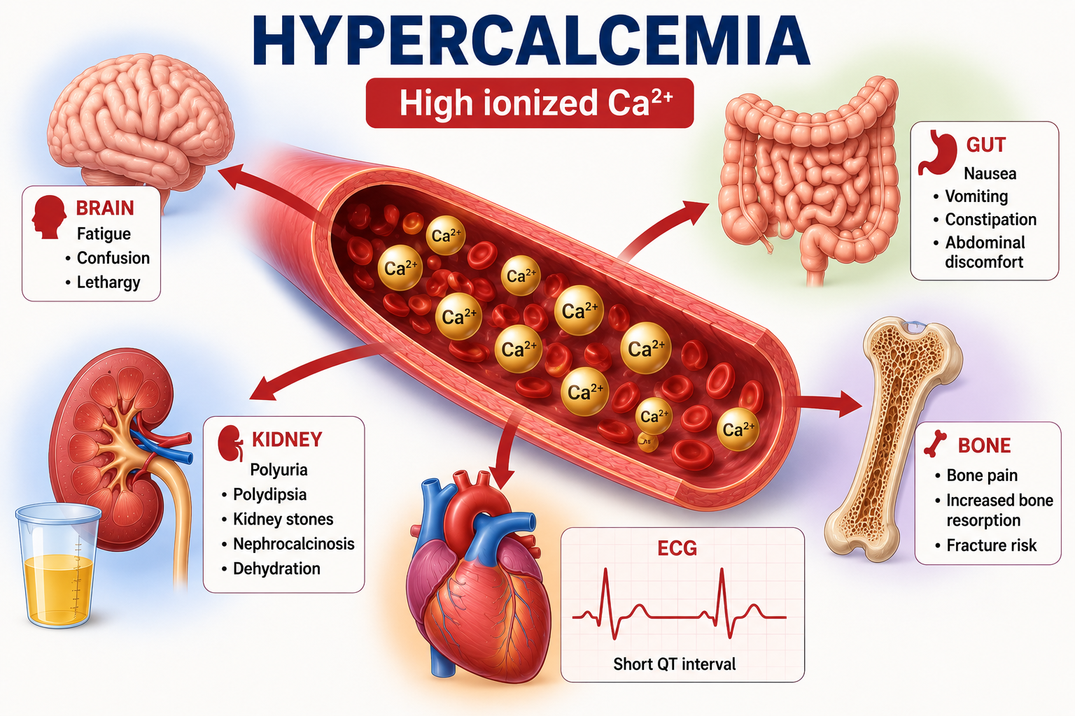
Figure 1. Hypercalcemia is a multisystem calcium disorder.
Learning Objectives
Define hypercalcemia and distinguish total from ionized calcium
Confirm that elevated calcium represents genuine hypercalcemia
Recognize symptoms, renal effects and ECG changes
Explain why hypercalcemia can cause polyuria and dehydration
Use PTH as the first major etiological branch
Recognize PTH-dependent and PTH-independent hypercalcemia
Understand primary hyperparathyroidism, tertiary hyperparathyroidism and FHH
Understand malignancy, vitamin D, granulomatous and medication-related mechanisms
Understand treatment principles without memorizing dosing protocols
Confirming Genuine Hypercalcemia
Total serum calcium includes ionized calcium, protein-bound calcium and complexed calcium. A significant proportion of protein-bound calcium is associated with albumin, so changes in protein concentration can influence total calcium without producing an equivalent change in ionized calcium.
An unexpected elevated total calcium should be interpreted with albumin, clinical context and ionized calcium when necessary. Dehydration and hemoconcentration can increase measured albumin and protein-bound calcium, so an isolated elevated total calcium result in a dehydrated patient may require reassessment after context is considered.
When total calcium interpretation is uncertain or accurate assessment of biologically active calcium is important, direct ionized calcium measurement can be useful. This is particularly relevant with albumin abnormalities, critical illness, acid-base disturbance or complex metabolic problems. For the physiology of calcium fractions and CaSR-PTH feedback, review Calcium Homeostasis Explained.
High total calcium↓Check albumin and hydration↓Repeat or confirm when appropriate↓Measure ionized calcium when needed↓Confirm genuine hypercalcemia
Symptoms of Hypercalcemia
Hypercalcemia can affect the kidneys, gastrointestinal tract, nervous system, skeletal muscle, cardiovascular system and bone. Symptoms become more likely as hypercalcemia becomes more severe or more acute.
Neurological: fatigue, lethargy, reduced concentration, weakness and confusion; severe cases may cause marked altered mental status, stupor or coma.
Gastrointestinal: anorexia, nausea, vomiting, constipation and abdominal discomfort.
Muscular: muscle weakness, contrasting with the increased neuromuscular excitability of hypocalcemia.
Renal: polyuria, polydipsia, dehydration, nephrolithiasis, nephrocalcinosis in selected chronic cases and deterioration in renal function.
The clinical effect depends not only on the absolute calcium concentration but also on speed of rise, duration, hydration, renal function, underlying cause and other metabolic abnormalities.
Why Hypercalcemia Causes Polyuria
Hypercalcemia can impair the kidney's concentrating ability and reduce responsiveness to vasopressin, producing a nephrogenic-DI-like concentrating defect that causes polyuria and water loss.
High calcium can interfere with renal responsiveness to vasopressin and with the medullary concentrating mechanism. This can resemble nephrogenic diabetes insipidus physiology, without implying that every patient with hypercalcemia has diabetes insipidus. The DI framework is explained in how nephrogenic diabetes insipidus impairs urine concentration.
Ca2+ rises↓Renal concentrating ability falls↓Polyuria↓Water loss↓Dehydration↓GFR may fall and renal calcium clearance may decline↓Hypercalcemia can worsen
This cycle is clinically important because volume depletion can amplify the calcium abnormality. Polyuria can also contribute to water deficit, a physiology discussed in Hypernatremia Explained.
Kidney and Bone Effects
Persistent hypercalcemia and/or hypercalciuria can increase the risk of renal stone formation, particularly in chronic disorders such as primary hyperparathyroidism.
Skeletal manifestations depend strongly on the cause. Conditions with excessive bone resorption can produce bone pain, reduced bone mineral density and fractures in more advanced disease. Not every patient with hypercalcemia has bone disease.
ECG Changes in Hypercalcemia
The classic ECG change in hypercalcemia is shortening of the QT interval, mainly due to shortening of the ST segment.
Increased extracellular calcium alters the ventricular action potential and shortens its plateau phase. Therefore hypercalcemia shortens ST duration and shortens the QT interval.
Severe calcium disturbances can affect cardiac conduction and rhythm, including bradyarrhythmias, conduction abnormalities and other rhythm disturbances, but not every patient with hypercalcemia develops an arrhythmia. This is the opposite classic ECG direction from hypocalcemia, which prolongs QT.
Figure 2. Hypercalcemia can affect kidney, brain, gut and ECG physiology.
How PTH Guides Diagnosis
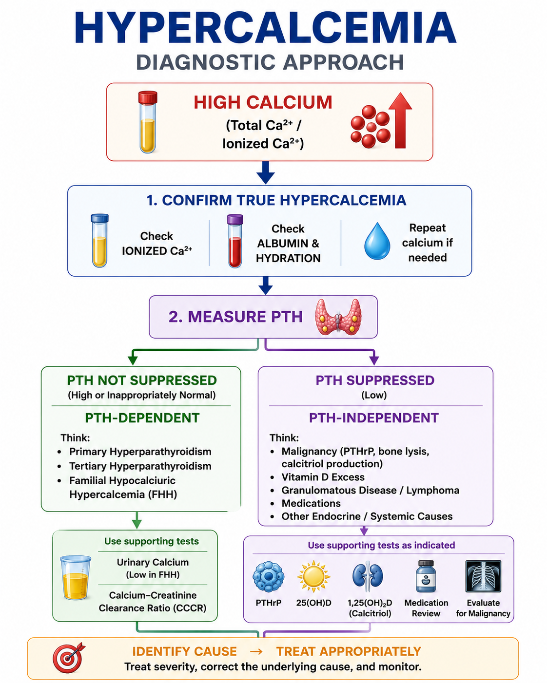
PTH should normally be suppressed when calcium is high. Elevated or inappropriately normal PTH suggests PTH-dependent hypercalcemia, while suppressed PTH suggests a non-PTH cause.
This is the central diagnostic concept. Normally, high calcium increases CaSR activation and suppresses PTH secretion. If PTH is within the reference range during confirmed hypercalcemia, it may still be inappropriately normal or inappropriately unsuppressed.
Hypercalcemia↓Check PTH↓PTH elevated or inappropriately normal: PTH-dependent↓PTH suppressed: PTH-independent
Figure 3. The PTH split is the backbone of hypercalcemia diagnosis.
PTH-Dependent Hypercalcemia
PTH-dependent hypercalcemia means calcium is high but PTH is elevated or inappropriately normal. Think particularly about primary hyperparathyroidism, tertiary hyperparathyroidism, familial hypocalciuric hypercalcemia and lithium-associated physiology where appropriate.
Then assess phosphate, renal function, urinary calcium, medication history, CKD history and family or lifelong calcium history where relevant.
Primary Hyperparathyroidism
Primary hyperparathyroidism is a major cause of hypercalcemia. The fundamental abnormality is autonomous or inappropriate PTH secretion despite elevated calcium.
The typical pattern is calcium high, PTH high or inappropriately normal and phosphate often low because PTH increases renal phosphate excretion. Even if PTH is not numerically above the laboratory reference interval, failure to suppress PTH is abnormal in hypercalcemia.
Excessive PTH can contribute to hypercalcemia through increased renal calcium conservation, increased calcitriol production, increased intestinal calcium availability indirectly and increased skeletal calcium mobilization through altered bone remodelling.
Tertiary Hyperparathyroidism
Long-standing secondary hyperparathyroidism, particularly in advanced chronic kidney disease, can eventually become autonomous. The parathyroid glands continue producing excessive PTH even when the original stimulus no longer adequately explains the secretion.
The patient may have markedly elevated PTH with hypercalcemia. Renal disease and previous long-standing secondary hyperparathyroidism provide important clinical context.
Familial Hypocalciuric Hypercalcemia
Familial hypocalciuric hypercalcemia, or FHH, is an important differential diagnosis when hypercalcemia occurs with PTH not suppressed. It is usually caused by altered calcium sensing, most commonly involving the calcium-sensing receptor pathway.
Because calcium sensing is altered, the body effectively behaves as though a higher calcium concentration is acceptable. The typical phenotype is mild hypercalcemia, PTH normal or mildly elevated/inappropriately normal, and relatively low urinary calcium excretion.
Primary hyperparathyroidism and FHH can both show high calcium with PTH not suppressed, but their management differs substantially. Parathyroid surgery does not correct the underlying calcium-sensing abnormality of FHH.
Urinary calcium assessment and the calcium-creatinine clearance ratio can help distinguish FHH from primary hyperparathyroidism. Very low CCCR supports FHH, whereas higher urinary calcium clearance supports primary hyperparathyroidism, but there is overlap. Kidney function, vitamin D status, calcium intake and medications such as thiazides can influence urinary calcium.
Important Limitation
CCCR is a diagnostic aid, not an absolute standalone test. Genetic assessment may be appropriate when the diagnosis remains uncertain.
PTH-Independent Hypercalcemia
If calcium is high and PTH is appropriately suppressed, the parathyroid glands are responding normally. The cause lies elsewhere.
Major categories include malignancy, vitamin D-related mechanisms, granulomatous disease, selected medications, immobilization in appropriate contexts and selected endocrine disorders. The next investigations should be selected according to clinical context, not ordered indiscriminately.
Malignancy-Associated Hypercalcemia
Malignancy is an important cause of clinically significant hypercalcemia, particularly when the calcium elevation is relatively acute, substantial or symptomatic. PTH should generally be suppressed.
The three important mechanisms are PTH-related peptide, osteolytic bone disease and increased calcitriol production in selected malignancies.
PTHrP
PTH-related peptide is produced by some tumors and can activate PTH receptors, causing hypercalcemia while the patient's endogenous PTH becomes suppressed.
PTHrP can produce PTH-like effects including increased renal calcium conservation, increased bone resorption and renal phosphate wasting. The pattern can include calcium high, PTH suppressed, PTHrP elevated and phosphate often reduced.
Osteolytic Hypercalcemia
Malignancy can also increase calcium through local or widespread bone destruction and resorption, releasing calcium from skeletal stores. This mechanism is particularly relevant in malignancies with substantial bone involvement.
Calcitriol-Producing Malignancy
Selected malignancies, particularly some lymphomas, can increase production of calcitriol. This increases intestinal calcium absorption and contributes to hypercalcemia. The expected PTH response is suppression.
Feature
Primary Hyperparathyroidism
Malignancy-Related Hypercalcemia
PTH
Elevated or inappropriately normal
Usually suppressed
Course
Often chronic or incidentally detected
Can be more acute or severe
PTHrP
Not the mechanism
May be elevated
Phosphate
Often low
Variable
Main mechanism
Excess PTH
PTHrP, bone resorption, calcitriol or other tumor effects
The PTH result is usually the most useful first discriminator.
Vitamin D and Hypercalcemia
Excess vitamin D activity can increase intestinal calcium absorption and contribute to hypercalcemia. This may occur through excessive vitamin D intake or inappropriate calcitriol production. PTH should generally become suppressed.
Excessive vitamin D exposure can increase calcium absorption sufficiently to cause hypercalcemia. Evaluation should consider 25-hydroxyvitamin D when vitamin D excess is suspected. Do not use calcitriol as a routine test for every patient with hypercalcemia.
Granulomatous Disease
Activated macrophages in some granulomatous diseases can express 1-alpha-hydroxylase and convert 25(OH)D to calcitriol. This production is less tightly regulated by the normal renal endocrine feedback system.
Granulomatous inflammation↓Extrarenal calcitriol production increases↓Intestinal calcium absorption increases↓Hypercalcemia↓PTH suppressed
This mechanism can occur in conditions such as sarcoidosis and selected infectious granulomatous diseases.
Medication-Related Hypercalcemia
Medication and supplement history should be reviewed in unexplained hypercalcemia. Relevant examples include thiazide diuretics, lithium, excessive calcium intake in appropriate contexts, excessive vitamin D exposure and vitamin A excess in selected situations.
Thiazide diuretics reduce urinary calcium excretion and can contribute to hypercalcemia or reveal previously unrecognized disorders such as primary hyperparathyroidism. Lithium can alter calcium-sensing/PTH physiology and may be associated with hyperparathyroid-type hypercalcemia.
Other Causes
Prolonged immobilization can increase bone resorption in selected patients, particularly when skeletal turnover is high. PTH should generally be suppressed.
Increased bone turnover in hyperthyroidism can occasionally produce hypercalcemia, but the calcium abnormality is usually not the dominant feature of the thyroid disorder. Adrenal insufficiency is a less common cause associated with hypercalcemia through multiple mechanisms and should be considered when supported by the wider clinical picture.
Diagnostic Approach to Hypercalcemia
Start with high total calcium. Ask whether the result is reproducible, what the albumin is, whether the patient is dehydrated and whether ionized calcium is needed. Do not start an extensive malignancy or endocrine work-up before establishing that the abnormality is real.
Immediately assess for significant dehydration, marked polyuria, vomiting, altered mental status, severe weakness, acute kidney injury, cardiac abnormalities or other features suggesting severe symptomatic hypercalcemia. Severe symptomatic disease requires treatment and investigation in parallel.
High calcium↓Confirm genuine hypercalcemia↓Assess severity↓Check PTH↓If PTH not suppressed: PTH-dependent pathway↓If PTH suppressed: PTH-independent pathway↓Use targeted tests and clinical context
Calcium
PTH
Additional Clue
Major Pattern
High
High or inappropriately normal
Phosphate often low
Primary hyperparathyroidism
High
Not suppressed
Urinary calcium relatively low
FHH
High
Markedly high
Long-standing CKD/secondary HPT
Tertiary hyperparathyroidism
High
Suppressed
PTHrP high
Humoral malignancy
High
Suppressed
Bone malignancy
Osteolytic hypercalcemia
High
Suppressed
25(OH)D markedly elevated
Vitamin D excess
High
Suppressed
Calcitriol high
Granulomatous disease or selected lymphoma
These are diagnostic patterns, not standalone diagnoses. Interpret them with clinical context, renal function, medications and appropriate confirmatory testing.
Treatment of Hypercalcemia
Management depends on calcium severity, symptoms, rate of rise, hydration, renal function and underlying mechanism. The major goals are to correct clinically important volume depletion, lower calcium when urgently necessary, reduce ongoing calcium release or absorption when appropriate, treat the underlying cause and monitor for treatment complications.
Mild, stable, asymptomatic hypercalcemia does not automatically require aggressive emergency calcium-lowering therapy. Management focuses on confirming the abnormality, establishing the cause, correcting reversible contributors, maintaining appropriate hydration and treating the underlying disease.
Significant symptomatic or severe hypercalcemia can require urgent treatment, particularly with neurological impairment, significant dehydration, renal dysfunction, major gastrointestinal symptoms or cardiac abnormalities.
Severe Symptomatic Hypercalcemia
IV Fluid
Many patients with significant hypercalcemia are volume depleted because of polyuria, reduced intake and sometimes vomiting. Volume depletion decreases renal calcium clearance and can worsen hypercalcemia. Restoring intravascular volume is an important initial treatment principle when volume depletion is present.
In appropriate patients, volume restoration is generally performed using isotonic saline. This restores extracellular volume, improves renal perfusion and increases renal calcium excretion. Fluid therapy must be individualized in patients with heart failure, significant kidney disease or other conditions limiting fluid tolerance.
Loop Diuretics
Routine loop diuretics should not be taught as standard calcium-lowering treatment. They may have a role when fluid overload requires management after appropriate volume restoration.
Calcitonin
Calcitonin can lower calcium relatively rapidly and can be useful while slower treatments begin working. Its major limitation is tachyphylaxis, so its calcium-lowering effect diminishes with repeated administration. Calcitonin is mainly short-term bridging therapy rather than definitive long-term treatment.
Antiresorptive Therapy
IV bisphosphonates inhibit osteoclast-mediated bone resorption and are particularly important in malignancy-associated hypercalcemia when increased bone resorption contributes significantly. Examples used clinically include zoledronic acid and pamidronate. They do not lower calcium as rapidly as calcitonin; their effect develops over days rather than hours, but lasts longer.
Denosumab inhibits RANKL-mediated osteoclast activity and can be useful in selected patients with hypercalcemia associated with malignancy, particularly when antiresorptive treatment is required, bisphosphonate therapy is inadequate or unsuitable, or renal impairment affects treatment choice.
Glucocorticoids and Dialysis
Glucocorticoids are not universal therapy for hypercalcemia. They are particularly useful in selected vitamin D or calcitriol-mediated hypercalcemia, such as granulomatous disease and selected lymphomas.
Dialysis may be considered in selected patients with severe refractory hypercalcemia, significant renal failure limiting calcium clearance, inability to tolerate fluid administration or insufficient response to other treatments. This is specialist-level emergency management.
Treat the Underlying Cause
Primary hyperparathyroidism may require parathyroid surgery when appropriate indications are met, but this article does not reproduce full surgical-indication guidance. Malignancy requires acute control of hypercalcemia when necessary and treatment of the underlying malignant process.
Vitamin D excess requires stopping inappropriate excess vitamin D exposure and managing the hypercalcemia according to severity, without instructing patients to discontinue medically prescribed therapy without clinical review. Granulomatous disease treatment targets the underlying inflammatory or infectious disorder and excessive calcitriol-mediated calcium absorption when present. Medication-related hypercalcemia requires review of contributing medications and supplements through appropriate clinical assessment.
Worked Clinical Cases
Case 1: Primary Hyperparathyroidism Pattern
A patient has repeatedly elevated calcium, upper-normal PTH and low phosphate. PTH is not truly normal, because during hypercalcemia PTH should be suppressed. This suggests PTH-dependent hypercalcemia, with primary hyperparathyroidism as an important possibility.
Case 2: Malignancy/PTHrP Pattern
A patient has relatively rapid symptomatic hypercalcemia, weight loss and known or suspected malignancy. Calcium is high, PTH is suppressed and PTHrP is elevated. This supports humoral hypercalcemia of malignancy.
Case 3: Calcitriol-Mediated Hypercalcemia
A patient has high calcium, suppressed PTH and elevated calcitriol with clinical evidence of granulomatous disease. Extrarenal calcitriol production can increase intestinal calcium absorption and cause hypercalcemia.
Case 4: FHH Pattern
A young adult has mild persistent hypercalcemia, no significant symptoms, similar calcium results documented previously, PTH not suppressed and relatively low urinary calcium. Consider familial hypocalciuric hypercalcemia.
Case 5: Severe Hypercalcemia With Dehydration
A patient presents with marked thirst, polyuria, vomiting, confusion and dehydration with significantly elevated calcium. Volume depletion is both a consequence and an amplifier of severe hypercalcemia.
Case 6: Tertiary Hyperparathyroidism
A patient with long-standing advanced CKD and prolonged secondary hyperparathyroidism develops high calcium with markedly elevated PTH. Consider tertiary hyperparathyroidism in the appropriate clinical context.
Case 7: Thiazide Exposure
A patient has mild hypercalcemia while taking a thiazide. The medication may contribute to reduced urinary calcium excretion or reveal previously unrecognized primary hyperparathyroidism.
Common Mistakes
Misconception: High total calcium always means high ionized calcium. Reality: interpret albumin and clinical context.
Misconception: Normal PTH excludes primary hyperparathyroidism. Reality: during hypercalcemia, PTH should be suppressed.
Misconception: High calcium should cause high PTH. Reality: normal physiology is Ca2+ rises and PTH falls.
Misconception: All hypercalcemia is caused by hyperparathyroidism. Reality: PTH-independent causes include malignancy, vitamin D mechanisms and other causes.
Misconception: Malignancy hypercalcemia causes high endogenous PTH. Reality: endogenous PTH should usually be suppressed.
Misconception: PTHrP and PTH are the same test. Reality: PTHrP is a distinct peptide.
Misconception: Low urine calcium proves FHH. Reality: urinary calcium and CCCR are aids with overlap and confounders.
Misconception: Every patient with hypercalcemia needs aggressive IV treatment. Reality: treatment intensity depends on severity, symptoms and mechanism.
Misconception: Loop diuretics are routinely required. Reality: routine forced diuresis is not the standard approach.
Misconception: Calcitonin is definitive long-term therapy. Reality: it works rapidly but is limited by tachyphylaxis.
Misconception: Bisphosphonates work immediately. Reality: they usually take longer to produce the major calcium-lowering effect.
Misconception: Hypercalcemia prolongs QT. Reality: the classic association is short QT.
One-Minute Revision
First confirm high calcium using albumin, hydration and ionized calcium when needed.
Once genuine hypercalcemia is confirmed, check PTH.
PTH high or not suppressed means PTH-dependent hypercalcemia.
PTH-dependent causes include primary hyperparathyroidism, FHH and tertiary hyperparathyroidism.
Suppressed PTH means PTH-independent hypercalcemia.
PTH-independent causes include malignancy, vitamin D/calcitriol mechanisms, granulomatous disease and medications.
Hypercalcemia can cause polyuria, dehydration, constipation, weakness, confusion, stones and short QT.
Severe disease requires severity-based treatment while the mechanism is investigated.
Calcitonin is fast but short-lived; antiresorptive therapy is slower but longer-lasting.
Key Clinical Pearls
Once genuine hypercalcemia is confirmed, ask whether PTH is suppressed. That single question separates most cases into PTH-dependent versus PTH-independent hypercalcemia.
Ionized calcium is the biologically active fraction.
Hypercalcemia can impair renal concentrating ability.
Dehydration can worsen hypercalcemia by reducing renal calcium clearance.
The classic ECG association is QT shortening.
A numerically normal PTH may be pathologically inappropriate in hypercalcemia.
Hypercalcemia is an abnormally elevated concentration of calcium in the circulation. Clinically, elevation of ionized calcium is particularly important because ionized calcium is the biologically active fraction.
What are the symptoms of hypercalcemia?
Hypercalcemia may cause fatigue, weakness, constipation, nausea, polyuria, polydipsia, dehydration, kidney stones and confusion. Severe cases can cause marked neurological, renal and cardiac abnormalities.
What are the main causes of hypercalcemia?
The most useful first division is PTH-dependent versus PTH-independent. PTH-dependent causes include primary and tertiary hyperparathyroidism and FHH. PTH-independent causes include malignancy, vitamin D-related disorders, granulomatous disease, selected medications and other systemic disorders.
What should PTH be in hypercalcemia?
PTH should normally be suppressed when calcium is elevated. Therefore a PTH concentration within the laboratory reference range may still be abnormal if it is not appropriately suppressed.
What does high calcium with high PTH suggest?
Confirmed hypercalcemia with elevated or inappropriately normal PTH indicates PTH-dependent hypercalcemia. Primary hyperparathyroidism is an important cause. FHH and tertiary hyperparathyroidism should also be considered in appropriate contexts.
What does high calcium with low PTH suggest?
High calcium with suppressed PTH indicates PTH-independent hypercalcemia. Potential causes include malignancy, vitamin D excess, calcitriol-mediated disease, medications and other systemic disorders.
Why does hypercalcemia cause polyuria?
Hypercalcemia can impair the kidney concentrating ability and reduce responsiveness to vasopressin, producing nephrogenic-DI-like physiology that causes polyuria and polydipsia.
What ECG change occurs in hypercalcemia?
The classic ECG association is shortening of the QT interval, primarily due to shortening of the ST segment.
What is PTHrP?
PTH-related peptide is produced by some tumors and can activate PTH receptors, causing increased calcium, phosphate wasting and increased bone resorption while endogenous PTH is suppressed.
What is FHH?
Familial hypocalciuric hypercalcemia is usually an inherited disorder of calcium sensing that causes lifelong mild hypercalcemia, nonsuppressed PTH and relatively low urinary calcium.
Why is urine calcium checked in hypercalcemia?
When calcium is high and PTH is not suppressed, urinary calcium can help distinguish primary hyperparathyroidism from familial hypocalciuric hypercalcemia, although overlap occurs.
How is severe hypercalcemia treated?
Management may include correction of volume depletion with isotonic fluid when appropriate, rapidly acting calcium-lowering therapy such as calcitonin in selected cases, longer-acting antiresorptive treatment and treatment directed at the underlying mechanism.
Why is calcitonin not used long term?
Calcitonin acts relatively quickly, but its effect decreases with repeated use because of tachyphylaxis. It is mainly useful as short-term therapy while slower treatments take effect.
Key Take-Home Messages
Hypercalcemia becomes much easier to diagnose when approached physiologically. First ask whether the hypercalcemia is genuine by considering total calcium, albumin, hydration and ionized calcium when necessary.
Then ask whether PTH is suppressed. High calcium with PTH high or inappropriately normal means PTH-dependent hypercalcemia. Think about primary hyperparathyroidism, FHH and tertiary hyperparathyroidism. High calcium with suppressed PTH means PTH-independent hypercalcemia. Consider malignancy, vitamin D or calcitriol mechanisms, granulomatous disease, medications and other context-specific causes.
Confirm↓Assess severity↓PTH↓Mechanism↓Treat
Treatment has two dimensions: control dangerous hypercalcemia and correct the mechanism causing it.
Medical Education Disclaimer
This article is intended for medical education only. It explains diagnostic and treatment principles, not patient-specific treatment. Severe hypercalcemia requires local protocols, monitoring and appropriately qualified clinical supervision.