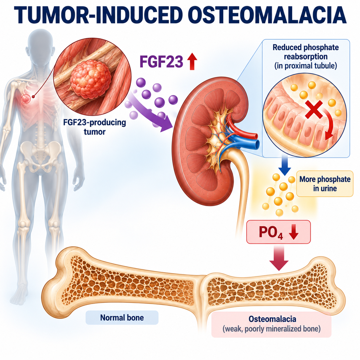
Tumor-induced osteomalacia, or TIO, is a rare acquired disorder in which a tumor produces excessive amounts of the phosphaturic hormone fibroblast growth factor 23 (FGF23).
The excess FGF23 causes the kidneys to waste phosphate and suppresses the expected calcitriol response. Persistent hypophosphatemia then reduces the phosphate available for normal skeletal mineralization, eventually producing osteomalacia.
The typical patient may present with progressive bone pain, proximal muscle weakness, difficulty walking, fatigue, insufficiency or fragility fractures, and declining physical function. Because these manifestations are nonspecific and because serum phosphate is not always measured routinely, the diagnosis can be delayed.
TIO at a Glance
The key diagnostic mistake is to begin searching for a tumor simply because serum phosphate is low. Biochemistry comes first; localization comes second.
What Is Tumor-Induced Osteomalacia?
Tumor-induced osteomalacia is a rare acquired paraneoplastic syndrome in which a tumor, most commonly a phosphaturic mesenchymal tumor, produces excessive FGF23, causing renal phosphate wasting, chronic hypophosphatemia and defective skeletal mineralization.
Do not describe TIO simply as a tumor causing low phosphate. The full pathophysiology includes endocrine FGF23 excess, renal phosphate wasting, altered calcitriol physiology and skeletal mineralization failure.
What Is a Phosphaturic Mesenchymal Tumor?
A phosphaturic mesenchymal tumor is the characteristic tumor underlying most cases of TIO. These tumors are often small, slow-growing lesions of bone or soft tissue that produce FGF23.
They can occur throughout the body. This explains an important clinical problem: the metabolic effects can be severe even when the tumor itself is tiny.
The patient's major problem may therefore be bone pain, weakness, fractures and hypophosphatemia rather than symptoms from the tumor mass itself.
Molecular abnormalities involving FN1-FGFR1 and FN1-FGF1 fusion genes have been identified in a substantial subset of phosphaturic mesenchymal tumors. This pathology detail does not replace the clinical biochemical diagnostic pathway.
How FGF23 Causes TIO
FGF23 acts on renal phosphate handling. Tumor-derived FGF23 reduces proximal tubular sodium-phosphate cotransporter activity, lowering renal phosphate reabsorption.
Therefore the low phosphate in TIO is caused principally by inappropriate renal phosphate wasting.
Why TIO Causes Renal Phosphate Wasting
TmP/GFR estimates renal tubular phosphate reabsorptive capacity relative to filtration. Excess FGF23 reduces this capacity.
Serum PO₄ low plus TmP/GFR low means that the kidney is failing to conserve phosphate appropriately despite hypophosphatemia.
This is a central clue to TIO, but it is not specific to TIO. Other causes include inherited FGF23 disorders, PTH-mediated phosphaturia, proximal tubular disorders, selected medications and other acquired conditions.
For the calculation framework, review Renal Phosphate Wasting Explained.
Why Calcitriol Is Low in TIO
FGF23 also alters vitamin D metabolism. It suppresses renal 1α-hydroxylase activity and promotes pathways involved in calcitriol catabolism.
Normally, hypophosphatemia should stimulate physiological mechanisms favouring increased calcitriol. In FGF23 excess, phosphate is low but calcitriol fails to rise appropriately.
For background, see Vitamin D Metabolism Explained.
How Hypophosphatemia Causes Osteomalacia
Persistent phosphate depletion reduces mineral availability for newly formed osteoid. Osteoid mineralization becomes impaired, producing osteomalacia.
FGF23 excess is the endocrine mechanism. Renal phosphate wasting is the renal consequence. Hypophosphatemia is the biochemical consequence. Osteomalacia is the skeletal consequence.
Review Osteomalacia Explained for the bone-mineralization endpoint.
What Are the Symptoms of TIO?
TIO often presents gradually. Important manifestations include diffuse bone pain, musculoskeletal pain, proximal muscle weakness, reduced exercise tolerance, difficulty rising or climbing stairs, gait disturbance, progressive difficulty walking, fatigue, fragility or insufficiency fractures, height loss or skeletal deformity in advanced disease.
These symptoms are not specific. They should trigger serum phosphate assessment when unexplained or progressive, especially if osteomalacia or recurrent fragility fractures are suspected.
Why Is TIO Often Missed?
TIO can be missed because symptoms are nonspecific, serum phosphate may not be included in routine laboratory panels, the tumor may be clinically silent, and the skeletal consequence can distract from the cause.
Bone pain and weakness may be attributed to degenerative disease, rheumatological conditions, neurological disorders, osteoporosis or nonspecific musculoskeletal pain.
A patient may be labelled as having osteoporosis because DXA shows low bone mineral density or fractures are present. But low BMD does not distinguish osteoporosis from osteomalacia.
When Should TIO Be Suspected?
A useful clinical trigger is an adult with otherwise unexplained progressive bone pain, proximal weakness, impaired walking, recurrent insufficiency or fragility fractures, or suspected osteomalacia, especially when serum phosphate is persistently low.
Then ask the decisive physiological question: is phosphate being lost through the kidney?
First Steps: Confirm Hypophosphatemia and Renal Loss
Do not diagnose TIO from a single unexplained phosphate result. Establish whether hypophosphatemia is genuine, whether it persists, the clinical context, renal function, nutritional context, medications, possible redistribution and other potential causes.
The normal response to hypophosphatemia is renal phosphate conservation. Continued renal phosphate loss despite low phosphate indicates renal phosphate wasting.
For formal investigation of chronic renal phosphate wasting, appropriately timed fasting serum and paired urine measurements are useful.
Laboratory Findings in TIO
In a patient with suspected TIO, the important biochemical evaluation includes serum phosphate, serum creatinine, urine phosphate, urine creatinine, TmP/GFR, total or bone-specific ALP, calcium, intact PTH, 25-OH D, 1,25-(OH)₂D when available, and FGF23 where clinically available and appropriate.
| Test | Characteristic Pattern |
|---|---|
| Serum phosphate | Low |
| TmP/GFR | Low |
| Calcium | Normal or slightly low |
| 25-OH D | Normal or low |
| 1,25-(OH)₂D | Low or inappropriately normal |
| ALP | High |
| PTH | Normal or high |
| FGF23 | High or inappropriately normal |
This is a characteristic pattern rather than a requirement that every patient must show every abnormality. Coexisting conditions such as nutritional vitamin D deficiency can modify the biochemical picture.
ALP and Calcium in TIO
Active osteomalacia increases osteoblastic activity associated with defective mineralization, so ALP may rise substantially. ALP elevation is not specific for TIO, and total ALP can come from other tissues, especially liver. Bone-specific ALP can sometimes help when source clarification is needed.
Do not teach that TIO must cause hypocalcemia. Calcium is often normal or sometimes slightly reduced. Normal serum calcium does not exclude severe phosphate-depletion osteomalacia.
When Should FGF23 Be Measured?
Do not start every hypophosphatemia investigation with FGF23. The logical sequence is low phosphate, renal phosphate wasting demonstrated, calcitriol low or inappropriately normal, and then FGF23 measurement where clinically available.
Intact FGF23 is preferred where available. FGF23 assays are not interchangeable, and interpretation depends on assay characteristics, renal function, medications, phosphate intake and physiological context.
During hypophosphatemia, FGF23 should be suppressed. A value within the general laboratory reference interval can still be inappropriately normal or inappropriately non-suppressed.
TIO Is a Diagnosis of Physiological Context
A patient does not have TIO merely because phosphate is low, FGF23 is detectable, osteomalacia is present or a mass is seen on imaging.
Differential Diagnosis of TIO
The main alternatives should be taught by mechanism: inherited FGF23-mediated disorders, PTH-mediated phosphate wasting, proximal tubular dysfunction, nonrenal hypophosphatemia, selected medications and other acquired renal tubular disorders.
Do not create an enormous rare-disease list. The practical task is to avoid calling every phosphate-wasting syndrome TIO.
TIO vs XLH
| Feature | TIO | XLH |
|---|---|---|
| Nature | Acquired | Inherited |
| Typical recognition | Adult | Often childhood |
| Previous childhood skeletal development | Usually normal | May show rickets or deformity |
| Family history | Usually absent | May be present |
| PO₄ | Low | Low |
| Renal phosphate wasting | Yes | Yes |
| FGF23-mediated | Yes | Yes |
| TmP/GFR | Low | Low |
| Tumor localization | Required when TIO suspected | Not part of routine XLH diagnosis |
| Genetic testing | Used when hereditary disease needs exclusion | Central diagnostic role |
| Definitive mechanism | FGF23-producing tumor | PHEX-related FGF23 dysregulation |
Age and family history are strong clues but not absolute rules. TIO can rarely occur in younger patients, and inherited disease may remain undiagnosed until adulthood.
Review FGF23 Disorders Explained for the broader inherited-versus-acquired framework, and X-Linked Hypophosphatemia Explained for the inherited PHEX-related branch.
TIO vs Hyperparathyroidism, Fanconi Syndrome and Vitamin D Deficiency
PTH-mediated phosphaturia
Both FGF23 and PTH are phosphaturic. Renal phosphate wasting does not prove FGF23 excess. Low PO₄ with low TmP/GFR, high calcium and inappropriately high PTH should make PTH-mediated phosphaturia more important.
Fanconi syndrome
Generalized proximal tubular dysfunction can also cause renal phosphate wasting. Look for inappropriate loss of other proximal tubular solutes, such as glucose in the appropriate serum-glucose context, bicarbonate, uric acid, amino acids or low-molecular-weight proteins.
Vitamin D deficiency
Vitamin D deficiency can produce osteomalacia, but its mechanism differs. The primary abnormality is low 25-OH D, often leading to secondary hyperparathyroidism. TIO is driven by FGF23 excess causing direct renal phosphate wasting and suppression of calcitriol physiology.
Low 25-OH D does not exclude TIO. Vitamin D insufficiency or deficiency is common and can coexist with TIO.
Physical Examination
Do not treat TIO as a laboratory-only diagnosis. Examination should assess gait, proximal muscle function, skeletal deformity, height and body proportions where relevant, areas of bone tenderness, dental or craniofacial clues that might suggest inherited disease, and palpable masses.
When Should Tumor Localization Begin?
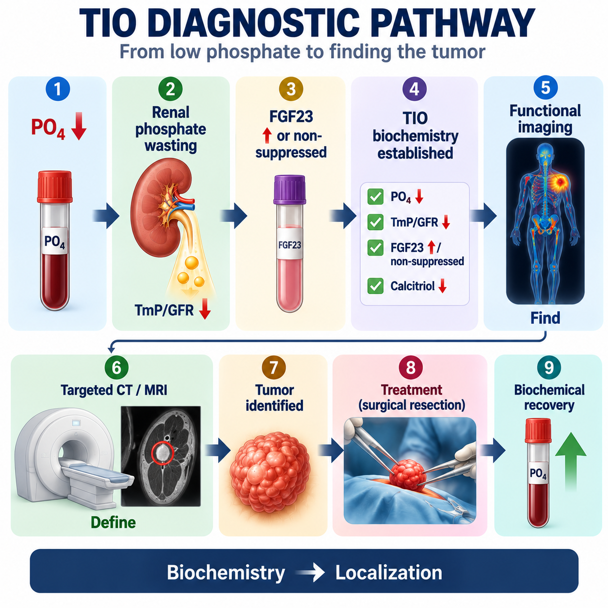
First establish hypophosphatemia, renal phosphate wasting, FGF23-mediated physiology and a clinical or biochemical diagnosis of TIO. Only then should the pathway move to tumor localization.
This sequence avoids chasing incidental lesions unrelated to the biochemical disease.
Functional Imaging for TIO
Once TIO is biochemically established, functional imaging comes first. The reason is that phosphaturic mesenchymal tumors commonly express somatostatin receptors.
Somatostatin-receptor PET/CT using suitable DOTA-based tracers is preferred where available. Where unavailable, specialist alternatives can include somatostatin-receptor scintigraphy or FDG-PET in selected circumstances.
The student-level concept is simple: functional imaging asks where biologically suspicious tumor activity is located.
CT and MRI in TIO
Once functional imaging identifies a candidate site, targeted MRI or CT is used to characterize the lesion anatomically.
These modalities help determine precise tumor location, size, bone versus soft-tissue involvement, relation to surrounding structures and the surgical approach.

What Is Venous FGF23 Sampling?
Venous FGF23 sampling is a specialist localization technique in which FGF23 concentrations from selected venous territories are compared to help determine whether a suspected lesion is the source of excess FGF23.
It can be useful particularly when two or more plausible lesions exist and the clinician needs to determine which lesion is causative. It is not a routine first-line localization test and is less useful when imaging has not identified a suspicious candidate lesion.
Should the Tumor Be Biopsied First?
Do not teach routine biopsy as the first confirmation step. Phosphaturic mesenchymal tumors require careful multidisciplinary planning because definitive treatment depends on complete excision, and unnecessary or poorly planned procedures can complicate subsequent management.
Biopsy decisions belong to the specialist multidisciplinary localization and surgical pathway. This article should not be used to create a universal biopsy rule.
Diagnostic Algorithm
- Measure serum phosphate in unexplained bone pain, weakness, fractures or suspected osteomalacia.
- If phosphate is low, confirm persistent hypophosphatemia and assess renal context.
- Use paired serum and urine phosphate and creatinine to assess renal phosphate handling and TmP/GFR.
- If the kidney is conserving appropriately, investigate nonrenal hypophosphatemia.
- If renal phosphate wasting is present, assess calcium, PTH, ALP, 25-OH D, 1,25-(OH)₂D and proximal tubular function.
- If FGF23 physiology is suspected, measure FGF23 where available and interpret it in context.
- If the pattern is FGF23-mediated, decide whether inherited disease or acquired TIO best fits.
- After TIO is biochemically established, use functional imaging followed by targeted MRI or CT.
- If competing lesions remain, venous FGF23 sampling may help in selected specialist cases.
- Proceed through specialist surgical planning and complete resection when feasible.
How Is TIO Treated?
The fundamental treatment principle is to remove the source of excess FGF23 when possible. Complete tumor resection is the preferred definitive treatment when feasible.
This distinguishes TIO from inherited FGF23 disorders such as XLH, where there is no single tumor source to remove.
Surgical Treatment
Complete resection with appropriate margins matters because incomplete removal can leave an active source of FGF23. Persistent or recurrent biochemical abnormalities after surgery should prompt evaluation for residual disease, recurrence, incorrect lesion attribution or another explanation for the biochemical phenotype.
The biochemical abnormality can improve faster than the skeletal consequences. Bone healing and functional recovery may take time, so persistent musculoskeletal symptoms need to be interpreted differently from persistent biochemical disease.
What If the Tumor Cannot Be Found or Resected?
Some patients have no identifiable tumor despite appropriate localization, an anatomically unresectable lesion, unacceptable surgical risk, or persistent/recurrent disease. These patients require medical management of the phosphate-wasting disorder under specialist supervision.
Conventional medical therapy aims to compensate for phosphate loss and abnormal active-vitamin-D physiology. It may use oral phosphate and active vitamin D under specialist supervision. Do not infer a universal dose from this article.
What Is the Role of Burosumab?
Burosumab is a monoclonal antibody directed against FGF23.
Do not write that every patient with TIO should receive burosumab. If the responsible tumor can be completely resected, surgery remains definitive treatment. Burosumab is relevant particularly when the tumor cannot be identified or cannot be completely resected, within appropriate regulatory and specialist treatment contexts.
Follow-Up After Treatment
Follow-up depends on whether the patient has successful resection, persistent disease, unresectable disease, an unidentified tumor, conventional medical therapy or FGF23-directed treatment.
Important follow-up domains can include serum phosphate, calcium, creatinine and renal function, PTH, ALP, urine calcium where appropriate, skeletal recovery, pain, mobility and function, and recurrence of biochemical phosphate wasting.
Do not present one imaging or monitoring interval as universally applicable to every TIO patient.
Recurrent or Persistent TIO
If the biochemical disorder persists or returns after treatment, consider incomplete resection, recurrence, another active lesion, malignant or metastatic disease in rare cases, incorrect attribution of the original lesion, or an alternative diagnosis.
Do not assume recurrent bone pain alone proves recurrent TIO. Re-establish the biochemical phenotype.
Worked Clinical Cases
Case 1: Low phosphate but no renal wasting
A patient has weakness and low serum phosphate, but renal phosphate handling demonstrates strong conservation. TIO is not supported by the available evidence. Investigate nonrenal mechanisms such as reduced intake, malabsorption or redistribution.
Case 2: Renal phosphate wasting
Persistent low phosphate with low TmP/GFR and high ALP first establishes renal phosphate wasting, not TIO. The mechanism still needs to be identified.
Case 3: FGF23-mediated pattern
Low phosphate, low TmP/GFR, calcium not pointing to PTH-mediated disease, low-normal calcitriol and FGF23 within the laboratory reference interval can still support FGF23-mediated disease because FGF23 should be suppressed during hypophosphatemia.
Case 4: TIO versus XLH
A previously healthy adult with recent progressive bone pain, proximal weakness, insufficiency fractures and FGF23-mediated renal phosphate wasting should raise concern for TIO. Childhood rickets, lifelong phosphate wasting or family history would shift attention toward XLH or inherited disease.
Case 5: Wrong imaging sequence
A whole-body tumor scan after one low phosphate result reverses the diagnostic pathway. Confirm hypophosphatemia, prove renal wasting, establish FGF23 physiology and then localize the tumor.
Case 6: Two candidate lesions
When established TIO has two plausible imaging lesions, selective or regional venous FGF23 sampling may help distinguish the source in selected specialist cases.
Case 7: Successful resection
After complete resection, renal phosphate wasting improves and serum phosphate normalizes. Biochemical recovery supports that the removed lesion was the pathological FGF23 source.
Case 8: Low 25-OH D
Low 25-OH D does not exclude TIO. Vitamin D deficiency is common and can coexist with an FGF23-mediated renal phosphate-wasting disorder.
Common Diagnostic Mistakes
- Assuming low phosphate equals TIO.
- Assuming osteomalacia equals TIO.
- Assuming renal phosphate wasting equals TIO.
- Assuming FGF23 must be above the reference range.
- Assuming normal calcium excludes severe TIO.
- Assuming low 25-OH D excludes TIO.
- Finding a tumor first, then checking phosphate.
- Assuming any lesion on imaging is the culprit tumor.
- Treating CT/MRI and functional imaging as if they answer the same question.
- Using venous FGF23 sampling as a routine screening test.
- Treating TIO and XLH as the same disease.
- Assuming burosumab removes the tumor.
- Preferring medical therapy over complete resection when a curative operation is feasible.
TIO in One Minute
Golden Rules
- TIO is an acquired FGF23-mediated renal phosphate-wasting disorder.
- Serum phosphate should be considered in unexplained bone pain, weakness and fragility fractures.
- Low phosphate alone does not diagnose TIO.
- First prove renal phosphate wasting.
- Then establish an FGF23-mediated pattern.
- FGF23 may be inappropriately normal rather than frankly elevated.
- Low or inappropriately normal calcitriol is an important clue.
- TIO is biochemically established before tumor localization.
- Functional imaging finds; targeted CT/MRI defines.
- Complete tumor resection is definitive treatment when feasible.
- Biochemical correction helps confirm successful treatment.
Frequently Asked Questions
Key Take-Home Messages
Tumor-induced osteomalacia is easiest to understand when the diagnostic sequence is kept strict. Start with bone pain, weakness, fractures or suspected osteomalacia, then measure serum phosphate.
If phosphate is low, do not immediately search for a tumor. Ask what the kidney is doing. A normal kidney should conserve phosphate. If TmP/GFR is low despite hypophosphatemia, renal phosphate wasting is present.
That still does not prove TIO. Next ask why the kidney is wasting phosphate. Consider PTH, proximal tubular disease and FGF23. If the pattern becomes low PO₄, low TmP/GFR, FGF23 high or inappropriately normal, and 1,25-(OH)₂D low or inappropriately normal, FGF23-mediated hypophosphatemia is likely.
Now ask whether the disease is inherited or acquired. A lifelong or childhood phenotype should raise concern for inherited disorders such as XLH. A previously healthy adult who develops progressive bone pain, proximal weakness, insufficiency fractures and FGF23-mediated renal phosphate wasting should raise concern for TIO.
Only then should the pathway move to tumor localization: functional imaging to find the biologically suspicious lesion, then MRI or CT to define its anatomy. Complete surgical resection is the preferred definitive treatment when feasible.
This article is intended for medical education only. It explains TIO physiology and diagnostic reasoning, not patient-specific medical advice, treatment eligibility, medication dosing, imaging protocols, monitoring intervals or procedural instructions.