X-linked hypophosphatemia, abbreviated XLH, is the most common inherited form of chronic FGF23-mediated hypophosphatemia.
It results from pathogenic variants involving PHEX and is inherited in an X-linked dominant pattern.
The central biochemical abnormality is not simply “low phosphate.” Instead, PHEX dysfunction leads to inappropriately increased FGF23 activity, which causes renal phosphate wasting and an impaired calcitriol response. The resulting chronic phosphate deficiency interferes with skeletal mineralization.
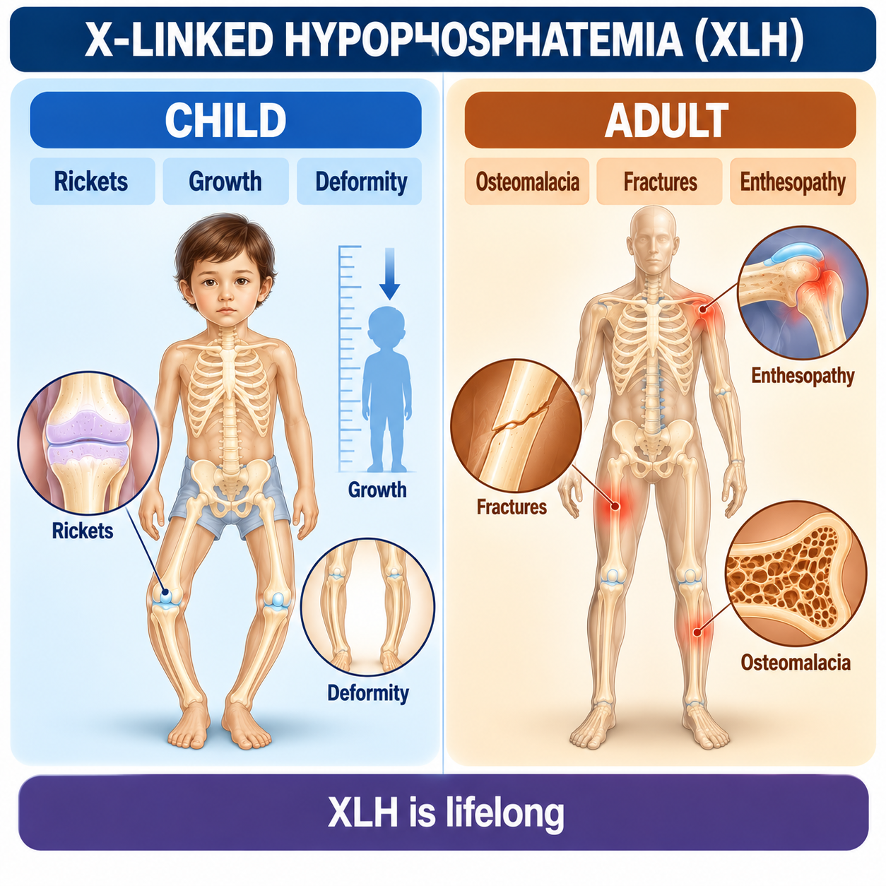
During childhood, when growth plates remain open, this produces rickets and impaired skeletal growth. In adults, defective mineralization can manifest as osteomalacia, while patients can also develop lifelong complications such as pain, enthesopathy, osteoarthritis, fractures or pseudofractures, reduced mobility and dental disease.
XLH is not simply a childhood rickets disorder. It is a lifelong inherited disorder of phosphate and mineral metabolism.
XLH at a Glance

What Is X-Linked Hypophosphatemia?
X-linked hypophosphatemia is an inherited PHEX-related disorder characterized by inappropriate FGF23 activity, renal phosphate wasting, chronic hypophosphatemia and impaired skeletal mineralization.
The disease affects both children and adults. The phenotype changes with age, but the underlying phosphate-regulatory abnormality persists.
Why the Old Name Can Be Misleading
Historically, disorders such as XLH were sometimes described using terms such as vitamin D-resistant rickets or hypophosphatemic rickets. These terms can obscure the actual physiology.
XLH is not primarily caused by failure to obtain enough nutritional vitamin D. The fundamental pathway is PHEX → FGF23 dysregulation → renal phosphate wasting. Therefore this article preferentially uses X-linked hypophosphatemia rather than treating “vitamin D-resistant rickets” as the main disease definition.
What Is PHEX?
PHEX stands for Phosphate-Regulating Endopeptidase Homolog, X-Linked. It is encoded by the PHEX gene on the X chromosome.
Pathogenic PHEX variants disrupt normal phosphate-regulatory physiology and result in inappropriate FGF23 activity.
Do not teach that PHEX simply “breaks down FGF23.” That is an oversimplification. The clinically important relationship is: loss of normal PHEX function → inappropriate FGF23 excess/activity. The exact molecular pathway linking PHEX dysfunction to FGF23 regulation is more complex than direct degradation.
How PHEX and FGF23 Are Connected
FGF23 is a bone-derived phosphaturic hormone that regulates renal phosphate handling and vitamin D metabolism. Its two major effects relevant to XLH are reduced renal phosphate reabsorption and reduced calcitriol availability. Review FGF23 Disorders Explained for how FGF23 controls phosphate and calcitriol.
Two major renal consequences follow:
Both contribute to impaired mineralization.

How FGF23 Causes Renal Phosphate Wasting
Normally, hypophosphatemia should cause the kidney to conserve phosphate. But in XLH, persistent FGF23 activity reduces proximal tubular phosphate reabsorption.
Serum PO₄ is low while the kidney continues losing phosphate. This is inappropriate renal phosphate wasting and is central to the diagnosis.
Because renal phosphate reabsorptive capacity is reduced, TmP/GFR falls inappropriately for the hypophosphatemia. PO₄ ↓ plus TmP/GFR ↓ supports renal phosphate wasting — but this does not by itself diagnose XLH. Other causes include TIO, other inherited FGF23 disorders, PTH-mediated phosphaturia and proximal tubular disorders.
For the calculation framework, review Renal Phosphate Wasting Explained: TRP, FEPO₄, TmP/GFR, FGF23 and Hypophosphatemia.
Why Calcitriol Is Inappropriately Low in XLH
Normally, PO₄ ↓ should promote a physiological increase in calcitriol. But FGF23 suppresses the expected renal calcitriol response.
The important word is inappropriate. A value does not have to fall below the population reference range to be physiologically abnormal.
For background, see Vitamin D Metabolism Explained.
Why XLH Causes Rickets
In a growing child, phosphate depletion plus abnormal calcitriol physiology reduce mineral availability, impairing growth-plate and osteoid mineralization.
This explains why childhood XLH can affect growth, limb alignment, gait and skeletal architecture.
Why XLH Causes Osteomalacia
Once growth plates have closed, rickets cannot newly develop in the same way. However, phosphate depletion can continue to impair mineralization of newly formed osteoid.
Rickets involves the growing skeleton and growth plate. Osteomalacia refers to defective mineralization of adult bone. An individual with childhood XLH may have residual deformities from previous rickets and active adult osteomalacia.
Review Osteomalacia Explained for how chronic phosphate depletion impairs adult bone mineralization.
XLH Is a Lifelong Disease
“The rickets ends when the child stops growing, therefore XLH is over.” This is wrong. The underlying PHEX–FGF23 disorder persists.
Adults can therefore experience persistent hypophosphatemia, osteomalacia, bone pain, muscle symptoms, pseudofractures, fractures, enthesopathy, osteoarthritis, stiffness, impaired mobility, spinal complications and dental disease.
The phenotype changes. The disease does not simply disappear.
How Is XLH Inherited?
XLH follows X-linked dominant inheritance. This means both males and females can be affected. Do not use outdated assumptions that an X-linked disorder necessarily affects only males.
Affected father
An affected father transmits his X chromosome to all daughters and his Y chromosome to all sons. Therefore, assuming the father carries the pathogenic PHEX variant on his X chromosome, daughters inherit that X while sons do not.
Heterozygous affected mother
Each pregnancy has approximately a 50% probability of inheriting the affected X chromosome, regardless of sex.
Individual genetic counselling should use the confirmed family genotype and clinical context rather than relying only on simplified inheritance diagrams.
Can XLH Occur Without Family History?
Family history is very useful. Ask about childhood bowed legs, short stature, unexplained rickets, corrective orthopaedic surgery, chronic bone/joint pain, dental abscesses, known hypophosphatemia and diagnosed XLH.
De novo PHEX variants occur, previous generations may have mild or unrecognized disease, and family history can be incomplete.
XLH in Children
Clinical manifestations vary. Potential childhood features include rickets, lower-limb deformity, genu varum, genu valgum, impaired linear growth, disproportionate short stature, abnormal gait, bone pain, delayed motor function in some children, dental disease and craniofacial abnormalities in some patients. Do not imply every child has all features.
Lower-Limb Deformity
Weight-bearing can make lower-limb deformities increasingly apparent, with patterns including genu varum or genu valgum, and more complex deformities in some patients. These result from abnormal mineralization and mechanical loading of the growing skeleton. Do not reduce XLH to “bow legs”; the phenotype is broader.
Growth
Growth impairment is an important childhood manifestation. The mechanism is multifactorial but includes abnormal growth-plate mineralization and skeletal disease. Poor growth plus rickets plus hypophosphatemia should prompt investigation of phosphate physiology rather than automatically assuming nutritional vitamin D deficiency.
Craniofacial and Other Childhood Features
Some patients can develop craniofacial abnormalities, including complications related to abnormal skull growth. XLH is a systemic skeletal disorder, and craniofacial complications can occur in selected patients and require specialist assessment.
XLH in Adults
Adults can present with chronic musculoskeletal pain, bone pain, muscle weakness or fatigue, stiffness, impaired mobility, osteomalacia, pseudofractures, insufficiency fractures, residual lower-limb deformity, enthesopathy, osteoarthritis, spinal disease and dental problems.
Adult XLH is not simply residual childhood deformity. There can be ongoing metabolic bone disease plus acquired structural complications.
Dental Disease in XLH
Dental manifestations are clinically important and can occur even when teeth appear externally normal. Patients can develop spontaneous dental abscesses, recurrent dental infection, periodontal problems and abnormalities related to dentin/mineralization.
Recurrent unexplained dental abscesses can be a clue to XLH. Dental disease should not be treated as a trivial secondary feature.
Enthesopathy and Osteoarthritis
Enthesopathy refers to pathological changes at sites where tendons or ligaments attach to bone. In XLH, enthesopathy can become increasingly important in adulthood and contribute to stiffness, pain, reduced range of motion and impaired mobility. It may progress even when childhood rickets is no longer the dominant manifestation.
Adults may develop premature or substantial degenerative joint disease, reflecting a combination of longstanding skeletal deformity, abnormal mechanical loading, enthesopathy and chronic musculoskeletal disease. Therefore adult disability is not explained solely by the current serum phosphate concentration.
Pseudofractures
Chronic osteomalacia can produce pseudofractures / Looser-type insufficiency lesions. These represent mineralization failure rather than necessarily high-energy traumatic fractures.
Laboratory Findings in XLH
| Test | Typical XLH Pattern |
|---|---|
| Serum phosphate | ↓ for age |
| TmP/GFR | ↓ / inappropriate renal wasting |
| FGF23 | ↑ or inappropriately non-suppressed |
| 1,25-(OH)₂D | Low or inappropriately normal |
| Calcium | Usually normal |
| ALP | Often ↑ with active rickets/osteomalacia |
| PTH | Often normal, but may vary |
| 25-OH D | Variable; deficiency can coexist |
| Renal function | Usually not the primary cause of phosphaturia |
No single laboratory value diagnoses XLH. The diagnosis comes from the phenotype, renal phosphate-wasting pattern and genetic/clinical context.
Why Pediatric Phosphate Reference Ranges Matter
Serum phosphate is physiologically higher during childhood and varies with age. A phosphate value that looks “normal” using an adult range may be abnormally low for a child.
Interpret serum phosphate against an age-appropriate pediatric reference interval. The same principle applies when interpreting renal phosphate handling.
When Should XLH Be Suspected?
Family history is supportive, not mandatory.
How Is XLH Diagnosed?
Step 1 — Confirm Hypophosphatemia
Confirm that serum phosphate is genuinely low for age. Consider timing, fasting status when relevant, acute illness, recent intake, redistribution and renal function. Do not diagnose XLH from one isolated low phosphate value.
Step 2 — Prove Renal Phosphate Wasting
A patient with low phosphate should normally conserve phosphate. Use paired serum/urine studies and TmP/GFR when indicated. If PO₄ ↓ and TmP/GFR is inappropriately ↓, renal phosphate wasting is established.
Step 3 — Identify the Mechanism
Once renal wasting is established, ask why phosphate is being lost. Major mechanisms: FGF23-mediated (XLH, TIO, other inherited FGF23 disorders), PTH-mediated (hyperparathyroid physiology) and proximal tubular dysfunction (Fanconi-type disorders and other tubulopathies). Renal phosphate wasting ≠ XLH by itself.
Step 4 — FGF23 Physiology
If the pattern suggests an FGF23-mediated disorder, FGF23 measurement can help where available. Hypophosphatemia should suppress FGF23. Therefore FGF23 within the population reference range can still be inappropriately non-suppressed and support FGF23-mediated disease. Do not use a universal numerical cutoff.
Step 5 — Clinical Phenotype
Ask: did disease begin in childhood? Was there rickets? Was growth impaired? Were lower-limb deformities present? Is there a family history? Are there characteristic dental problems? Are adult enthesopathy/osteoarthritis or childhood skeletal abnormalities present? A lifelong phenotype strongly supports an inherited disorder.
Role of PHEX Genetic Testing
Where available, PHEX genetic testing should be used to confirm XLH in a clinically compatible patient. Genetic confirmation is valuable for diagnostic certainty, differentiating inherited disorders, family counselling, identifying affected relatives and future reproductive counselling. Do not invent a specific commercial panel.
What If PHEX Testing Is Negative?
A negative PHEX result in a patient with genuine FGF23-mediated renal phosphate wasting does not automatically mean the biochemical diagnosis was wrong. Consider technical/genetic limitations, alternative inherited FGF23 disorders and acquired FGF23-mediated disease.
Other Inherited FGF23 Disorders
Other genes associated with inherited hypophosphatemic disorders include, among others, FGF23, DMP1 and ENPP1.
PHEX-negative FGF23-mediated hypophosphatemia requires diagnostic reconsideration, not abandonment of physiology.
XLH vs TIO
| Feature | XLH | TIO |
|---|---|---|
| Nature | Inherited | Acquired |
| Mechanism | PHEX-related FGF23 dysregulation | FGF23-producing tumor |
| Typical onset | Childhood/lifelong | Usually acquired later |
| Childhood rickets | Common | Usually absent |
| Growth abnormality | Can occur | Usually absent before disease onset |
| Family history | May be present | Usually absent |
| PO₄ | ↓ | ↓ |
| TmP/GFR | ↓ | ↓ |
| FGF23-mediated | Yes | Yes |
| Calcitriol | Inappropriately low/normal | Inappropriately low/normal |
| PHEX testing | Relevant | Not the primary explanation |
| Tumor localization | No | Yes, after biochemical diagnosis |
| Definitive treatment | Lifelong disease management | Tumor resection when feasible |
Age and family history guide the differential but are not absolute. De novo XLH can occur, and inherited disease can occasionally remain undiagnosed until adulthood.
Review Tumor-Induced Osteomalacia Explained for the major acquired FGF23-mediated differential diagnosis.
XLH vs Vitamin D Deficiency
Both can cause rickets, but the physiology differs.
| Feature | XLH | Nutritional Vitamin D Deficiency |
|---|---|---|
| Primary mechanism | FGF23-mediated renal PO₄ wasting | Inadequate vitamin D availability |
| 25-OH D | May be normal | Usually ↓ |
| Renal PO₄ wasting | Primary feature | Often secondary to PTH if present |
| PTH | Variable | Often ↑ when physiologically significant |
| Calcium | Usually normal | Normal or ↓ depending on severity |
| FGF23 | Inappropriately active | Not primary driver |
| PHEX | May have pathogenic variant | Not causal |
Vitamin D deficiency can coexist with XLH. Do not assume one excludes the other.
See Vitamin D Deficiency Explained for the differential diagnosis of low serum phosphate alongside vitamin D status.
XLH vs Fanconi Syndrome
XLH primarily produces an FGF23-driven phosphate-handling abnormality. Fanconi syndrome causes generalized proximal tubular dysfunction. Look for glycosuria inappropriate for serum glucose, bicarbonate wasting, uric acid wasting, aminoaciduria, low-molecular-weight proteinuria and other proximal tubular abnormalities.
Isolated/hormonal PO₄ wasting → think regulatory mechanisms. PO₄ wasting + multiple solute losses → think proximal tubule. This is a reasoning aid rather than an absolute diagnostic rule.
XLH vs Hyperparathyroidism
PTH and FGF23 are both phosphaturic hormones. Therefore both can produce PO₄ ↓ plus renal phosphate wasting. But in XLH, FGF23 is the main driver, calcium is usually normal, and an inherited/lifelong phenotype may be present. In primary hyperparathyroid physiology, PTH is inappropriately elevated, hypercalcemia is an important clue, and FGF23 is not the primary explanation.
TODO: Link Primary Hyperparathyroidism article after publication.
XLH Diagnostic Algorithm
Treatment Goals in XLH
XLH treatment is not simply about making one phosphate result normal. Goals include improving rickets, improving mineralization, reducing bone pain, reducing pseudofractures/fractures, improving physical function, supporting growth in children, limiting skeletal deformity, improving dental outcomes, reducing disease burden and minimizing treatment complications.
Treatment should therefore be disease- and patient-centred rather than laboratory-number-centred.
Conventional Phosphate and Active Vitamin-D Therapy
Two broad treatment strategies exist:
Historically, XLH has been managed with multiple-dose oral phosphate supplementation plus active vitamin D, such as calcitriol or alfacalcidol depending on local practice. Do not provide universal doses.
Why Phosphate Alone Is Not the Whole Solution
Oral phosphate attempts to replace phosphate being lost because of persistent FGF23-driven renal wasting. But the underlying hormonal abnormality remains. Phosphate administration can also influence PTH and mineral physiology. Therefore conventional treatment requires careful specialist monitoring rather than simply “give phosphate until the serum phosphate is normal.”
Why Active Vitamin D Is Used
FGF23 suppresses normal calcitriol physiology. Therefore active vitamin D has historically been combined with phosphate to support calcium/phosphate absorption, mineralization and management of the hormonal consequences of conventional therapy. Again, do not provide a universal dose.
Limitations of Conventional Therapy
Conventional therapy can improve rickets and mineralization but does not directly correct the underlying FGF23 excess. Treatment burden can also be substantial. Potential complications requiring monitoring include hyperparathyroidism, hypercalciuria, nephrocalcinosis and renal effects. Therefore therapy must be individualized and monitored.
What Is Burosumab? How Does It Work?
Burosumab is a fully human monoclonal antibody that binds and inhibits FGF23. It targets the central hormonal abnormality of XLH.
Together, the mineralization environment improves. This is fundamentally different from simply replacing phosphate.
| Conventional Therapy | Burosumab |
|---|---|
| Replaces phosphate + provides active vitamin D | Targets FGF23 |
| Does not directly remove FGF23 excess | Neutralizes FGF23 activity |
| Requires frequent oral treatment | Administration is treatment-protocol dependent |
| Can improve mineralization | Can improve phosphate physiology/mineralization |
| Requires biochemical/renal monitoring | Requires biochemical and disease-specific monitoring |
Do not imply that every patient should automatically receive one or the other. Treatment depends on age, disease manifestations, previous treatment, availability, regulatory approval, contraindications and specialist assessment.
Current treatment frameworks do not use concurrent oral phosphate/active vitamin-D therapy with burosumab as a casual combination strategy. Do not create a regimen combining them, and do not provide transition timing or doses in this general educational article.
Treatment in Children
The broad objectives are to heal active rickets, improve mineralization, improve growth trajectory where possible, reduce pain, improve function, limit progressive deformity, support dental health and reduce long-term skeletal burden.
Modern treatment frameworks give an important role to burosumab in pediatric XLH, particularly in active disease. However, treatment selection belongs to specialist care. Do not create universal eligibility criteria.
Treatment in Adults
Adult treatment should focus on clinically meaningful active disease. Relevant manifestations include bone pain, osteomalacia, pseudofractures, fractures, impaired mobility/function and biochemical evidence of active mineralization disturbance.
Not every adult complication is reversible simply by correcting serum phosphate. For example, established enthesopathy and structural osteoarthritis may not disappear because phosphate physiology improves. This distinction is important when setting treatment expectations.
Orthopaedic Management
Some patients have substantial lower-limb deformity or other structural skeletal abnormalities. Medical treatment improves metabolic bone physiology but does not automatically correct every established mechanical deformity. Selected patients may require specialist orthopaedic assessment in addition to metabolic treatment. Do not provide operative thresholds.
Dental Management
XLH requires ongoing dental attention, emphasizing preventive dental care, early recognition of infection, management of recurrent abscesses, periodontal assessment and collaboration between dental and metabolic teams. Dental disease is part of XLH — not an unrelated coincidence.
Long-Term Monitoring
Nephrocalcinosis is an important monitoring issue, particularly in patients receiving conventional phosphate/active-vitamin-D treatment. This does not mean XLH itself always causes nephrocalcinosis — renal complications need to be interpreted in the context of disease, treatment, urinary calcium, phosphate therapy, active vitamin-D exposure and kidney function.
PTH abnormalities can develop in XLH, particularly in relation to chronic phosphate therapy and mineral metabolism, including secondary hyperparathyroid physiology or more persistent/autonomous hyperparathyroid states in selected patients. PTH should not be ignored during long-term management.
Depending on age, treatment and disease state, follow-up can include serum phosphate, calcium, ALP, PTH, creatinine/renal function, 25-OH D, urinary calcium, growth in children, limb alignment, radiographic rickets where clinically indicated, pain, mobility/function, fractures/pseudofractures, renal imaging where appropriate, dental assessment and musculoskeletal complications. Do not hardcode one universal monitoring interval.
Treatment success should not be judged solely by whether phosphate entered the normal range. Assess skeletal disease, rickets healing, ALP, pain, fractures, physical function, growth, complications and treatment safety — particularly in adults with longstanding structural disease.
Transition From Pediatric to Adult Care
XLH should not fall out of follow-up when growth stops.
The transition should preserve information about genotype, childhood treatment, orthopaedic history, growth/deformity, dental disease, renal complications, previous nephrocalcinosis, treatment response and current metabolic disease.
Worked Clinical Cases
Case 1: Child With Rickets
A 4-year-old has bowed legs, abnormal gait, impaired growth, phosphate low for age, ALP elevated, calcium normal and renal phosphate wasting. The first conclusion is not “vitamin D deficiency” — instead, hypophosphatemic rickets with renal phosphate wasting needs evaluation. If FGF23 is inappropriately active and the phenotype is compatible, XLH becomes an important diagnosis.
Case 2: Adult Reference Range Error
A child has a phosphate result that falls inside the laboratory's displayed adult reference interval, but the value is below the expected range for age. Error: “phosphate is normal.” Correct interpretation: pediatric phosphate must be interpreted using an age-appropriate range. Lesson: children are not small adults.
Case 3: Strong Family History
A child has hypophosphatemic rickets, renal phosphate wasting and an affected mother with childhood bowed legs and chronic hypophosphatemia. An inherited disorder is strongly suggested. Appropriate biochemical assessment plus PHEX genetic testing can confirm XLH.
Case 4: No Family History
A child has typical hypophosphatemic rickets, renal phosphate wasting, FGF23-mediated physiology and no known affected relatives. Error: “XLH is impossible.” Correct reasoning: de novo PHEX disease can occur. Lesson: no family history does not exclude XLH.
Case 5: Adult XLH
A 42-year-old has a history of childhood bowed legs, corrective surgery in adolescence, chronic hypophosphatemia, renal phosphate wasting, dental abscess history, increasing stiffness, enthesopathy and bone pain. This is compatible with adult XLH, with both longstanding structural complications and potentially active metabolic bone disease. Lesson: XLH does not end after childhood.
Case 6: XLH or TIO?
A 52-year-old has entirely normal childhood growth, no childhood skeletal deformity, no family history, new progressive bone pain, proximal weakness, insufficiency fractures, renal phosphate wasting and an FGF23-mediated biochemical pattern. This acquired phenotype should raise strong concern for TIO rather than automatically diagnosing XLH. Lesson: the timeline is diagnostic information.
Case 7: Fanconi Pattern
A patient has hypophosphatemia, renal phosphate wasting, glycosuria despite non-elevated serum glucose, bicarbonate loss and additional proximal tubular abnormalities. This is more consistent with generalized proximal tubular dysfunction than isolated XLH.
Case 8: Hypercalcemia
A patient has phosphate ↓, renal phosphate wasting, calcium ↑ and PTH inappropriately ↑. Think PTH-mediated phosphaturia before attributing the phosphate loss to XLH.
Case 9: Normal Phosphate After Treatment but Persistent Stiffness
An adult with longstanding XLH receives appropriate metabolic treatment. Phosphate physiology improves, but substantial joint stiffness and enthesopathy remain. Error: “treatment failed because symptoms remain.” Correct reasoning: longstanding enthesopathy, osteoarthritis and deformity may represent structural disease that does not immediately reverse with metabolic correction. Lesson: biochemical correction and structural recovery are not identical.
Common Mistakes
XLH in One Minute
Golden Rules
- XLH is a lifelong inherited FGF23-mediated phosphate-wasting disorder.
- PHEX dysfunction leads to inappropriate FGF23 activity.
- FGF23 causes renal phosphate wasting and suppresses the expected calcitriol response.
- Children require age-specific phosphate interpretation.
- Low phosphate alone does not diagnose XLH.
- Renal phosphate wasting must be established.
- A normal-range FGF23 may still be inappropriately non-suppressed.
- No family history does not exclude XLH.
- Rickets is the growing-skeleton manifestation; osteomalacia can occur in adults.
- XLH does not disappear when growth stops.
- Burosumab targets FGF23 rather than merely replacing phosphate.
- Treatment success must be assessed clinically as well as biochemically.
Clinical Pearls
- XLH is the commonest inherited FGF23-mediated hypophosphatemic disorder.
- It is PHEX-related.
- It is X-linked dominant.
- Both males and females can be affected.
- De novo disease occurs.
- Family history is useful but not essential.
- FGF23 drives renal phosphate wasting.
- TmP/GFR is reduced inappropriately.
- FGF23 also suppresses calcitriol physiology.
- Calcitriol can be low or inappropriately normal.
- Serum calcium is usually normal.
- ALP commonly rises with active rickets.
- Pediatric phosphate ranges are age-dependent.
- Adult ranges must not be applied blindly to children.
- Childhood XLH causes hypophosphatemic rickets.
- Lower-limb deformity can occur.
- Growth can be impaired.
- Dental abscesses can be an important clue.
- XLH continues into adulthood.
- Adult osteomalacia can occur.
- Pseudofractures can occur.
- Enthesopathy is an important adult complication.
- Osteoarthritis and stiffness can contribute substantially to disability.
- TIO is an important adult differential.
- Normal childhood development with newly acquired disease argues against typical lifelong XLH.
- Fanconi syndrome causes broader proximal tubular losses.
- Hyperparathyroidism can also cause phosphaturia.
- PHEX testing can confirm XLH.
- PHEX-negative disease may require evaluation for other inherited or acquired causes.
- Conventional therapy uses phosphate plus active vitamin D.
- Conventional therapy does not neutralize FGF23.
- Burosumab blocks FGF23.
- Burosumab improves renal phosphate conservation.
- Nephrocalcinosis is an important monitoring issue.
- PTH requires monitoring during long-term management.
- Dental care is part of XLH management.
- Orthopaedic care may be necessary for established deformity.
- Adult structural disease may not completely reverse with biochemical treatment.
- XLH requires multidisciplinary management.
- XLH requires lifelong follow-up.
Frequently Asked Questions
Key Take-Home Messages
X-linked hypophosphatemia becomes much easier to understand when it is approached as an FGF23 disorder rather than simply “a form of rickets.”
Start with PHEX. A pathogenic PHEX variant causes abnormal regulation of FGF23, which remains excessively active. FGF23 then acts on the kidney: renal phosphate reabsorption falls, urinary phosphate loss rises, and serum phosphate falls. At the same time, the calcitriol response falls. Therefore the mineralization environment becomes abnormal.
In a growing child, rickets develops. After growth plates close, osteomalacia can persist or develop. But XLH is broader than either diagnosis. Over a lifetime, patients may also develop skeletal deformity, dental disease, pseudofractures, enthesopathy, osteoarthritis, pain and impaired mobility. Therefore XLH does not end when childhood ends.
When diagnosing the disorder, start with: is phosphate low for age? Then: is the kidney inappropriately wasting phosphate? If yes, why? If the pattern indicates FGF23-mediated renal phosphate wasting, ask: inherited or acquired? Childhood rickets, growth abnormalities, lifelong skeletal disease and family history favour inherited disease, but no family history does not exclude XLH. Confirm where possible with PHEX genetic testing.
Treatment then follows two major physiological strategies. Conventional therapy attempts to compensate for the consequences of FGF23 excess using phosphate plus active vitamin D, whereas targeted treatment with burosumab acts upstream by blocking FGF23.
The final memory pathway: PHEX → FGF23 ↑ → renal PO₄ wasting → hypophosphatemia → impaired mineralization, with rickets in children and osteomalacia plus lifelong musculoskeletal disease in adults.
This article is intended for medical education only. It explains XLH physiology and diagnostic reasoning, not patient-specific medical advice, treatment eligibility, medication dosing, monitoring intervals or procedural instructions.