Primary hyperparathyroidism, abbreviated PHPT, is a disorder in which one or more parathyroid glands secrete PTH inappropriately relative to the serum calcium concentration.
The classical biochemical presentation is hypercalcemia with elevated PTH or inappropriately normal PTH. The second situation is especially important: normally, Ca²⁺ ↑ should strongly suppress PTH ↓. Therefore a PTH concentration that remains within the laboratory's population reference interval can still be pathological when calcium is elevated.
High calcium + PTH not suppressed = PTH-dependent hypercalcemia. Primary hyperparathyroidism is the major diagnosis in this situation, but important alternatives — particularly familial hypocalciuric hypercalcemia (FHH) — must be considered before treatment.
Modern PHPT is often discovered incidentally rather than through the dramatic classical presentation of severe bone and renal disease. Nevertheless, the skeleton and kidneys remain the two major target organs that must be assessed after diagnosis.
PHPT at a Glance

What Is Primary Hyperparathyroidism?
Parathyroid hormone is an 84-amino-acid peptide hormone produced by the parathyroid glands that plays a central role in extracellular calcium regulation and also influences phosphate and vitamin D metabolism. The dominant physiological regulator is ionized calcium, acting through the calcium-sensing receptor (CaSR) on parathyroid cells.
In PHPT, one or more glands secrete PTH autonomously or inappropriately relative to calcium, producing a self-sustaining cycle of hypercalcemia and inappropriate PTH activity.
How Calcium Normally Controls PTH
Under normal physiology, Ca²⁺ ↓ reduces CaSR stimulation, so PTH secretion rises to restore extracellular calcium. Conversely, Ca²⁺ ↑ increases CaSR activation, so PTH secretion falls.
Hypercalcemia should suppress PTH. This physiological expectation is the foundation of PHPT diagnosis.
For the complete normal calcium–PTH feedback loop, including calcitonin and the roles of vitamin D and the kidney, see Calcium Homeostasis Explained.
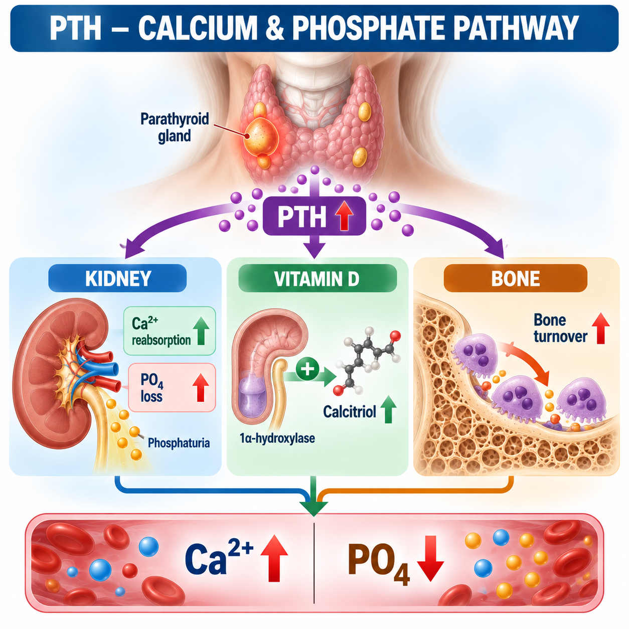
What Does PTH Do to the Kidney, Phosphate and Vitamin D?
Calcium
PTH promotes renal calcium conservation, particularly through effects on distal nephron calcium handling. Overall, the tendency to conserve urinary calcium increases — although urinary calcium in PHPT can still be substantial because the filtered calcium load is increased. Do not teach that PTH always causes low urinary calcium; that is incorrect.
Phosphate
PTH decreases proximal tubular phosphate reabsorption.
This is PTH-mediated renal phosphate wasting, and it links this article directly to the phosphate cluster. See Phosphate Homeostasis Explained for normal renal phosphate regulation and Renal Phosphate Wasting Explained for how TmP/GFR identifies inappropriate renal phosphate loss.
Vitamin D
PTH stimulates renal 1α-hydroxylase, which promotes conversion toward calcitriol (1,25-(OH)₂D) when renal function and substrate availability permit. Calcitriol then promotes intestinal calcium and phosphate absorption. See Vitamin D Metabolism Explained for the full calcitriol synthesis pathway.
PTH vs FGF23
| Effect | PTH | FGF23 |
|---|---|---|
| Renal PO₄ reabsorption | ↓ | ↓ |
| Phosphaturia | ↑ | ↑ |
| Serum PO₄ | Tends ↓ | ↓ |
| Calcitriol production | ↑ | ↓ |
| Major physiological role | Calcium defense | Phosphate regulation |
| Major disease example | PHPT | XLH / TIO |
Both waste phosphate, but PTH pushes calcitriol up while FGF23 pushes calcitriol down. Do not imply calcitriol must actually be elevated in every PHPT patient; renal function, vitamin D substrate and other regulatory factors modify the measured concentration.
See FGF23 Disorders Explained for how FGF23 differs from PTH in phosphate and calcitriol regulation.
How PTH Affects Bone
PTH receptors are expressed on cells of the osteoblast lineage rather than PTH simply acting as a direct osteoclast stimulant. Persistent PTH excess promotes signalling that increases osteoclast formation and bone resorption through osteoblast/osteocyte-mediated pathways such as RANKL. Therefore chronic PHPT can increase bone turnover and cause skeletal loss.
What Causes Primary Hyperparathyroidism?
Most sporadic PHPT results from a single parathyroid adenoma. A smaller proportion results from multigland hyperplasia. Less commonly, double adenomas/multigland disease or parathyroid carcinoma occur. Do not imply every patient has one adenoma.
Primary vs Secondary vs Tertiary Hyperparathyroidism
| Disorder | Main Driver | Calcium Tendency | PTH |
|---|---|---|---|
| Primary HPT | Autonomous/inappropriate parathyroid secretion | ↑ | ↑ / inappropriate normal |
| Secondary HPT | Physiological response to chronic stimulus | Normal/↓ commonly | ↑ |
| Tertiary HPT | Autonomous secretion after longstanding secondary HPT | ↑ | Often markedly ↑ |
Common secondary causes include vitamin D deficiency, CKD, inadequate calcium intake, malabsorption and selected medications. This distinction becomes especially important when evaluating high PTH with normal calcium.
For the detailed physiology of chronic PTH stimulation, see Secondary Hyperparathyroidism Explained and, in the context of chronic kidney disease, CKD-MBD Explained.
Laboratory Findings in PHPT
| Test | Typical PHPT Pattern |
|---|---|
| Serum calcium | ↑ |
| PTH | ↑ or inappropriately normal |
| Serum phosphate | Low / low-normal commonly |
| 25-OH D | Variable |
| 1,25-(OH)₂D | Variable; can be increased |
| ALP | Normal or ↑ depending on skeletal activity |
| Urinary calcium | Variable |
| Renal function | Variable |
No single phosphate, ALP, vitamin-D or urinary-calcium pattern is required for PHPT. The central biochemical relationship is hypercalcemia with PTH that is elevated or fails to suppress appropriately.
Why Phosphate Is Low
Low phosphate can support PTH physiology, but normal phosphate does not exclude PHPT — phosphate is supportive, not diagnostic.
Alkaline Phosphatase
ALP reflects, among other sources, osteoblastic activity. In modern mild PHPT, ALP may be normal; in more active skeletal disease, ALP may increase. Normal ALP does not exclude PHPT.
Vitamin D in PHPT
25-OH vitamin D should be measured. Vitamin D deficiency can coexist with PHPT and can increase PTH stimulation, worsen skeletal disease, complicate interpretation and influence urinary calcium. Do not assume that vitamin D deficiency explaining the high PTH means PHPT is impossible — if the patient is hypercalcemic, the entire calcium–PTH relationship still requires explanation. See Vitamin D Deficiency Explained for vitamin D deficiency as a cause of secondary hyperparathyroidism.
Can PTH Be Normal in Primary Hyperparathyroidism?
The Fifth International Workshop defines hypercalcemic PHPT as elevated albumin-adjusted serum calcium in the presence of elevated or inappropriately normal intact PTH on two occasions at least two weeks apart.
Suppose Ca²⁺ is high and PTH is mid-reference range. A common error is: “PTH is normal, therefore the hypercalcemia is not due to the parathyroids.” Wrong. The correct physiological question is: what should PTH be when calcium is high? Answer: suppressed.
Just as low phosphate should suppress FGF23, high calcium should suppress PTH. High Ca + normal-range PTH can mean inappropriately normal PTH and therefore PTH-dependent hypercalcemia.
How Is PHPT Diagnosed? Confirming the Calcium
Do not diagnose PHPT from an unexplained single calcium result. Assess repeat calcium, albumin, laboratory context, medications and hydration/clinical context. Albumin-adjusted total calcium is commonly used.
Ionized calcium is particularly useful when total calcium is difficult to interpret, albumin/protein abnormalities complicate interpretation, or normocalcemic PHPT is being considered. The Workshop specifically recommends ionized calcium when normocalcemic PHPT is under consideration.
PTH-Dependent vs PTH-Independent Hypercalcemia
Once hypercalcemia is confirmed, measure PTH.
Initial PHPT Evaluation
Once PHPT is suspected/diagnosed, biochemical evaluation should include adjusted total calcium, ionized calcium if appropriate, intact PTH, serum phosphorus, 25-OH vitamin D and creatinine/renal function. The Fifth Workshop additionally recommends formal skeletal and renal assessment after diagnosis.
Clinical Presentations
Symptomatic PHPT
Overt renal or skeletal complications.
Asymptomatic PHPT
Typically discovered through biochemical testing without overt classical symptoms. Patients may subsequently be found to have target-organ involvement despite being considered clinically “asymptomatic.”
Normocalcemic PHPT
A distinct biochemical phenotype requiring strict exclusion of secondary causes (discussed below).
Classical PHPT
Historically severe PHPT could present with nephrolithiasis, nephrocalcinosis, fractures, severe skeletal disease, osteitis fibrosa cystica and substantial hypercalcemia. Modern biochemical screening has shifted many diagnoses toward milder/asymptomatic disease.
Renal Manifestations
The kidneys are one of the two principal target organs. Important manifestations include nephrolithiasis, nephrocalcinosis and reduced renal function in some patients. Hypercalciuria can contribute to stone risk, but stone formation is multifactorial.
“High PTH causes low urinary calcium, so PHPT cannot cause stones.” The increased filtered calcium load can result in substantial urinary calcium despite PTH-mediated renal calcium conservation.
Skeletal Manifestations
Chronic PTH excess increases bone turnover. Potential consequences include reduced BMD, osteoporosis, fragility fractures, vertebral fractures and severe classical skeletal disease in advanced cases. PHPT classically has important effects on cortical bone, which is one reason the distal one-third radius is included in recommended DXA assessment.
Osteitis Fibrosa Cystica
A classical severe skeletal manifestation of hyperparathyroidism, with high-turnover bone disease, subperiosteal resorption, cystic skeletal lesions and brown tumors. Modern PHPT is often diagnosed before this stage. Do not describe brown tumors as true neoplastic tumors — they represent reactive lesions associated with severe hyperparathyroid bone disease.
How Should PHPT Be Evaluated? DXA and Renal Assessment
The Fifth Workshop recommends three-site DXA, including lumbar spine, hip and distal one-third radius, plus assessment for vertebral fracture with vertebral fracture assessment (VFA) or vertebral radiography, with trabecular bone score where available.
Recommended renal evaluation includes eGFR or preferably creatinine clearance, 24-hour urinary calcium, biochemical stone-risk assessment where appropriate, and imaging for nephrolithiasis/nephrocalcinosis. This assesses target-organ involvement and also contributes to management decisions.
PHPT vs Familial Hypocalciuric Hypercalcemia (FHH)
FHH can mimic PHPT because both may show Ca ↑ with PTH not suppressed. But FHH is fundamentally different: it is commonly caused by altered calcium sensing, most often involving CASR, with less common related genetic mechanisms.
In classic FHH, altered calcium sensing changes the body's perceived calcium set-point. Patients can have mild lifelong hypercalcemia with PTH normal or mildly elevated and characteristically relatively low urinary calcium excretion. This contrasts with many patients with PHPT.
Calcium/Creatinine Clearance Ratio (CCCR)
using appropriately matched units and urine/serum measurements. The Fifth Workshop notes that CCCR <0.01 raises suspicion for FHH, particularly in younger patients or those with a family history of hypercalcemia.
Do not teach “CCCR <0.01 = FHH and CCCR >0.01 = PHPT.” There is overlap between FHH and PHPT. Urinary calcium can also be affected by vitamin D deficiency, reduced calcium intake, CKD, thiazide treatment and other clinical factors. CCCR is a clue, not an absolute verdict. When FHH remains clinically plausible, family assessment and/or appropriate genetic evaluation may be needed.
Why FHH Matters Before Surgery
PHPT can be cured by parathyroidectomy. But FHH represents an altered systemic calcium-sensing set-point, so routine parathyroid surgery generally does not correct the underlying physiology of typical FHH. Mistaking FHH for PHPT can lead to unnecessary surgery.
When to Think Particularly About FHH
Suspicion increases with younger age, lifelong or longstanding mild hypercalcemia, family history of hypercalcemia, relatively low urinary calcium, low CCCR and absence of a typical progressive PHPT phenotype.
For the complete FHH-side physiology, CCCR interpretation, confounders and genetic testing, see Familial Hypocalciuric Hypercalcemia Explained.

Medications: Lithium and Thiazides
Important medication contexts include lithium and thiazide diuretics. Both can complicate the biochemical evaluation of hypercalcemia and PTH. A careful medication history is therefore mandatory before concluding that the biochemical phenotype is straightforward sporadic PHPT.
What Is Normocalcemic Primary Hyperparathyroidism?
Normocalcemic primary hyperparathyroidism is characterized by persistently elevated PTH despite repeatedly normal adjusted total calcium and normal ionized calcium, after secondary causes of elevated PTH have been excluded. The Fifth Workshop requires elevated PTH on at least two occasions over 3–6 months, with normal adjusted and ionized calcium and exclusion of secondary causes.
Before diagnosing it, consider and appropriately exclude vitamin D deficiency, CKD/reduced renal function, inadequate calcium intake, malabsorption, hypercalciuria, medications affecting PTH/mineral metabolism and other relevant secondary causes. Normal Ca + high PTH ≠ automatic normocalcemic PHPT.
| Feature | Secondary HPT | Normocalcemic PHPT |
|---|---|---|
| Calcium | Usually normal/low | Normal |
| PTH | ↑ | ↑ |
| Secondary stimulus | Present | Must be excluded |
| Vitamin D deficiency | Common cause | Must be addressed/excluded |
| CKD | Common cause | Must be excluded as explanation |
| Diagnosis | Physiological response | Diagnosis of exclusion |
For the full diagnostic work-up, secondary-cause exclusion sequence, natural history and management approach, see Normocalcemic Primary Hyperparathyroidism Explained.
Genetic PHPT
Most typical adult sporadic PHPT does not require broad genetic testing. However, inherited syndromes become more important in younger patients, multigland disease, strong family history and syndromic clinical features. The Fifth Workshop recommends considering genetic evaluation in patients younger than 30, with multigland disease, and/or with family history of hypercalcemia or syndromic disease. Possible syndromic contexts include MEN-related disorders and other hereditary hyperparathyroid syndromes. This is not a genetics catalogue — specialist referral guides further testing.
Diagnostic Algorithm
Why Parathyroid Imaging Does Not Diagnose PHPT
Parathyroid imaging does not diagnose PHPT. PHPT is a biochemical diagnosis. Imaging is performed after the decision for surgery to localize the abnormal gland(s) and facilitate operative planning. The Fifth Workshop explicitly states that preoperative imaging is not recommended for diagnostic purposes.
Preoperative Localization
For patients proceeding to surgery, localization options include high-resolution neck ultrasound, technetium-99m sestamibi-based imaging, and contrast-enhanced 4D CT in appropriate contexts. The exact approach depends on local expertise, prior surgery, anatomy, renal/contrast considerations, expected multigland disease and surgeon preference. This is not an imaging protocol article.
A Negative Scan Does Not Rule Out PHPT
Because diagnosis is biochemical, negative localization imaging ≠ no PHPT. A patient can have definite biochemical PHPT even when imaging fails to localize a lesion. This is a major exam and clinical teaching point.
When Is Parathyroidectomy Recommended?
Parathyroidectomy is the definitive treatment for PHPT. Successful surgery removes the pathological source of inappropriate PTH secretion. The Fifth Workshop recommends surgery as the curative treatment for appropriate patients and notes that parathyroidectomy is an option for all patients when patient and physician agree and there are no contraindications.
Symptomatic Disease
Patients with overt PHPT-related renal or skeletal complications generally have a clear reason for definitive surgical treatment when appropriate.
Current Surgical Criteria for Asymptomatic PHPT
According to the Fifth International Workshop, surgery is recommended when one or more of the following is present:
- Serum calcium >1.0 mg/dL (0.25 mmol/L) above the upper limit of normal
- Skeletal involvement: vertebral fracture by VFA or vertebral X-ray, OR T-score ≤−2.5 at any site
- Renal involvement: eGFR or creatinine clearance <60 mL/min, OR nephrolithiasis/nephrocalcinosis on imaging, OR hypercalciuria (approximately >250 mg/day in women or >300 mg/day in men)
- Age <50 years (sufficient by itself)
These are guideline criteria rather than a substitute for individualized surgical assessment. Parathyroidectomy remains an option even in patients who do not meet a listed criterion if patient and clinician agree and there are no contraindications.
Do not imply that a younger patient necessarily has more severe disease today — the age criterion reflects long anticipated lifetime exposure to PHPT and potential cumulative risk.
The guideline recommends that surgery be performed by an experienced parathyroid surgeon, because gland localization can vary, multigland disease occurs, recurrent laryngeal nerves and surrounding structures require careful management, and operative strategy depends on disease context.
What Happens After Successful Surgery?
Bone density commonly improves after successful surgery, although recovery varies by site and patient. Renal stone risk can also improve, although prior structural disease and other stone-risk factors may remain relevant.
Hungry Bone Syndrome
After removal of a strong chronic PTH stimulus, some patients — particularly those with substantial high-turnover skeletal disease — can develop prolonged postoperative hypocalcemia because mineral is rapidly taken up by recovering bone. This is hungry bone syndrome: high-turnover bone suddenly loses PTH drive → mineral uptake into bone increases → serum Ca can fall substantially. No management protocol is provided here.
Medical Management: Patients Who Do Not Have Surgery
Not every patient undergoes parathyroidectomy. Reasons may include no guideline indication, patient preference, operative contraindications, substantial surgical risk, or other individualized considerations. These patients require structured monitoring, and selected patients may benefit from medical therapy targeted to the main treatment goal.
Do Not Severely Restrict Calcium
A common mistake is: “calcium is high, therefore stop dietary calcium.” Routine severe calcium restriction is not recommended. Inadequate calcium intake can further stimulate PTH. The Fifth Workshop recommends calcium intake consistent with normal nutritional recommendations rather than aggressive restriction.
Vitamin D Replacement
Vitamin D deficiency should not simply be ignored because calcium is elevated. The Fifth Workshop recommends maintaining 25-OH vitamin D above 30 ng/mL while remaining below the laboratory's upper normal range, with cautious supplementation where indicated. Correct vitamin D deficiency carefully rather than avoiding vitamin D because calcium is high. No universal replacement dose is provided here.
What Is Cinacalcet?
Cinacalcet is a calcimimetic that increases the sensitivity of the calcium-sensing receptor to extracellular calcium. This suppresses PTH secretion and can reduce serum calcium in PHPT. The Fifth Workshop recommends cinacalcet when reduction of serum calcium is indicated in patients being managed medically.
Cinacalcet does not cure PHPT and does not remove the abnormal parathyroid gland. Its principal therapeutic strength is calcium control, not serving as the principal therapy for increasing BMD.
Bone-Directed Treatment
For patients managed without surgery who require improvement in BMD, antiresorptive treatment such as bisphosphonates or denosumab may be used in appropriate patients. The Fifth Workshop specifically recognizes alendronate or denosumab for increasing BMD when there are no contraindications. No universal doses are provided here.
Cinacalcet → calcium. Antiresorptive therapy → bone. Parathyroidectomy → disease source. Combination strategies may be considered in selected patients with both problems, but neither medical therapy should be confused with surgical cure.
Monitoring Without Surgery
For patients who do not undergo parathyroidectomy, the Fifth Workshop recommends:
- Annually: serum calcium, 25-OH vitamin D, creatinine clearance (preferably) or appropriate renal assessment, with PTH as clinically indicated.
- Skeleton: three-site DXA every 1–2 years unless BMD is normal and individualized circumstances support another approach; vertebral imaging/VFA/TBS when clinically indicated.
- Renal: abdominal imaging when clinically indicated; 24-hour urinary calcium when clinically indicated.
These intervals are included because they are explicitly provided by the current international guideline.
When to Reconsider Surgery During Monitoring
Calcium becomes consistently >1 mg/dL above the upper limit; a low-trauma fracture occurs; a kidney stone develops; BMD falls significantly to T-score ≤−2.5; or creatinine clearance significantly declines.
Nonclassical Symptoms
Patients may report fatigue, cognitive complaints, mood symptoms, reduced quality of life and nonspecific musculoskeletal symptoms. However, the causal relationship and predictability of improvement after surgery are less straightforward than for classical renal and skeletal disease. The Fifth Workshop states that evidence is insufficient to recommend surgery specifically to improve neurocognitive function, quality of life or cardiovascular indices. Avoid statements such as “PHPT definitely causes depression and surgery will cure it.”
Worked Clinical Cases
Case 1: Classic PHPT
Repeated Ca ↑, PTH ↑, phosphate low, renal function preserved. This is PTH-dependent hypercalcemia; the combination strongly supports primary hyperparathyroidism after relevant mimics are considered. Lesson: high Ca + high PTH is inappropriate physiology.
Case 2: “Normal” PTH
Repeated calcium clearly elevated; PTH in the middle of the laboratory reference interval. Error: “PTH is normal, so this cannot be PHPT.” Correct reasoning: with hypercalcemia, PTH should be suppressed. The result is inappropriately normal and represents PTH-dependent hypercalcemia.
Case 3: Suppressed PTH
Calcium ↑, PTH markedly suppressed. This does not fit ordinary PHPT physiology — investigate PTH-independent hypercalcemia instead. Lesson: PTH is the first major branching test in hypercalcemia.
Case 4: PHPT vs FHH
A 24-year-old has longstanding mild hypercalcemia, PTH not suppressed, father and sister with mild hypercalcemia, very low urinary calcium, CCCR <0.01. FHH should be strongly considered. Do not send directly to parathyroid surgery without resolving this differential. Lesson: FHH must be considered before potentially unnecessary surgery.
Case 5: Kidney Stone
Patient has confirmed PHPT and renal imaging shows nephrolithiasis. This represents renal target-organ involvement and is a guideline indication for surgery in an appropriate surgical candidate.
Case 6: Osteoporosis
Patient has confirmed PHPT and T-score −2.7 at an appropriate DXA site. This meets the Fifth Workshop skeletal criterion (T-score ≤−2.5) for recommending parathyroidectomy.
Case 7: Normal Calcium + High PTH + Vitamin D Deficiency
Total and ionized calcium normal, PTH ↑, 25-OH D markedly low. Error: “normocalcemic PHPT.” Correct interpretation: first consider secondary hyperparathyroidism from vitamin D deficiency. Normocalcemic PHPT requires exclusion of secondary causes.
Case 8: Normal Calcium + High PTH + CKD
Calcium normal, PTH ↑, significantly impaired renal function. CKD-related secondary hyperparathyroidism must be considered. Do not label this normocalcemic PHPT simply because calcium is normal.
Case 9: Positive Sestamibi Without Biochemical PHPT
A neck scan is interpreted as showing a possible parathyroid lesion, but calcium and PTH are normal with no established biochemical PHPT. Error: “scan proves parathyroid disease.” Correct reasoning: imaging does not diagnose PHPT; it is for preoperative localization after biochemical diagnosis and a decision for surgery.
Case 10: Negative Imaging
Repeatedly elevated calcium, inappropriately elevated PTH, FHH appropriately excluded, clear biochemical PHPT — but ultrasound and sestamibi fail to localize a lesion. Error: “no adenoma was seen, therefore there is no PHPT.” Correct interpretation: PHPT remains a biochemical diagnosis; negative localization imaging does not negate it.
Common Mistakes
PHPT in One Minute
Golden Rules
- PTH must always be interpreted in relation to calcium.
- Hypercalcemia should suppress PTH.
- Therefore normal-range PTH can be abnormal during hypercalcemia.
- High calcium + PTH not suppressed = PTH-dependent hypercalcemia.
- PHPT is a biochemical diagnosis.
- Parathyroid imaging localizes disease; it does not diagnose it.
- PTH causes renal phosphate wasting.
- PTH and FGF23 both promote phosphaturia, but their effects on calcitriol differ.
- FHH must be considered before parathyroidectomy.
- CCCR helps but is not perfectly diagnostic.
- Normal calcium + high PTH requires exclusion of secondary hyperparathyroidism before diagnosing normocalcemic PHPT.
- The skeleton and kidneys are the major classical target organs.
- Parathyroidectomy is the definitive treatment.
Clinical Pearls
- PHPT is fundamentally inappropriate PTH secretion relative to calcium.
- Hypercalcemia should suppress PTH.
- Therefore a normal PTH may be pathologically inappropriate.
- PTH is the first major branching test in confirmed hypercalcemia.
- Suppressed PTH points away from PHPT.
- PTH promotes renal calcium conservation.
- PHPT can nevertheless cause hypercalciuria.
- PTH reduces renal phosphate reabsorption.
- Serum phosphate is therefore often low.
- Normal phosphate does not exclude PHPT.
- PTH stimulates renal calcitriol synthesis.
- FGF23 suppresses calcitriol.
- Both PTH and FGF23 are phosphaturic.
- Bone turnover increases in PHPT.
- Cortical bone is particularly relevant.
- Distal one-third radius belongs in PHPT DXA assessment.
- Kidney stones are a classical complication.
- Nephrocalcinosis can occur.
- Renal function matters to surgical assessment.
- FHH can closely mimic PHPT.
- CCCR <0.01 raises suspicion for FHH.
- CCCR is not perfectly discriminatory.
- Family history is valuable when FHH is suspected.
- Vitamin D deficiency can alter urinary calcium.
- CKD can complicate urinary calcium interpretation.
- Imaging is not diagnostic.
- Imaging should follow biochemical diagnosis and the decision for surgery.
- Negative imaging does not exclude PHPT.
- Parathyroidectomy is definitive therapy.
- Surgery can be appropriate even in asymptomatic disease.
- Age <50 is a current guideline indication.
- Stones/nephrocalcinosis are surgical indications.
- Osteoporosis or vertebral fracture can be surgical indications.
- Reduced renal function can be a surgical indication.
- Severe calcium elevation can be a surgical indication.
- Calcium should not routinely be severely restricted.
- Vitamin D deficiency should be managed cautiously.
- Cinacalcet mainly lowers calcium.
- Antiresorptive therapy mainly targets BMD.
- Normocalcemic PHPT is a diagnosis of exclusion.
- Secondary hyperparathyroidism is much more common than casually diagnosed normocalcemic PHPT.
- Hungry bone syndrome can occur after successful surgery in high-turnover disease.
Frequently Asked Questions
Key Take-Home Messages
Primary hyperparathyroidism becomes much easier to diagnose when PTH is interpreted physiologically rather than simply against its laboratory reference range.
Start with calcium. If Ca²⁺ ↑, ask what PTH should be. The normal answer is suppressed. Therefore Ca²⁺ ↑ + PTH ↑ is abnormal. But equally important, Ca²⁺ ↑ + PTH “normal” can also be abnormal because PTH has failed to suppress. Thus hypercalcemia + non-suppressed PTH → PTH-dependent hypercalcemia.
Now distinguish PHPT from important alternatives, particularly FHH, using clinical history, age, family history, urinary calcium physiology, CCCR where appropriate and genetic assessment when indicated.
If PHPT is established, do not order imaging to prove the diagnosis. Instead: diagnosis = biochemistry and imaging = localization. Next determine what PHPT has done to the patient — assess bone with appropriate skeletal evaluation, and kidney with renal function, urinary calcium assessment and stone/nephrocalcinosis evaluation.
Then determine management. If definitive treatment is appropriate, parathyroidectomy removes the pathological source. If surgery is not performed, monitor the disease and treat specific problems: cinacalcet for hypercalcemia, antiresorptive therapy for low BMD — but neither should be confused with surgical cure.
Finally, remember the relationship to the phosphate cluster: PTH ↑ and FGF23 ↑ can both cause renal phosphate wasting, but PTH stimulates calcitriol while FGF23 suppresses calcitriol. The final diagnostic memory rule: high Ca → check PTH → if PTH is not suppressed, think PTH-dependent hypercalcemia → distinguish PHPT from FHH → assess bone + kidney → decide on surgery.
This article is intended for medical education only. It explains PHPT physiology and diagnostic reasoning, not patient-specific medical advice, surgical eligibility, medication dosing, monitoring intervals or procedural instructions.