Hypercalcemia is not a diagnosis. It is a biochemical abnormality produced by several different physiological mechanisms. The most useful first question is not “which disease causes high calcium?” It is:
Is the hypercalcemia PTH-dependent or PTH-independent? Before even asking that, the clinician must confirm that the patient truly has an elevated biologically relevant calcium concentration — total serum calcium is influenced by albumin concentration, protein binding, pH and hydration state, so an unexpectedly high total calcium result may occasionally represent pseudo-hypercalcemia rather than true elevation of ionized calcium.
Once true hypercalcemia is established, PTH becomes the central branching test. This approach dramatically narrows the differential and prevents indiscriminate testing.
The Central Diagnostic Pathway
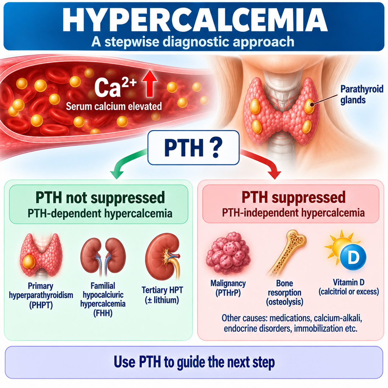
HIGH Ca → CHECK PTH. PTH not suppressed = PTH-dependent. PTH suppressed = PTH-independent. This is the single most important diagnostic decision in this article.
What Is Hypercalcemia? Why PTH Is the First Branching Test
Hypercalcemia means serum calcium is above the laboratory-specific reference range. Because reference intervals vary between laboratories, no single universal “normal calcium” value should be used as the sole definition; typical total calcium ranges around 8.5–10.5 mg/dL and ionized calcium around 1.16–1.31 mmol/L, but laboratory-specific ranges should always be used.
This article is the diagnostic map for the AtMedStu calcium/bone cluster — it does not repeat every detail of the linked articles. For symptoms, ECG changes and a general overview of hypercalcemia, see Hypercalcemia Explained. For normal calcium regulation, see Calcium Homeostasis Explained.
Normally: Ca²⁺ ↑ → CaSR activation ↑ → PTH secretion ↓. Therefore hypercalcemia should suppress PTH. If PTH remains elevated, high-normal or mid-normal when calcium is clearly elevated, it may be inappropriately non-suppressed, and the hypercalcemia is therefore PTH-dependent. PTH is the most useful initial test for separating PTH-dependent from PTH-independent hypercalcemia, and PHPT and malignancy together account for the large majority of cases.
Is the Calcium Really High?
Total Calcium vs Ionized Calcium
Serum calcium exists in three main forms: protein-bound calcium (mainly albumin-bound, roughly 45%), complexed calcium (bound to anions such as phosphate or citrate, roughly 10%), and ionized calcium — the biologically active fraction, roughly 45%. The key clinical principle is: total Ca is not the same as ionized Ca.
Why Albumin Matters
When albumin rises, total calcium may rise even though ionized calcium remains normal. Similarly, low albumin may lower measured total calcium without necessarily lowering ionized calcium proportionately. Total calcium must therefore be interpreted in context.
Dehydration or hemoconcentration can increase albumin concentration, producing high total calcium while ionized calcium is normal. This phenomenon is pseudo-hypercalcemia.
Albumin-Corrected Calcium
A common formula is corrected Ca = measured Ca + 0.8 × (4.0 − albumin in g/dL). However, do not teach this as perfect — albumin-correction algorithms may have poor accuracy. Albumin-adjusted calcium can help contextualize total calcium, but when the result is clinically discordant, protein abnormalities are substantial, or pH is significantly altered, direct ionized calcium measurement is preferable.
When Ionized Calcium Is Particularly Useful
Consider direct ionized calcium measurement when albumin is markedly abnormal, substantial protein abnormalities exist, acid-base disturbance is important, dehydration/hemoconcentration may distort total calcium, total calcium does not fit the clinical picture, or pseudo-hypercalcemia is suspected.
Why pH Matters
Hydrogen ions compete with calcium for albumin binding. In acidosis, albumin binds less calcium and ionized calcium tends to increase. In alkalosis, albumin binds more calcium and ionized calcium tends to decrease. Thus pH can alter ionized Ca without a proportional change in total calcium.
First Step: Confirm True Hypercalcemia
Before building a differential, repeat/confirm as appropriate. Assess total calcium, albumin, ionized calcium if needed, hydration, renal function, medication/supplement history and clinical context. Do not launch a malignancy work-up from one unexplained borderline total calcium value without confirming the abnormality.
The First Diagnostic Test: PTH
The next test after confirmation is intact PTH, interpreted against serum calcium — not simply compared with its laboratory reference interval. Ask: is PTH appropriately suppressed for this calcium?
| PTH Pattern | Interpretation |
|---|---|
| PTH elevated | PTH-dependent |
| PTH upper-normal / mid-normal during hypercalcemia | Potentially inappropriately normal → still PTH-dependent |
| PTH low / suppressed | PTH-independent |
This branch determines the rest of the diagnostic work-up.
PTH-Dependent Hypercalcemia
The main categories are primary hyperparathyroidism, familial hypocalciuric hypercalcemia, tertiary hyperparathyroidism, lithium-associated hypercalcemia, and less common genetic/syndromic or rare PTH-mediated causes. This section is a map, not a full re-derivation — see the linked articles for depth.
Primary Hyperparathyroidism
Typical biochemical relationship: Ca ↑ + PTH ↑ or inappropriately normal, often with PO₄ low/low-normal. Possible complications include nephrolithiasis, osteoporosis, fractures and renal impairment. See Primary Hyperparathyroidism Explained for PTH-dependent hypercalcemia due to PHPT in full.
Remember the one-line rule: high Ca should suppress PTH. Therefore high Ca + “normal” PTH may actually be inappropriately normal PTH — still PTH-dependent physiology.
Familial Hypocalciuric Hypercalcemia (FHH)
Typical clues: mild hypercalcemia, longstanding/lifelong pattern, family history, PTH normal/mildly high, relatively low urinary calcium, and a low CCCR often present — but CCCR is supportive, not absolute. See Familial Hypocalciuric Hypercalcemia Explained for distinguishing PHPT from FHH in full, and Primary Hyperparathyroidism Explained for the comparison table.
| Feature | PHPT | FHH |
|---|---|---|
| Hypercalcemia | Yes | Yes |
| PTH | ↑ / inappropriate normal | Normal/mild ↑ |
| Urinary Ca | Variable/often higher | Relatively low |
| CCCR | Often higher | Often low |
| Family history | Usually absent | Often present |
| Onset | Acquired | Lifelong |
| Stones/bone disease | More typical | Usually uncommon |
| Surgery | Can cure | Usually not corrective |
Tertiary Hyperparathyroidism
Tertiary hyperparathyroidism occurs when longstanding secondary hyperparathyroidism progresses to autonomous PTH secretion. Typical contexts include advanced CKD, longstanding renal secondary hyperparathyroidism, and selected post-transplant states. Biochemistry can show Ca ↑ + PTH markedly ↑; clinical context is essential. See CKD-MBD Explained for tertiary hyperparathyroidism and renal mineral disease in full.
Lithium and Thiazides
Lithium alters CaSR–PTH physiology and may produce Ca ↑ + PTH not suppressed. Lithium-associated hypercalcemia therefore belongs conceptually in the PTH-dependent branch rather than simply in a generic medication list.
Thiazides reduce renal calcium clearance. However, sustained hypercalcemia in an otherwise intact calcium-regulatory system is not expected in most patients — thiazide-associated hypercalcemia may instead reveal or “unmask” mild underlying PHPT. Do not automatically blame the thiazide if PTH remains inappropriate.
PTH-Independent Hypercalcemia
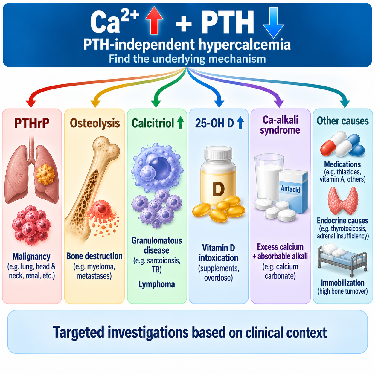
If PTH is suppressed, the parathyroids are responding appropriately — therefore another mechanism is driving calcium upward. This branch is organized by mechanism: PTHrP-mediated, osteolytic/cytokine-mediated bone resorption, calcitriol-mediated, vitamin D intoxication, calcium-alkali syndrome, medication-related, endocrine causes, immobilization/high bone turnover, and other rare disorders.
Malignancy-Associated Hypercalcemia
Malignancy is a major cause of PTH-suppressed hypercalcemia. Mechanisms include PTHrP, local osteolysis/cytokines, calcitriol production, and rare ectopic hormone mechanisms. Do not teach malignancy hypercalcemia = PTHrP only — several mechanisms exist.
Humoral Hypercalcemia of Malignancy
The classic mechanism: tumor → PTHrP ↑ → PTH1 receptor activation → bone resorption ↑ + renal Ca conservation ↑ + phosphate reabsorption ↓ → Ca ↑, PO₄ ↓, while PTH is suppressed. PTHrP produces many PTH-like effects through the shared receptor.
Commonly associated tumor groups include squamous-cell carcinomas (especially lung/head-neck and several other sites), renal cell carcinoma, bladder carcinoma, ovarian carcinoma, and selected breast and other cancers — this is not an exhaustive memorization list. The main teaching concept: PTHrP = humoral malignancy mechanism.
When to Measure PTHrP
Do not order PTHrP in every hypercalcemic patient. Consider it when Ca ↑ + PTH suppressed and malignancy is known, clinically suspected, or the pattern suggests humoral hypercalcemia. PTHrP is a second-line mechanism test, not the first test after calcium.
Osteolytic Hypercalcemia and Multiple Myeloma
Some cancers raise calcium through local bone destruction and osteoclast activation rather than primarily through circulating PTHrP. Examples include multiple myeloma and extensive skeletal metastases. Pattern: Ca ↑, PTH suppressed, and PTHrP may be normal.
When hypercalcemia is accompanied by anemia, renal dysfunction, bone pain, lytic lesions, or an elevated total protein/protein gap, consider plasma-cell disease. Appropriate tests may include, depending on context: serum protein electrophoresis, urine protein electrophoresis, serum free light chains, CBC, renal function, and skeletal imaging.
Calcitriol-Mediated Hypercalcemia
Pathway: 1,25-(OH)₂D ↑ → intestinal Ca absorption ↑ → Ca ↑ → PTH suppressed. Two important categories: granulomatous disease and lymphoma.
Granulomatous Disease
Activated macrophages in granulomas can express extra-renal 1α-hydroxylase, which converts 25-OH D to 1,25-(OH)₂D outside the usual renal regulatory system. This can raise calcitriol, increasing calcium absorption and causing hypercalcemia. Examples include sarcoidosis, tuberculosis, selected fungal infections and other granulomatous inflammatory disorders. Do not diagnose granulomatous disease from calcitriol alone — use clinical context.
Lymphoma
Some lymphomas can produce excess calcitriol or stimulate calcitriol production. Therefore Ca ↑ + PTH suppressed + 1,25-(OH)₂D ↑ should raise suspicion for lymphoma or granulomatous disease.
25-OH D vs 1,25-(OH)₂D
| Mechanism | 25-OH D | 1,25-(OH)₂D |
|---|---|---|
| Vitamin D intoxication | Markedly ↑ commonly | Variable |
| Granulomatous disease | Often normal/variable | ↑ |
| Lymphoma calcitriol mechanism | Variable | ↑ |
| Nutritional vitamin D deficiency | ↓ | Variable |
Memory rule: 25-OH D → stores/excess intake. 1,25-(OH)₂D → active hormone mechanism. See Vitamin D Metabolism Explained for 25-OH D and calcitriol physiology in full.
Vitamin D Intoxication
Excess vitamin D exposure can produce 25-OH D ↑ → intestinal calcium absorption ↑ → Ca ↑ → PTH suppressed. Causes can include excessive supplements, prescribing/dispensing errors, inappropriate high-dose use, and selected industrial/fortification errors. No universal toxicity threshold is given here.
25-OH D is the main test for vitamin D status and intoxication; 1,25-(OH)₂D is useful when a calcitriol-mediated mechanism is suspected. See Vitamin D Deficiency Explained for interpretation of vitamin D testing in full.

Calcium-Alkali Syndrome
Modern calcium-alkali syndrome commonly results from excessive intake of calcium plus absorbable alkali, often calcium carbonate-containing products. Classic biochemical triad: hypercalcemia + metabolic alkalosis + renal dysfunction.
The traditional term is milk-alkali syndrome, but calcium-alkali syndrome better reflects many modern cases involving calcium carbonate supplements or antacids.
Medication-Related Hypercalcemia
Potential medication-related contexts include thiazides, lithium, vitamin D, vitamin A, teriparatide, abaloparatide, selected SGLT2 inhibitor cases, denosumab withdrawal in certain settings, and other uncommon agents. These mechanisms differ substantially — do not put all drugs into one mechanism.
Vitamin A
Excess vitamin A can increase bone resorption and contribute to PTH-suppressed hypercalcemia. Ask about supplements, retinoid exposure and unusual high-dose use.
Teriparatide / Abaloparatide
Teriparatide can transiently raise serum calcium after administration; abaloparatide can also produce hypercalcemic effects, commonly transient. Do not diagnose PHPT from a poorly timed calcium sample without considering these drugs.
Denosumab Withdrawal
A rebound increase in osteoclastic activity after denosumab discontinuation can cause hypercalcemia, particularly described in children, high-turnover skeletal disease, and selected adults — an advanced medication pearl rather than a major diagnostic branch.
SGLT2 Inhibitors
Rare reversible hypercalcemia has been reported with SGLT2 inhibitors, often with additional risk factors such as dehydration, high calcium intake, thiazides or underlying PHPT. Do not present SGLT2 inhibitors as a common cause — this association is case-report level and often multifactorial.
Immobilization and Endocrine Causes
Immobilization
Prolonged immobilization can increase bone resorption, especially in individuals with high baseline bone turnover. Pattern: Ca ↑, PTH suppressed. More likely in younger patients, major skeletal disease, prolonged immobility, and high bone-turnover conditions.
Hyperthyroidism
Thyrotoxicosis can produce hypercalcemia through increased bone turnover, typically with PTH suppressed. Hypercalcemia is usually not the dominant presenting feature. Test thyroid function when clinical context suggests thyrotoxicosis, not as an automatic test in everyone.
Adrenal Insufficiency
Adrenal insufficiency can rarely cause PTH-independent hypercalcemia through complex mechanisms that may involve volume depletion, altered renal calcium handling and skeletal effects. Consider it only when compatible clinical features exist.
Other Endocrine Causes
Less common endocrine associations include pheochromocytoma, VIPoma, and acromegaly in selected contexts. These are not routine first-line targets — test based on clinical suspicion.
Which Second-Line Tests Should Be Ordered?
| Mechanism | Example | Key Clue |
|---|---|---|
| PTHrP | Squamous malignancy | PTH suppressed, PTHrP ↑ |
| Osteolysis | Myeloma/metastases | Bone lesions |
| Calcitriol ↑ | Sarcoid/lymphoma | 1,25D ↑ |
| Vitamin D excess | Supplement toxicity | 25-OH D ↑ |
| Calcium-alkali | Calcium carbonate | Alkalosis + renal dysfunction |
| Drug | Vitamin A etc. | Exposure history |
| Immobilization | Prolonged immobility | High bone turnover |
| Thyrotoxicosis | Hyperthyroidism | TSH suppressed |
| Adrenal insufficiency | Addisonian context | Clinical endocrine clues |
Do not order everything. Choose tests based on history + exam + mechanism. Potential targeted tests include PTHrP, 25-OH D, 1,25-(OH)₂D, CBC, renal function, SPEP/UPEP, serum free light chains, TSH, cortisol/adrenal testing and imaging. The article's central teaching is targeted second-line testing, not shotgun investigation.
When Each Test Makes Sense
PTHrP — order when PTH suppressed and malignancy/humoral mechanism is plausible; not when PTH is clearly elevated. 25-OH D — measure when supplement toxicity is possible, vitamin D status matters clinically, or unexplained suppressed-PTH hypercalcemia exists; a markedly elevated level supports vitamin D excess. 1,25-(OH)₂D — consider when PTH suppressed and lymphoma or granulomatous disease is suspected, or calcium is unexplained despite other testing; elevated calcitriol directs the investigation toward extra-renal calcitriol production. Myeloma studies (SPEP/UPEP/free light chains) — order when hypercalcemia is accompanied by anemia, renal dysfunction, bone pain, lytic lesions, recurrent infections or abnormal proteins, not universally.
Clinical History Checklist
Ask specifically about: supplements (calcium, vitamin D, vitamin A); drugs (thiazides, lithium, osteoporosis medications, other relevant agents); malignancy (known cancer, unexplained weight loss, bone pain, masses, constitutional symptoms); granulomatous disease (sarcoidosis, TB exposure, chronic respiratory/infectious disease); endocrine symptoms (thyrotoxicosis, adrenal insufficiency); and immobilization (prolonged bed rest, spinal injury, major neurological disease).
Complete Diagnostic Algorithm
Parathyroid ultrasound/sestamibi does not diagnose the cause of hypercalcemia. Do calcium → PTH → biochemical diagnosis before parathyroid localization, when surgery is appropriate.
Why Shotgun Testing Is Poor Practice
Do not order PTHrP, calcitriol, 25-OH D, SPEP, UPEP, CT chest/abdomen/pelvis, parathyroid imaging and endocrine panels simultaneously in every patient. Instead: PTH first, then mechanism-guided testing. This saves time, cost and false-positive investigations.
How Severe Is the Hypercalcemia?
Severity depends not only on how high the calcium is, but also how fast it rose. A patient with slowly developing chronic hypercalcemia may tolerate a level that would cause marked symptoms if reached rapidly. Acute and severe hypercalcemia is more symptomatic, while chronic mild disease may be relatively well tolerated.
Symptoms of Acute Hypercalcemia
Potential manifestations include anorexia, nausea, vomiting, polyuria, polydipsia, dehydration, weakness, confusion, reduced consciousness, renal impairment and cardiac conduction changes — including confusion progressing to stupor/coma and a shortened QT interval. Severe acute hypercalcemia can become a medical emergency.
Confusion/altered consciousness, severe dehydration, persistent vomiting, acute kidney injury, marked weakness, cardiac arrhythmia/ECG abnormality, or rapidly rising/markedly elevated calcium. Do not make one laboratory number the only determinant of urgency.
Practical Severity Ranges
Use cautiously and clearly as general clinical orientation: mild, often <12 mg/dL depending on laboratory and clinical context; more significant, approximately 12–14 mg/dL, often more symptomatic if acute; severe, >14 mg/dL, commonly associated with significant symptoms and generally requiring urgent management. Most patients are symptomatic above approximately 14 mg/dL, and acute therapy becomes increasingly important in the 12–14 mg/dL range depending on context. Do not use these numbers without considering symptoms, rate of rise and clinical condition.
Always assess calcium level, rate of rise, symptoms, renal function, volume status, ECG/cardiac status when indicated, and the underlying cause.
Acute Management Principles
This section is kept brief because a separate article should own acute treatment in depth. Core principles: restore volume when appropriate; stop contributing agents; reduce calcium when severe/symptomatic; treat the cause. No universal doses are given here.
Fluids — hypercalcemia often causes nephrogenic diuresis, polyuria and dehydration, so volume repletion with appropriate IV fluid is often foundational, but fluid choice and rate must account for cardiac function, renal function and volume status.
Calcitonin can produce rapid short-term calcium reduction and may be useful in severe hypercalcemia while slower therapies take effect, but tachyphylaxis develops — the Endocrine Society recommends limiting calcitonin in hypercalcemia of malignancy to roughly 48–72 hours.
For hypercalcemia of malignancy, IV bisphosphonate or denosumab are major antiresorptive therapies; the Endocrine Society recommends one of these for adults with hypercalcemia of malignancy and conditionally favors denosumab over IV bisphosphonate. This exact recommendation should not be generalized to every nonmalignant hypercalcemia.
Glucocorticoids are especially relevant when hypercalcemia is calcitriol-mediated, such as lymphoma or granulomatous disease — the Endocrine Society recommends glucocorticoids as first-line treatment for calcitriol-mediated hypercalcemia of malignancy, with antiresorptives added when necessary.
Dialysis may be considered in selected patients with severe refractory hypercalcemia, major renal impairment, inability to tolerate volume therapy, or life-threatening disease — reserved for severe refractory cases or patients with renal insufficiency.
Worked Clinical Cases
Case 1: Classic PHPT
Ca repeatedly ↑, PTH ↑, phosphate low, chronic mild course. Interpretation: PTH-dependent hypercalcemia, most likely PHPT after FHH/medication context is addressed.
Case 2: High Ca + Normal PTH
Calcium clearly elevated, PTH sits mid-reference range. Error: “PTH normal, therefore not parathyroid.” Correct reasoning: PTH should be suppressed. Therefore PTH is inappropriately normal → PTH-dependent hypercalcemia.
Case 3: FHH
A 25-year-old has lifelong mild hypercalcemia, PTH normal, several relatives with high calcium, CCCR low. Interpretation: FHH strongly considered. Do not send directly for parathyroidectomy.
Case 4: PTHrP Malignancy
Rapid severe hypercalcemia, PTH suppressed, weight loss, lung mass, PTHrP elevated. Interpretation: humoral hypercalcemia of malignancy.
Case 5: Multiple Myeloma
Ca ↑, PTH suppressed, bone pain, anemia, renal impairment, lytic lesions, monoclonal protein. Interpretation: osteolytic/myeloma-associated hypercalcemia — PTHrP need not be elevated.
Case 6: Granulomatous Disease
Ca ↑, PTH suppressed, 25-OH D not markedly elevated, 1,25-(OH)₂D ↑, sarcoid-like clinical picture. Interpretation: calcitriol-mediated hypercalcemia.
Case 7: Vitamin D Intoxication
Ca ↑, PTH suppressed, 25-OH D markedly elevated, history of high-dose vitamin D use. Interpretation: vitamin D excess.
Case 8: Calcium-Alkali Syndrome
Ca ↑, PTH suppressed, metabolic alkalosis, AKI, heavy calcium carbonate use. Interpretation: calcium-alkali syndrome.
Case 9: Thiazide + High PTH
Ca ↑, taking thiazide, PTH inappropriately high. Error: “thiazide explains everything.” Correct reasoning: thiazides can reduce renal calcium clearance but may also unmask PHPT — evaluate the PTH-dependent pathway.
Case 10: Immobilization
A young patient with prolonged immobilization develops Ca ↑, PTH suppressed, no vitamin D excess, no malignancy evidence. Interpretation: immobilization-related bone resorption may be responsible.
Case 11: Pseudo-Hypercalcemia
Total Ca mildly ↑, albumin elevated during marked dehydration, ionized calcium normal. Interpretation: pseudo-hypercalcemia. Do not launch a PHPT/malignancy work-up based solely on the total calcium value.
Case 12: Lymphoma
Ca ↑, PTH suppressed, PTHrP normal, 1,25-(OH)₂D ↑, lymphadenopathy. Interpretation: calcitriol-mediated malignancy such as lymphoma should be considered.
Common Diagnostic Mistakes
Hypercalcemia in One Minute
Golden Rules
- Confirm that the hypercalcemia is real before explaining it.
- Ionized calcium is the biologically active fraction.
- Albumin-correction formulas are useful but imperfect.
- Hypercalcemia should suppress PTH.
- PTH is the first major branching test.
- Normal-range PTH can be abnormal when calcium is high.
- PHPT and FHH are both PTH-dependent.
- Malignancy hypercalcemia is not always PTHrP-mediated.
- PTHrP is a second-line test, not the first test.
- 25-OH D and 1,25-(OH)₂D answer different questions.
- Elevated calcitriol suggests lymphoma or granulomatous disease in the right context.
- Calcium-alkali syndrome combines hypercalcemia, alkalosis and renal dysfunction.
- Medication history matters.
- Do not diagnose PHPT with imaging.
- Severity depends on calcium level, symptoms, rate of rise, renal function and cause.
- Use mechanism-guided testing rather than ordering every possible test.
Frequently Asked Questions
Key Take-Home Messages
The diagnostic approach to hypercalcemia should not begin with a long list of diseases. It should begin with: is the calcium really high? Assess repeat calcium, albumin, hydration, and ionized calcium when needed.
Once true hypercalcemia is established, measure PTH. If PTH is not suppressed, the hypercalcemia is PTH-dependent, and the main question becomes PHPT vs FHH vs other PTH-related physiology. If PTH is suppressed, the hypercalcemia is PTH-independent — investigate by mechanism: PTHrP for humoral malignancy when appropriate, bone destruction for myeloma or extensive skeletal malignancy, 1,25-(OH)₂D for calcitriol-mediated disease such as lymphoma or granulomatous disease, 25-OH D for vitamin D excess, and consider calcium-alkali syndrome, drugs, endocrine disease and immobilization based on the history and clinical picture.
Do not order every test in every patient. The correct reasoning is: calcium → PTH → mechanism → targeted testing. Finally, never assess severity from one number alone — consider level, rate of rise, symptoms, kidney function, volume status and cause.
The final memory rule: HIGH Ca → CONFIRM → PTH → DEPENDENT OR INDEPENDENT → TARGET THE MECHANISM.
Hypercalcemia becomes diagnostically manageable once it is divided by physiology. First confirm that the calcium elevation is real. Then ask whether PTH has responded appropriately. A non-suppressed PTH points toward a PTH-dependent disorder such as PHPT or FHH; a suppressed PTH directs the investigation toward malignancy, vitamin D-related mechanisms, calcium-alkali syndrome, drugs and other non-parathyroid causes.
This article is intended for medical education only. It explains a diagnostic reasoning framework for hypercalcemia, not patient-specific medical advice, treatment doses, emergency protocols or monitoring intervals.