Low calcium should make PTH rise
Hypoparathyroidism is insufficient PTH activity causing hypocalcemia, commonly with hyperphosphatemia and low or inappropriately normal PTH.
Low Ca + low/inappropriately normal PTH + high PO₄ → hypoparathyroid physiology. Confirm the pattern, check magnesium and investigate the cause.
Primary hyperparathyroidism
PTH excessive → Ca ↑ → PO₄ ↓
Hypoparathyroidism
PTH deficient → Ca ↓ → PO₄ ↑
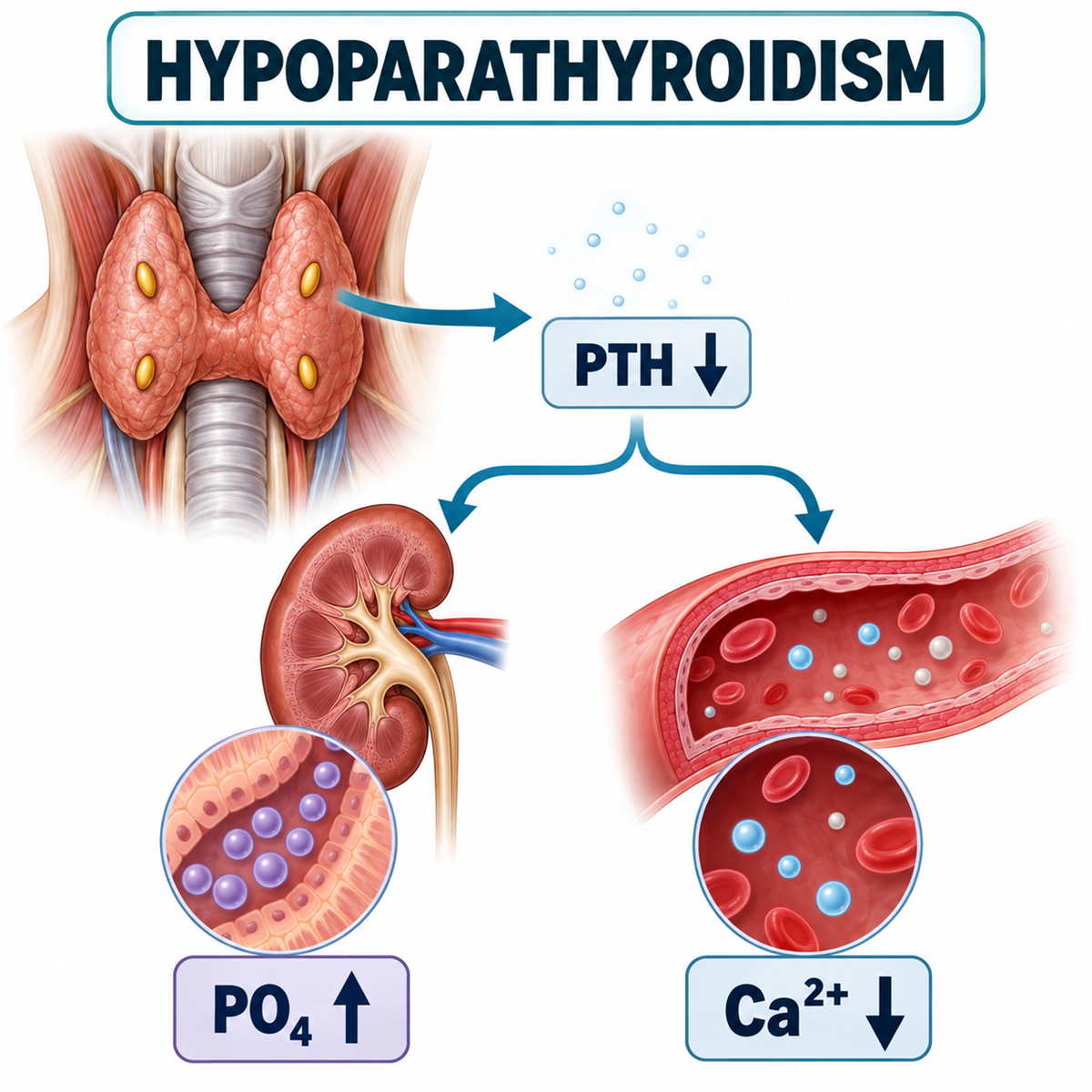
What Is Hypoparathyroidism?
Introduction
Hypoparathyroidism is a disorder in which insufficient biologically effective parathyroid hormone activity leads to impaired calcium homeostasis.
The classic biochemical pattern is:
HYPOCALCEMIAwith:
LOW OR INAPPROPRIATELY NORMAL PTHand commonly:
HYPERPHOSPHATEMIA.The key word is:
INAPPROPRIATELYbecause in hypocalcemia the normal physiological response is a substantial increase in PTH secretion.
Therefore a PTH result that lies inside the laboratory reference range can still be abnormal when serum calcium is low.
The 2025 ESE guideline recommends considering chronic hypoparathyroidism in patients with persistent hypocalcemia and PTH that is low or inappropriately normal. (OUP Academic)
Hypoparathyroidism at a glance
Calcium
↓PTH
↓ / inappropriately normalPhosphate
↑ commonlyCalcitriol
↓ / inappropriately lowUrinary calcium
Can be:
inappropriately high relative to serum calciumespecially during conventional treatment.
Major long-term problem
BALANCING SYMPTOM CONTROL AGAINST HYPERCALCIURIA / RENAL COMPLICATIONSHow PTH Controls Calcium, Phosphate and Calcitriol
Normal PTH physiology
Normally:
Ca²⁺ ↓CaSR activation ↓
PTH secretion ↑PTH then acts to restore extracellular calcium through:
Kidney
renal calcium reabsorption ↑Kidney
phosphate reabsorption ↓Kidney
1α-hydroxylase ↑ calcitriol ↑Intestine
indirectly:
calcium absorption ↑Bone
PTH-dependent calcium mobilization can increase.
What happens when PTH is absent?
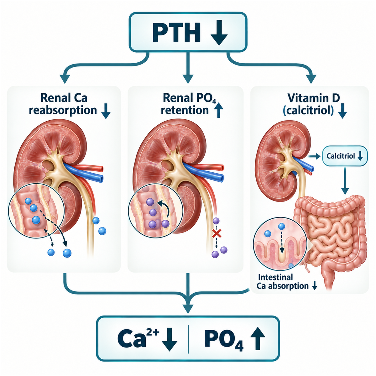
When PTH is deficient:
Kidney
calcium reabsorption ↓ urinary calcium loss tendency ↑Kidney
phosphate excretion ↓ phosphate retention ↑Kidney
1α-hydroxylase stimulation ↓ calcitriol ↓Intestine
calcium absorption ↓ SERUM Ca ↓Therefore:
PTH deficiency produces low calcium + high phosphateCore pathway
PTH ↓Kidney
Ca reabsorption ↓
PO₄ excretion ↓
calcitriol production ↓
Blood
Ca²⁺ ↓ PO₄ ↑Neuromuscular system
EXCITABILITY ↑symptoms of hypocalcemia.
Why phosphate rises
PTH normally promotes:
PHOSPHATURIAWithout PTH:
proximal tubular phosphate reabsorption ↑ urinary phosphate excretion ↓ serum phosphate ↑This is one of the most useful biochemical clues.
Memory rule:
PTH LOW → PO₄ HIGHwhile:
PTH HIGH → PO₄ LOWin many calcium disorders.
Why calcitriol falls
PTH stimulates renal:
1α-hydroxylaseWithout sufficient PTH:
calcitriol production fallsTherefore:
intestinal calcium absorption fallsThis is why chronic hypoparathyroidism is usually treated with:
ACTIVE VITAMIN Drather than relying only on ordinary vitamin D.
Can PTH Be Normal in Hypoparathyroidism?
Suppose:
Ca²⁺ is lowand:
PTH is mid-reference rangeA common error is:
“PTH is normal, therefore the parathyroids are functioning.”
Wrong.
The physiological question is:
WHAT SHOULD PTH BE DURING HYPOCALCEMIA?Answer:
HIGHTherefore:
LOW Ca + NORMAL-RANGE PTHmay represent:
INAPPROPRIATELY NORMAL PTHand is compatible with hypoparathyroidism. (OUP Academic)
Typical Laboratory Pattern and Diagnosis
Diagnostic pattern
| Test | Typical hypoparathyroidism |
|---|---|
| Serum calcium | ↓ |
| Ionized calcium | ↓ |
| PTH | ↓ or inappropriately normal |
| Phosphate | ↑ commonly |
| Magnesium | Variable |
| 25-OH D | Variable |
| 1,25-(OH)₂D | Low/inappropriately low |
| Creatinine/eGFR | Important for differential/monitoring |
| Urinary calcium | Variable; may be relatively high |
Confirm true hypocalcemia
Do not diagnose from one low total calcium result.
Assess:
- albumin-adjusted calcium;
- ionized calcium where needed;
- albumin;
- magnesium;
- renal function;
- acid-base context where relevant.
Endotext notes that ionized calcium can confirm true hypocalcemia when total calcium interpretation is uncertain. (NCBI)
First diagnostic branch
Once true hypocalcemia is established:
MEASURE PTHThen:
PTH elevated
The parathyroids are responding.
Think:
SECONDARY CAUSE / PTH RESISTANCEPTH low / inappropriate normal
Think:
HYPOPARATHYROIDISMComplete hypocalcemia–PTH algorithm
Ca²⁺ ↓ PTHPTH ↑
Think:
- vitamin D deficiency;
- CKD;
- malabsorption;
- hypocalcemia from phosphate excess;
- pseudohypoparathyroidism;
- other secondary causes.
PTH ↓ / inappropriate normal
CHECK MgSevere hypomagnesemia?
FUNCTIONAL HYPOPARATHYROIDISM POSSIBLEMagnesium acceptable
Review:
SURGERY AUTOIMMUNE GENETIC INFILTRATIVE / OTHER HYPOPARATHYROIDISMPostsurgical Hypoparathyroidism and the 12-Month Definition
Most common cause in adults
The most common cause of acquired hypoparathyroidism is:
NECK SURGERYincluding:
- thyroid surgery;
- parathyroid surgery;
- extensive neck surgery.
Endotext identifies surgical removal or injury of the parathyroid glands as the most common cause of primary hypoparathyroidism with hypocalcemia and hyperphosphatemia. (NCBI)
Postsurgical hypoparathyroidism
Mechanisms include:
- accidental gland removal;
- vascular compromise;
- direct injury;
- devascularization;
- postoperative edema;
- transient suppression/dysfunction.
Hypoparathyroidism after neck surgery can be:
TRANSIENTor:
CHRONICUpdated definition of chronic postsurgical hypoparathyroidism
This is an important current guideline update.
The 2025 revised ESE guideline defines postsurgical chronic hypoparathyroidism as:
PERSISTING MORE THAN 12 MONTHS AFTER SURGERYbecause recovery can occur later than previously appreciated. (OUP Academic)
The older six-month cutoff should not be used as the default definition of chronic postsurgical disease.
Why recovery can occur late
Residual parathyroid tissue may gradually recover.
Therefore:
PERSISTENT POSTOPERATIVE PTH DEFICIENCY DOES NOT ALWAYS MEAN IMMEDIATE PERMANENCEThe guideline even recommends periodic assessment of endogenous PTH function in chronic postsurgical disease because recovery can occasionally occur later. (OUP Academic)
Autoimmune, Genetic and Other Causes
Nonsurgical causes
Important categories include:
- autoimmune hypoparathyroidism;
- genetic hypoparathyroidism;
- syndromic disease;
- infiltrative/destructive disease;
- severe magnesium disturbance;
- rare radiation or iron/copper-related gland injury.
Autoimmune hypoparathyroidism
Autoimmune destruction of parathyroid tissue can occur:
- in isolation;
- as part of autoimmune polyglandular syndromes.
Clinical clues include:
- other autoimmune endocrine disease;
- candidiasis in APECED contexts;
- adrenal insufficiency;
- other autoimmune features.
Genetic hypoparathyroidism
Possible mechanisms include genes involving:
- PTH production;
- parathyroid development;
- calcium sensing;
- transcriptional regulation;
- syndromic chromosomal disorders.
Examples may include:
- activating CASR variants;
- GCM2;
- GNA11;
- 22q11.2 deletion;
- selected syndromic causes.
The 2025 ESE guideline recommends considering:
GENETIC TESTING / FAMILY SCREENINGin nonsurgical hypoparathyroidism without an obvious alternative cause. (OUP Academic)
Activating CaSR variants
This provides a useful contrast with FHH.
FHH
CaSR sensitivity ↓→ Ca ↑.
Activating CaSR disorders
CaSR sensitivity ↑PTH secretion suppressed at lower calcium
Ca ↓renal calcium wasting can increase.
Magnesium and Functional Hypoparathyroidism
Magnesium: critical diagnostic step
Magnesium is essential for:
- PTH secretion;
- PTH action.
Therefore severe:
HYPOMAGNESEMIAcan produce:
FUNCTIONAL HYPOPARATHYROIDISMEndotext lists magnesium depletion as a cause of functional hypoparathyroidism. (NCBI)
How low magnesium causes hypocalcemia
Severe magnesium deficiency can cause:
PTH secretion ↓and:
PTH resistance ↑Therefore:
Ca remains low despite treatmentuntil magnesium is corrected.
Memory rule:
REFRACTORY HYPOCALCEMIA → CHECK MAGNESIUMMagnesium excess
Severe hypermagnesemia can also suppress PTH secretion and contribute to hypocalcemia.
This is less common.
Hypoparathyroidism vs PTH Resistance, Vitamin D Deficiency and CKD
Pseudohypoparathyroidism
Pseudohypoparathyroidism is:
PTH RESISTANCEnot PTH deficiency.
Typical biochemical pattern:
Ca ↓ PO₄ ↑but:
PTH ↑This differentiates it from classical hypoparathyroidism.
Hypoparathyroidism vs pseudohypoparathyroidism
| Feature | Hypoparathyroidism | Pseudohypoparathyroidism |
|---|---|---|
| Calcium | ↓ | ↓ |
| Phosphate | ↑ | ↑ |
| PTH | ↓ / inappropriate normal | ↑ |
| Main defect | PTH deficiency | PTH resistance |
Memory rule:
LOW Ca + HIGH PO₄ → LOOK AT PTHVitamin D deficiency differential
Vitamin D deficiency also causes hypocalcemic physiology.
But typical response is:
PTH ↑because the parathyroid glands are functioning.
Therefore:
Vitamin D deficiency
Ca ↓/normal
PTH ↑
PO₄ often ↓ because PTH wastes phosphate
versus:
Hypoparathyroidism
Ca ↓
PTH ↓
PO₄ ↑
CKD differential
CKD may cause:
- calcitriol deficiency;
- phosphate retention;
- hypocalcemia;
- secondary hyperparathyroidism.
Thus:
PTH is usually elevatedrather than low.
If a CKD patient has:
low calcium + low PTHconsider:
- hypoparathyroidism;
- oversuppressed PTH;
- adynamic bone/mineral context;
- magnesium or medication issues.
Symptoms and Chronic Manifestations
Clinical symptoms of hypocalcemia
Symptoms depend on:
- degree of hypocalcemia;
- rate of fall;
- chronicity.
Possible manifestations include:
- perioral tingling;
- distal paresthesia;
- muscle cramps;
- carpopedal spasm;
- tetany;
- weakness;
- seizures;
- laryngospasm;
- bronchospasm;
- neuropsychiatric symptoms;
- QT prolongation.
The 2025 ESE guideline notes severe manifestations can include seizures, cardiac failure, bronchospasm and laryngospasm. (OUP Academic)
Chvostek and trousseau signs
Chvostek sign
Facial muscle contraction after tapping the facial nerve.
Trousseau sign
Carpal spasm induced by prolonged blood-pressure cuff inflation.
Neither sign is perfectly sensitive or specific.
Chronic manifestations
Longstanding hypoparathyroidism can be associated with:
- basal ganglia or intracranial calcification;
- cataracts;
- renal stones/nephrocalcinosis;
- reduced renal function;
- neurocognitive symptoms;
- impaired quality of life;
- abnormal bone remodeling.
These complications do not occur in every patient.
Why Kidney Complications Occur
Why renal complications occur
PTH normally:
promotes renal calcium conservationIn hypoparathyroidism this effect is lost.
Conventional treatment raises serum calcium using:
oral calcium + active vitamin Dbut does not fully restore PTH-mediated renal calcium conservation.
Therefore:
SERUM Ca may improvewhile:
URINARY Ca remains excessiveThis creates risk of:
- nephrolithiasis;
- nephrocalcinosis;
- renal dysfunction.
The fundamental treatment problem
THE GOAL IS NOT TO NORMALIZE CALCIUM AT ANY COSTWhy?
Because pushing serum calcium too high with conventional therapy can increase:
HYPERCALCIURIAand:
RENAL COMPLICATIONSThe 2025 ESE guideline recommends aiming for serum calcium in the:
LOWER PART OF THE REFERENCE RANGE OR SLIGHTLY BELOWwhile controlling symptoms and minimizing complications. (OUP Academic)
Chronic Treatment Goals and Who Should Receive Treatment
Treatment goals
According to the 2025 ESE guideline, management aims to:
- reduce hypocalcemic symptoms;
- improve quality of life;
- maintain calcium in low-normal/slightly below range;
- avoid hypercalcemia;
- keep phosphate controlled;
- normalize magnesium;
- maintain adequate vitamin D;
- avoid hypercalciuria;
- preserve renal function. (OUP Academic)
Who should receive treatment?
The 2025 ESE guideline recommends treating:
- patients with hypocalcemic symptoms;
and/or:
- adjusted calcium below 8.0 mg/dL (2.0 mmol/L), corresponding approximately to ionized calcium below 1.00 mmol/L.
It also suggests offering treatment to apparently asymptomatic patients between this threshold and the lower limit of normal to assess whether wellbeing improves. (OUP Academic)
These are guideline-based thresholds for chronic treatment, not an emergency dosing protocol.
Conventional chronic treatment
The conventional treatment framework is:
ACTIVE VITAMIN D ADEQUATE CALCIUM INTAKEwith:
CALCIUM SUPPLEMENTS WHEN REQUIREDplus:
MAGNESIUM / VITAMIN D OPTIMIZATIONand management of:
PHOSPHATE + URINARY CALCIUM
Active Vitamin D: Calcitriol and Alfacalcidol
Why active vitamin D is used
In hypoparathyroidism:
PTH ↓ renal 1α-hydroxylase stimulation ↓Therefore endogenous calcitriol production is impaired.
So treatment commonly uses:
CALCITRIOLor:
ALFACALCIDOLwhere available.
The 2025 ESE guideline recommends an activated vitamin D analogue when available. (OUP Academic)
Calcitriol
Calcitriol is:
1,25-(OH)₂Dthe active vitamin D hormone.
It increases:
intestinal calcium absorptionand helps maintain serum calcium.
Alfacalcidol
Alfacalcidol is:
1α-hydroxyvitamin DIt is converted in the liver to active vitamin D.
It does not require renal 1α-hydroxylation.
Availability varies by country.
Ordinary vitamin D still matters
Even if active vitamin D is used, patients should maintain adequate:
25-OH D STATUSThe revised ESE guideline suggests:
25-OH D >30 ng/mLas an adequate target. (OUP Academic)
This prevents concurrent nutritional vitamin D deficiency.
Calcium Intake, Supplements and Levothyroxine
Calcium intake
The revised ESE guideline suggests adult dietary elemental calcium intake around:
800–1000 mg/dayfor nonpregnant adults.
Supplements may be added if target calcium cannot be achieved with:
- active vitamin D;
- adequate dietary calcium.
If calcium supplementation exceeds about:
500 mg elemental calcium/daythe guideline suggests dividing it into smaller doses across the day. (OUP Academic)
Calcium carbonate vs calcium citrate
Calcium carbonate:
- requires gastric acid for optimal absorption;
- provides more elemental calcium per tablet.
Calcium citrate:
- is absorbed better in reduced gastric acidity;
- may be preferable after some bariatric procedures;
- may be useful with proton-pump inhibitor use/achlorhydria.
The revised guideline specifically notes calcium citrate may be preferable in patients after bariatric surgery because achlorhydria and malabsorption are common. (OUP Academic)
Important drug interaction
Calcium supplements can interfere with absorption of:
LEVOTHYROXINEThis matters because postsurgical hypothyroidism and hypoparathyroidism may coexist after thyroid surgery.
Do not take calcium supplements simultaneously with thyroid hormone replacement.
The ESE guideline specifically highlights this interaction. (OUP Academic)
Magnesium and Phosphate Management
Magnesium replacement
If magnesium is low:
CORRECT ITThe 2025 ESE guideline recommends measures to restore magnesium into the reference range. (OUP Academic)
This may be essential for:
- PTH secretion;
- PTH responsiveness;
- correction of hypocalcemia.
Phosphate management
The guideline recommends aiming for:
PHOSPHATE WITHIN THE REFERENCE RANGEbecause hyperphosphatemia can contribute to:
- calcium-phosphate precipitation;
- ectopic calcification concerns;
- treatment burden.
Management may include:
- dietary measures;
- adjustment of calcium treatment;
- adjustment of active vitamin D.
If persistent despite optimized conventional therapy, PTH replacement may be considered. (OUP Academic)
Calcium × phosphate concept
The main teaching concept is:
HIGH Ca + HIGH PO₄ INCREASES PRECIPITATION RISKTherefore avoid simultaneously overtreating calcium while phosphate remains high.
Hypercalciuria, Thiazides and Sodium Intake
Hypercalciuria
Conventional therapy can cause:
URINARY CALCIUM ↑The 2025 ESE guideline uses hypercalciuria values:
Men
>300 mg/24 hWomen
>250 mg/24 hor:
>4 mg/kg/24 hfor either sex. (OUP Academic)
Why urinary calcium matters
A patient may have:
serum calcium low-normalbut:
urinary calcium highTherefore serum calcium alone does not tell you whether treatment is safe.
Memory rule:
MONITOR BLOOD + URINEManagement of hypercalciuria
The revised ESE guideline suggests:
- reducing unnecessary calcium supplement burden;
- adjusting active vitamin D;
- reducing sodium intake;
- considering a thiazide;
- considering PTH replacement if hypercalciuria persists despite optimized conventional therapy. (OUP Academic)
Why thiazides help here
Thiazides:
reduce urinary calcium excretionThis can be useful in chronic hypoparathyroidism with problematic hypercalciuria.
This differs from their role in hypercalcemia diagnosis.
Sodium intake
High sodium intake increases urinary calcium excretion.
The 2025 ESE guideline suggests sodium restriction to:
<2.4 g sodium/dayequivalent to approximately:
<6 g salt/daywhen managing hypercalciuria. (OUP Academic)
Acute Symptomatic Hypocalcemia
Acute symptomatic hypocalcemia
Life-threatening manifestations can include:
- seizures;
- laryngospasm;
- bronchospasm;
- arrhythmia;
- heart failure;
- severe tetany.
These require:
URGENT IV CALCIUMand monitoring.
The revised ESE guideline includes IV calcium gluconate as preferred acute treatment in life-threatening hypocalcemia. (OUP Academic)
Acute calcium principle
IV CALCIUM GLUCONATE RAPID CORRECTION OF SYMPTOMATIC HYPOCALCEMIAfollowed by:
slower ongoing replacementas needed.
IV calcium is acute rescue treatment; ongoing chronic therapy follows the principles below.
Why calcium gluconate is commonly preferred
Compared with calcium chloride:
calcium gluconate is less damaging to peripheral tissue if extravasation occursCalcium chloride contains more elemental calcium but is more irritating and is generally reserved for selected monitored/central-access contexts.
Acute treatment must also correct magnesium
If severe hypomagnesemia is present:
CALCIUM MAY NOT CORRECT UNTIL Mg IS RESTOREDTherefore acute evaluation should include magnesium.
ECG
Hypocalcemia can prolong:
QT INTERVALSevere symptomatic cases may require ECG monitoring during IV calcium treatment.
PTH Replacement Therapy and Palopegteriparatide
PTH replacement therapy
The 2025 ESE guideline recommends:
PTH REPLACEMENT THERAPYfor chronic hypoparathyroidism when patients continue to have:
SIGNS OR SYMPTOMSdespite optimized conventional treatment with:
- active vitamin D;
- adequate calcium intake. (OUP Academic)
When to consider PTH replacement
The revised guideline suggests considering PTH replacement when one or more persist despite optimized conventional therapy:
- frequent calcium fluctuations;
- symptomatic hypocalcemia;
- impaired quality of life attributable to disease;
- eGFR <60 mL/min/1.73 m²;
- hypercalciuria;
- hyperphosphatemia. (OUP Academic)
This is a major update compared with older practice.
Why PTH replacement is physiologically attractive
Conventional treatment replaces:
CALCIUMand:
ACTIVE VITAMIN Dbut does not replace the hormone responsible for:
- renal calcium conservation;
- phosphate excretion;
- calcitriol regulation.
PTH replacement restores more of the missing physiology.
Therefore it can:
increase renal Ca conservation increase phosphate excretion reduce supplement burdenand improve biochemical stability.
Current PTH replacement landscape
The 2025 ESE guideline notes that older rhPTH(1-84) products were withdrawn from the market by the end of 2024.
It identifies:
PALOPEGTERIPARATIDEas an approved sustained PTH replacement option in jurisdictions including the EU and US at guideline publication. (OUP Academic)
Availability and reimbursement vary by jurisdiction and can change.
Palopegteriparatide
It is a long-acting prodrug that provides sustained release of:
PTH(1-34)The goal is more physiological PTH exposure across the day.
Clinical trial data have shown improvements in:
- serum calcium stability;
- urinary calcium;
- phosphate;
- supplement burden;
- some quality-of-life measures.
Long-term hard-outcome data remain limited. (OUP Academic)
Goal of PTH replacement
The revised ESE guideline suggests titrating PTH replacement toward:
stable calcium in target rangewhile reducing or eliminating:
active vitamin Dand:
calcium supplementswhen possible. (OUP Academic)
PTH replacement is not for every patient
A patient who is:
- asymptomatic;
- biochemically stable;
- with acceptable urinary calcium;
- preserved renal function;
- good quality of life;
may continue to do well with conventional treatment.
PTH REPLACEMENT = FOR INADEQUATELY CONTROLLED CHRONIC DISEASEnot automatic first-line replacement for everyone.
Long-Term Monitoring, Bone Health and Quality of Life
Monitoring
The 2025 ESE guideline recommends regular monitoring of:
- adjusted or ionized calcium;
- phosphate;
- magnesium;
- creatinine/eGFR;
- symptoms.
A typical suggested biochemical interval in stable chronic disease is:
every 3–6 monthswith individualized adjustment. (OUP Academic)
PTH monitoring
The revised guideline suggests:
PTH ABOUT ONCE YEARLYas appropriate,
particularly to assess for potential endogenous recovery in postsurgical disease. (OUP Academic)
24-hour urinary calcium
Urinary calcium should be assessed periodically and particularly when:
- treatment changes;
- stones/nephrocalcinosis suspected;
- renal function worsens;
- high supplement burden exists.
Renal monitoring
Assess:
- creatinine/eGFR;
- urinary calcium;
- history of nephrolithiasis;
- nephrocalcinosis;
- imaging when clinically indicated.
Long-term kidney complications are among the most important treatment-related concerns in chronic hypoparathyroidism.
Why bone density can be high
Chronic PTH deficiency reduces:
BONE TURNOVERTherefore BMD may be:
NORMAL OR HIGHThis does not necessarily mean bone quality is completely normal or that skeletal monitoring is irrelevant.
Do not equate high BMD with “excellent bone health.”
Quality of life
Patients with chronic hypoparathyroidism may have persistent:
- fatigue;
- cognitive symptoms;
- mood symptoms;
- neuromuscular symptoms;
- treatment burden;
even when calcium appears “acceptable.”
The 2025 ESE guideline explicitly includes:
QUALITY OF LIFEas a major management goal and as one potential indication for considering PTH replacement when impaired despite optimized treatment. (OUP Academic)
Complete Diagnostic Algorithm
PTH elevated
The parathyroid response is present.
- Vitamin D deficiency or malabsorption
- CKD or phosphate excess
- Pseudohypoparathyroidism/PTH resistance
- Other secondary causes
PTH low or inappropriately normal
Hypoparathyroid physiology → check magnesium.
Severely low or high magnesium: functional hypoparathyroidism is possible.
Magnesium acceptable: review neck surgery. Persistent disease more than 12 months after surgery supports chronic postsurgical hypoparathyroidism.
No surgical cause: assess autoimmune, genetic, infiltrative and other rare causes.
Interpret persistent biochemical findings with the clinical context. Urgent symptomatic hypocalcemia requires stabilization alongside investigation.
Complete Chronic Treatment Algorithm
- Assess symptoms, calcium, phosphate, magnesium and renal function.
- Use active vitamin D and adequate dietary calcium; add calcium supplements if required.
- During conventional treatment, target symptom control and low-normal or slightly low calcium; avoid overtreatment.
- Monitor urinary calcium, phosphate, magnesium and eGFR.
- Hypercalciuria: adjust calcium/active vitamin D, reduce sodium and consider a thiazide. Consider PTH replacement if the problem persists despite optimized treatment.
- Hyperphosphatemia: adjust diet and calcium/active vitamin D. Consider PTH replacement if persistent despite optimized treatment.
- Persistent symptoms, calcium fluctuations, disease-related poor quality of life or renal impairment: discuss PTH replacement with the endocrine team.
Worked Clinical Cases
Worked case 1: postsurgical hypoparathyroidism
Patient two days after total thyroidectomy:
- tingling;
- Ca ↓;
- phosphate ↑;
- PTH very low.
Interpretation
POSTSURGICAL HYPOPARATHYROIDISMLesson
The biochemical pattern is:
LOW Ca + LOW PTH + HIGH PO₄Case 2: “normal” PTH
Patient:
- Ca clearly low;
- PTH in lower-middle reference range.
Error
“PTH is normal.”
Correct reasoning
During hypocalcemia:
PTH SHOULD BE HIGHTherefore:
PTH IS INAPPROPRIATELY NORMALand supports hypoparathyroidism.
Case 3: vitamin D deficiency
Patient:
- Ca low-normal;
- phosphate low;
- PTH high;
- 25-OH D markedly low.
Interpretation
SECONDARY HYPERPARATHYROIDISMrather than primary hypoparathyroidism.
Case 4: pseudohypoparathyroidism
Patient:
- Ca ↓;
- phosphate ↑;
- PTH markedly ↑.
Interpretation
The parathyroids are responding.
Consider:
PTH RESISTANCErather than PTH deficiency.
Case 5: severe hypomagnesemia
Patient:
- Ca ↓;
- PTH low-normal;
- Mg profoundly ↓;
- calcium does not correct despite supplementation.
Interpretation
FUNCTIONAL HYPOPARATHYROIDISM FROM Mg DEFICIENCYLesson
CHECK Mg IN REFRACTORY HYPOCALCEMIACase 6: transient postsurgical disease
Patient remains on calcium/active vitamin D six months after thyroidectomy.
PTH begins to recover.
Lesson
Do not automatically label permanent disease too early.
Current ESE guidance uses:
>12 MONTHSto define chronic postsurgical hypoparathyroidism. (OUP Academic)
Case 7: hypercalciuria on conventional therapy
Patient:
- serum Ca low-normal;
- asymptomatic;
- 24-h urine calcium high;
- renal function preserved.
Error
“Serum calcium is fine, therefore treatment is perfect.”
Correct reasoning
URINARY CALCIUM MATTERSAdjust treatment to reduce renal risk.
Case 8: hyperphosphatemia
Patient:
- Ca low-normal;
- phosphate persistently high;
- taking high-dose active vitamin D/calcium.
Interpretation
Treatment is not optimally balancing mineral physiology.
Consider:
- dietary phosphate;
- treatment adjustment;
- PTH replacement in difficult persistent disease.
Case 9: poor QoL despite “normal” labs
Patient:
- calcium in target;
- phosphate acceptable;
- persistent fatigue/cognitive symptoms;
- high pill burden;
- frequent symptomatic fluctuations.
Interpretation
Biochemistry alone does not define good disease control.
This may be a scenario to discuss:
PTH REPLACEMENTwith an experienced endocrine team.
Case 10: CKD + hypercalciuria
Patient:
- chronic HypoPT;
- eGFR 48 mL/min/1.73 m²;
- persistent urinary calcium elevation despite optimized conventional therapy.
Interpretation
This is a guideline-supported context for considering:
PTH REPLACEMENT THERAPY (OUP Academic)Case 11: activating CaSR disorder
Young patient:
- lifelong hypocalcemia;
- low/inappropriate PTH;
- urinary calcium relatively high;
- family history.
Interpretation
Consider:
ACTIVATING CaSR-RELATED HYPOCALCEMIArather than acquired hypoparathyroidism.
Case 12: life-threatening hypocalcemia
Patient:
- severe symptomatic hypocalcemia;
- tetany;
- seizure;
- prolonged QT.
Management principle
URGENT IV CALCIUM ECG / ELECTROLYTE MONITORING CORRECT Mg IDENTIFY CAUSEDo not wait for oral therapy alone.
Common Mistakes
Mistake 1
Low calcium alone diagnoses hypoparathyroidism.
Wrong.
Mistake 2
Normal-range PTH excludes hypoparathyroidism.
Wrong.
Mistake 3
Low calcium should have normal PTH.
Wrong.
It should stimulate PTH.
Mistake 4
Phosphate should be low in hypoparathyroidism.
Wrong.
It is commonly high.
Mistake 5
Vitamin D deficiency and HypoPT have the same PTH pattern.
Wrong.
Mistake 6
Pseudohypoparathyroidism has low PTH.
Wrong.
PTH is high.
Mistake 7
Magnesium does not matter.
Wrong.
Mistake 8
Persistent postsurgical disease at 6 months is definitely permanent.
Outdated.
Current ESE guidance defines chronic postsurgical disease after >12 months. (OUP Academic)
Mistake 9
Treatment goal is high-normal calcium.
Wrong for most conventional treatment.
Mistake 10
Serum calcium alone determines treatment safety.
Wrong.
Mistake 11
Urinary calcium does not matter.
Wrong.
Mistake 12
Ordinary vitamin D replaces calcitriol physiology completely.
Wrong.
Mistake 13
Active vitamin D is unnecessary if 25-OH D is normal.
Wrong.
Mistake 14
Hyperphosphatemia is harmless.
Wrong.
Mistake 15
Thiazides always worsen hypoparathyroidism.
Wrong.
They may help reduce hypercalciuria.
Mistake 16
PTH replacement is experimental only.
Outdated.
Mistake 17
PTH replacement is mandatory for every patient.
Wrong.
Mistake 18
PTH replacement means calcium monitoring is unnecessary.
Wrong.
Mistake 19
High BMD means bone physiology is completely normal.
Wrong.
Mistake 20
Acute symptomatic hypocalcemia can be managed with oral calcium only.
Wrong.
Hypoparathyroidism in One Minute
One-minute revision
HYPOPARATHYROIDISM IN ONE MINUTE
Ca²⁺ ↓ PTH?PTH high
Think:
secondary HPT / PTH resistance
PTH low or inappropriate normal
HYPOPARATHYROIDISM PO₄ ↑Check:
Mg surgical history autoimmune/genetic causeChronic treatment:
ACTIVE VITAMIN D ADEQUATE CaTarget:
LOW-NORMAL CaMonitor:
URINE Ca + PO₄ + Mg + KIDNEYPoor control?
PTH REPLACEMENTGolden rules
Low calcium should increase PTH.
Therefore low calcium with normal-range PTH can still represent hypoparathyroidism.
Hypoparathyroidism classically causes low calcium and high phosphate.
PTH deficiency reduces renal calcium conservation.
PTH deficiency reduces phosphate excretion.
PTH deficiency reduces calcitriol production.
Magnesium deficiency can produce functional hypoparathyroidism.
Pseudohypoparathyroidism has high PTH, not low PTH.
Postsurgical hypoparathyroidism is the most common acquired adult cause.
Current ESE guidance defines chronic postsurgical disease as persisting >12 months.
The treatment goal is symptom control with calcium usually in the lower reference range or slightly below—not normalization at any cost.
Active vitamin D is central to conventional therapy.
Urinary calcium must be monitored.
Hyperphosphatemia matters.
Kidney complications are a major long-term concern.
PTH replacement is an important modern option when conventional treatment is inadequate.
Frequently Asked Questions
What is hypoparathyroidism?
What are the typical lab findings?
Can PTH be normal in hypoparathyroidism?
Why is phosphate high?
Why is calcitriol low?
What is the most common cause?
When is postsurgical hypoparathyroidism considered chronic?
Can magnesium deficiency cause hypoparathyroidism?
How is hypoparathyroidism different from pseudohypoparathyroidism?
What is the main chronic treatment?
What calcium level should be targeted?
Why not normalize calcium to high-normal?
What is the role of PTH replacement?
What is palopegteriparatide?
What should be monitored long term?
Key Take-Home Messages
Hypoparathyroidism becomes easier to recognize when PTH is interpreted against serum calcium.
Normally:
Ca²⁺ ↓should produce:
PTH ↑Therefore:
Ca²⁺ ↓ + PTH ↓or:
Ca²⁺ ↓ + PTH “NORMAL”is physiologically abnormal.
The next clue is:
PHOSPHATEBecause PTH normally promotes phosphate excretion:
PTH deficiency → PO₄ retention → PO₄ ↑At the same time:
renal calcium conservation ↓and:
calcitriol production ↓therefore:
SERUM CALCIUM FALLSThe diagnostic sequence is:
LOW Ca → CHECK PTH → IF LOW/INAPPROPRIATE, CHECK Mg + SURGICAL HISTORY + CAUSEThe most common adult cause is:
POSTSURGICAL HYPOPARATHYROIDISMbut severe magnesium deficiency, autoimmune disease and genetic disorders must also be considered.
Treatment should not aim to force serum calcium to the upper normal range.
Instead:
CONTROL SYMPTOMSwhile targeting:
LOW-NORMAL / SLIGHTLY LOW Caand preventing:
HYPERCALCIURIA HYPERPHOSPHATEMIA RENAL DAMAGEConventional therapy uses:
ACTIVE VITAMIN D + ADEQUATE CALCIUMBut when patients remain inadequately controlled despite optimized conventional therapy:
PTH REPLACEMENTcan restore more physiological calcium and phosphate handling.
The final memory rule is:
LOW Ca → LOW/INAPPROPRIATE PTH → HIGH PO₄ → HYPOPARATHYROIDISM → TREAT Ca/VITAMIN D BUT PROTECT THE KIDNEYSHypoparathyroidism is not simply a disease of low calcium. It is a loss of PTH physiology. The diagnosis depends on recognizing that PTH is inappropriately low during hypocalcemia, while successful long-term management requires more than correcting serum calcium—it requires balancing symptoms, phosphate, urinary calcium, renal health and, in selected patients, restoration of PTH itself.
This article explains diagnosis and treatment principles for adults. Individual treatment requires clinical assessment and local protocols. The guideline values shown are not universal prescriptions.